
Refractive surgery With easy explaination and diagrams For PG ophthalmology qith every detail Kanski dukes ak khurana
LASIK surgery flap diagram cornea
Refractive Surgery – Ophthalmology (Kanski / AK Khurana style)
Topic Breakdown
- Basic Principles – optics of ametropia, candidate selection criteria, preoperative workup (topography, pachymetry, keratoconus screening)
- Incisional Corneal Surgery – Radial Keratotomy (RK), Astigmatic Keratotomy (AK) — historical but PG-exam relevant
- Surface Ablation Techniques – PRK, LASEK, Epi-LASIK (technique + comparison table)
- LASIK – flap creation (microkeratome vs femtosecond laser), excimer ablation, step-by-step technique, nomogram basics
- SMILE (Small Incision Lenticule Extraction) – newest flapless technique, how it differs from LASIK
- Phakic Intraocular Lenses (ICL / PIOL) – for high myopia/hyperopia beyond laser range
- Refractive Lens Exchange (Clear Lens Extraction) – for presbyopia/high hyperopia
- Complications of Refractive Surgery – dry eye, DLK, flap striae/dislocation, infectious keratitis, ectasia, under/overcorrection, glare/halos
- Other/Adjunct procedures – Intacs (intrastromal corneal ring segments), conductive keratoplasty, presbyopia correction options
Quick Overview
Which part of Refractive Surgery do you want covered in full PG-exam detail first?
Refractive Surgery – Ophthalmology (Kanski / AK Khurana style)
Topic Breakdown
- Basic Principles – optics of ametropia, candidate selection criteria, preoperative workup (topography, pachymetry, keratoconus screening)
- Incisional Corneal Surgery – Radial Keratotomy (RK), Astigmatic Keratotomy (AK) — historical but PG-exam relevant
- Surface Ablation Techniques – PRK, LASEK, Epi-LASIK (technique + comparison table)
- LASIK – flap creation (microkeratome vs femtosecond laser), excimer ablation, step-by-step technique, nomogram basics
- SMILE (Small Incision Lenticule Extraction) – newest flapless technique, how it differs from LASIK
- Phakic Intraocular Lenses (ICL / PIOL) – for high myopia/hyperopia beyond laser range
- Refractive Lens Exchange (Clear Lens Extraction) – for presbyopia/high hyperopia
- Complications of Refractive Surgery – dry eye, DLK, flap striae/dislocation, infectious keratitis, ectasia, under/overcorrection, glare/halos
- Other/Adjunct procedures – Intacs (intrastromal corneal ring segments), conductive keratoplasty, presbyopia correction options
Quick Overview
You have access to a medical textbook library via query_library_db and read_file (and fetch_image), following the medical-library skill protocol (headings_fts and paragraphs_fts search, then read_file on block files with line offsets). TASK: Research "Refractive Surgery" topic for a postgraduate (PG) ophthalmology exam, specifically these subtopics, using Kanski's Clinical Ophthalmology (ISBN 9780443110993, chapter "Corneal and Refractive Surgery" - the REFRACTIVE PROCEDURES section appears after line ~4959 in block2.md, look for "Laser in situ keratomileusis" around line 5533), and any AK Khurana ophthalmology textbook or "Duke" ophthalmology textbook present in the library (search v_book_overview or headings_fts/paragraphs_fts for "Khurana" or ophthalmology speciality books), and Wills Eye Manual sections on refractive surgery complications found earlier (ISBN 9781975160753, block1.md around lines 6169-6478). Subtopics to cover in FULL DETAIL (a PG exam would need): 1. Basic principles of refractive surgery - optics of ametropia correction, goals, patient selection criteria, preoperative workup (corneal topography, pachymetry, screening for subclinical keratoconus, dry eye assessment, contact lens discontinuation) 2. Incisional corneal surgery: Radial Keratotomy (RK) and Astigmatic Keratotomy (AK) - principle, technique, indications (now largely historical), complications 3. Surface ablation techniques: PRK (Photorefractive Keratectomy), LASEK, Epi-LASIK - technique steps, differences between them, indications, advantages/disadvantages vs LASIK 4. LASIK (Laser In Situ Keratomileusis) - full step-by-step technique (flap creation via microkeratome vs femtosecond laser, excimer laser ablation of stromal bed, flap repositioning), indications, contraindications, advantages, and ALL complications (intraoperative: flap complications, buttonhole, free cap; postoperative: DLK/diffuse lamellar keratitis, epithelial ingrowth, striae, infectious keratitis, dry eye, ectasia, glare/halos, under/overcorrection) For each subtopic, extract as much specific textbook detail as possible (exact technique steps, specific numbers/parameters like ablation depth, flap thickness, residual stromal bed thresholds, complication rates/classification if given) with proper citations (book title + page number if available). Also use query_images_db or search relevant image captions for diagrams related to: LASIK flap, PRK, keratotomy, corneal ablation, refractive surgery complications like diffuse lamellar keratitis, corneal ectasia - and note down any useful image URLs with captions to include. Return a single detailed, well-organized markdown report covering all 4 subtopics above with citations and any relevant image URLs (markdown image syntax). Be thorough - this is for exam preparation, include as much authoritative detail as you found.
You have access to a medical textbook library via query_library_db and read_file (and fetch_image), following the medical-library skill protocol (headings_fts and paragraphs_fts search, then read_file on block files with line offsets). TASK: Research "Refractive Surgery" topic for a postgraduate (PG) ophthalmology exam, specifically these subtopics, using Kanski's Clinical Ophthalmology (ISBN 9780443110993, chapter "Corneal and Refractive Surgery" - look around line 5650 for "Small incision lenticule extraction (SMILE)" in block2.md, and continue reading further for phakic IOLs and lens-based procedures which likely follow), and any AK Khurana ophthalmology textbook or "Duke" ophthalmology textbook present in the library (search v_book_overview or headings_fts/paragraphs_fts for "Khurana" or ophthalmology speciality), and Wills Eye Manual sections on refractive surgery complications (ISBN 9781975160753, block1.md, search headings_fts for "phakic", "ectasia", "diffuse lamellar keratitis"). Subtopics to cover in FULL DETAIL (a PG exam would need): 1. SMILE (Small Incision Lenticule Extraction) - principle, technique steps, how it differs from LASIK (flapless), indications, advantages/disadvantages, complications 2. Phakic Intraocular Lenses (Phakic IOL / ICL - Implantable Collamer Lens) - types (anterior chamber angle-supported, iris-fixated, posterior chamber/ICL), indications (high myopia/hyperopia beyond laser correction range), preoperative requirements (anterior chamber depth, endothelial cell count), technique, complications (cataract, glaucoma, endothelial cell loss, pupillary block) 3. Refractive Lens Exchange (Clear Lens Extraction) - indications (high hyperopia, presbyopia, very high myopia unsuitable for other methods), technique, IOL choice considerations, risks (retinal detachment risk in high myopes) 4. Other/adjunct refractive procedures: Intacs (intrastromal corneal ring segments) for keratoconus, Conductive Keratoplasty, presbyopia-correcting procedures if mentioned 5. Overall/general complications of refractive surgery as a class - post-LASIK/PRK corneal ectasia (risk factors, detection via topography, percent tissue altered, Randleman criteria if present), diffuse lamellar keratitis (DLK) grading, infectious keratitis, dry eye syndrome, glare and halos, under/overcorrection and enhancement procedures, epithelial ingrowth For each subtopic extract specific textbook detail (technique steps, specific numeric parameters/thresholds, classification systems, complication rates) with citations (book title + page number). Also search for image captions/diagrams related to: phakic IOL, ICL implantable collamer lens, corneal ectasia, diffuse lamellar keratitis, Intacs corneal ring segments - note useful image URLs with captions. Return a single detailed, well-organized markdown report covering all 5 subtopics above with citations and relevant image URLs (markdown image syntax). Be thorough for exam preparation purposes.
Refractive Surgery – Ophthalmology (Kanski / AK Khurana style)
Topic Breakdown
- Basic Principles – optics of ametropia, candidate selection criteria, preoperative workup (topography, pachymetry, keratoconus screening)
- Incisional Corneal Surgery – Radial Keratotomy (RK), Astigmatic Keratotomy (AK) — historical but PG-exam relevant
- Surface Ablation Techniques – PRK, LASEK, Epi-LASIK (technique + comparison table)
- LASIK – flap creation (microkeratome vs femtosecond laser), excimer ablation, step-by-step technique, nomogram basics
- SMILE (Small Incision Lenticule Extraction) – newest flapless technique, how it differs from LASIK
- Phakic Intraocular Lenses (ICL / PIOL) – for high myopia/hyperopia beyond laser range
- Refractive Lens Exchange (Clear Lens Extraction) – for presbyopia/high hyperopia
- Complications of Refractive Surgery – dry eye, DLK, flap striae/dislocation, infectious keratitis, ectasia, under/overcorrection, glare/halos
- Other/Adjunct procedures – Intacs (intrastromal corneal ring segments), conductive keratoplasty, presbyopia correction options
Quick Overview
Which subtopic should I research in full exam-level detail first?
Which refractive surgery subtopic should I research in full exam-level detail first?
LASIK flap femtosecond laser cornea diagram
phakic intraocular lens implantable collamer lens diagram
corneal ectasia after LASIK topography
Refractive Surgery – PG Ophthalmology Notes (Kanski, Ch. 8)
1. Basic Concept & Why It Matters
- Unrealistic patient expectations
- Dry eye syndrome
- Unstable refractive error (still changing)
- Systemic disease: SLE, rheumatoid arthritis, immunocompromise
- Ocular disease: chronic blepharitis, prior HSV keratitis, glaucoma, macular degeneration
| Test | Why |
|---|---|
| Complete ocular exam | Baseline |
| Tear film osmolarity | Rule out dry eye |
| Corneal pachymetry | Residual stromal bed thickness, exclude mild keratoconus |
| Corneal topography | Exclude irregular astigmatism, keratoconus, forme fruste keratoconus (the #1 screen-out for ectasia risk) |
| Corneal OCT | Epithelial thickness map |
| Wavefront aberrometry | Corneal + whole-eye aberrations |
2. Techniques by Refractive Error (how Kanski organizes it)
A. Myopia
| Modality | Range/Notes |
|---|---|
| Surface ablation (PRK/LASEK) | Low-moderate myopia |
| LASIK | Moderate-high myopia (limited by corneal thickness) |
| SMILE | Myopia + myopic astigmatism |
| Clear lens exchange | Very high myopia; risk = retinal detachment |
| Phakic IOL (iris-claw "lobster claw", or posterior chamber ICL) | Very high myopia beyond laser range |
| Radial keratotomy (RK) | Historical only |
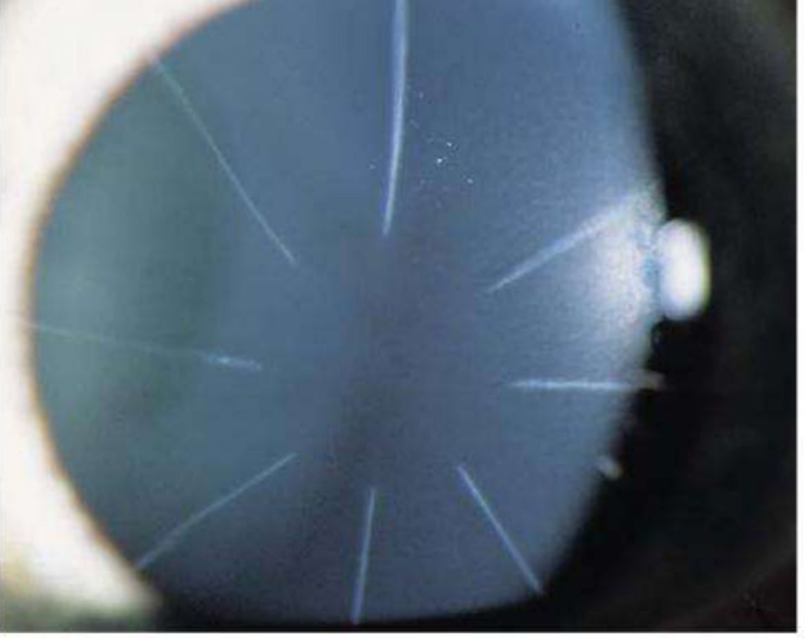
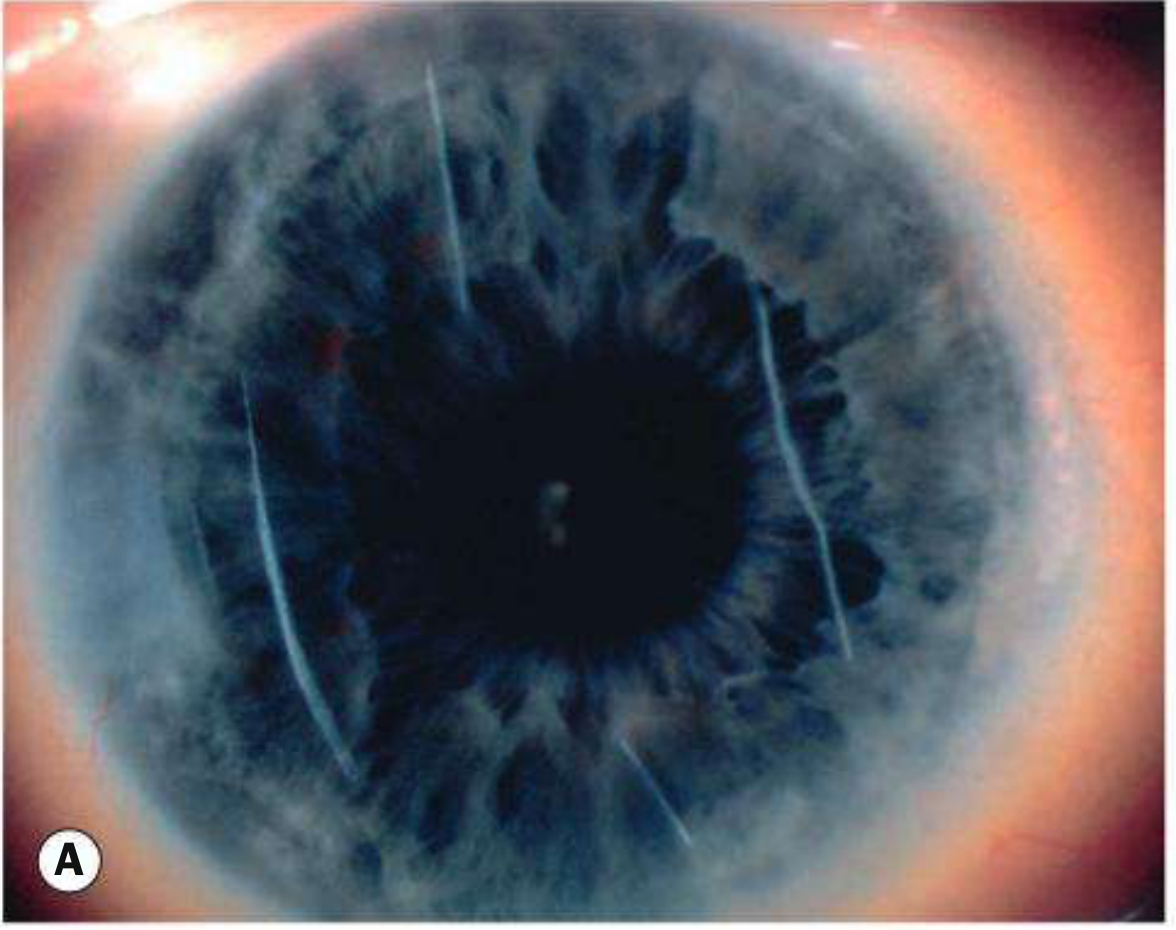
- Iris-claw ("lobster claw") implant: clipped onto iris at 3 and 9 o'clock. Complications: subluxation/dislocation, oval pupil, endothelial cell loss, cataract, pupillary-block glaucoma, retinal detachment.
- Posterior chamber phakic implant (ICL - Implantable Collamer Lens): sits behind iris, in front of natural lens, supported in ciliary sulcus; power range -3 D to -20.5 D. Complications: uveitis, pupillary block, endothelial cell loss, cataract, retinal detachment.

B. Hyperopia
- Surface ablation: low degrees only
- LASIK: up to +4 D
- Conductive keratoplasty (CK): radiofrequency energy applied via micro-needle probe in 1-2 peripheral rings → thermal stromal shrinkage → increased central corneal curvature. Disadvantages: early overcorrection, regression, induced astigmatism.
- Clear lens extraction / phakic implants for high hyperopia
C. Astigmatism
| Modality | Correction limit |
|---|---|
| Limbal relaxing incisions / arcuate keratotomy | Paired arcuate cuts in the steep meridian axis |
| PRK/LASEK | up to 3 D |
| LASIK | up to 5 D |
| Toric IOL | Any degree (lens surgery) - risk of postoperative rotation off-axis |
| Conductive keratoplasty | Adjunct |
D. Presbyopia (least satisfactory outcomes)
- Clear/refractive/presbyopic lens exchange (CLE/RLE/PreLEx) with multifocal, trifocal, EDOF, or accommodating IOLs
- Monovision: dominant eye set for distance, other eye for near - needs neuro-adaptation
- Conductive keratoplasty (can add mild multifocality)
- PresbyLASIK: alters corneal asphericity centre-to-periphery to create a multifocal corneal profile
- Intracorneal inlays (placed in a stromal pocket or under a flap) - complications include decentration, dry eye, keratolysis, vascularization, opacification, epithelial ingrowth
- Scleral expansion surgery - largely abandoned (inconsistent results)
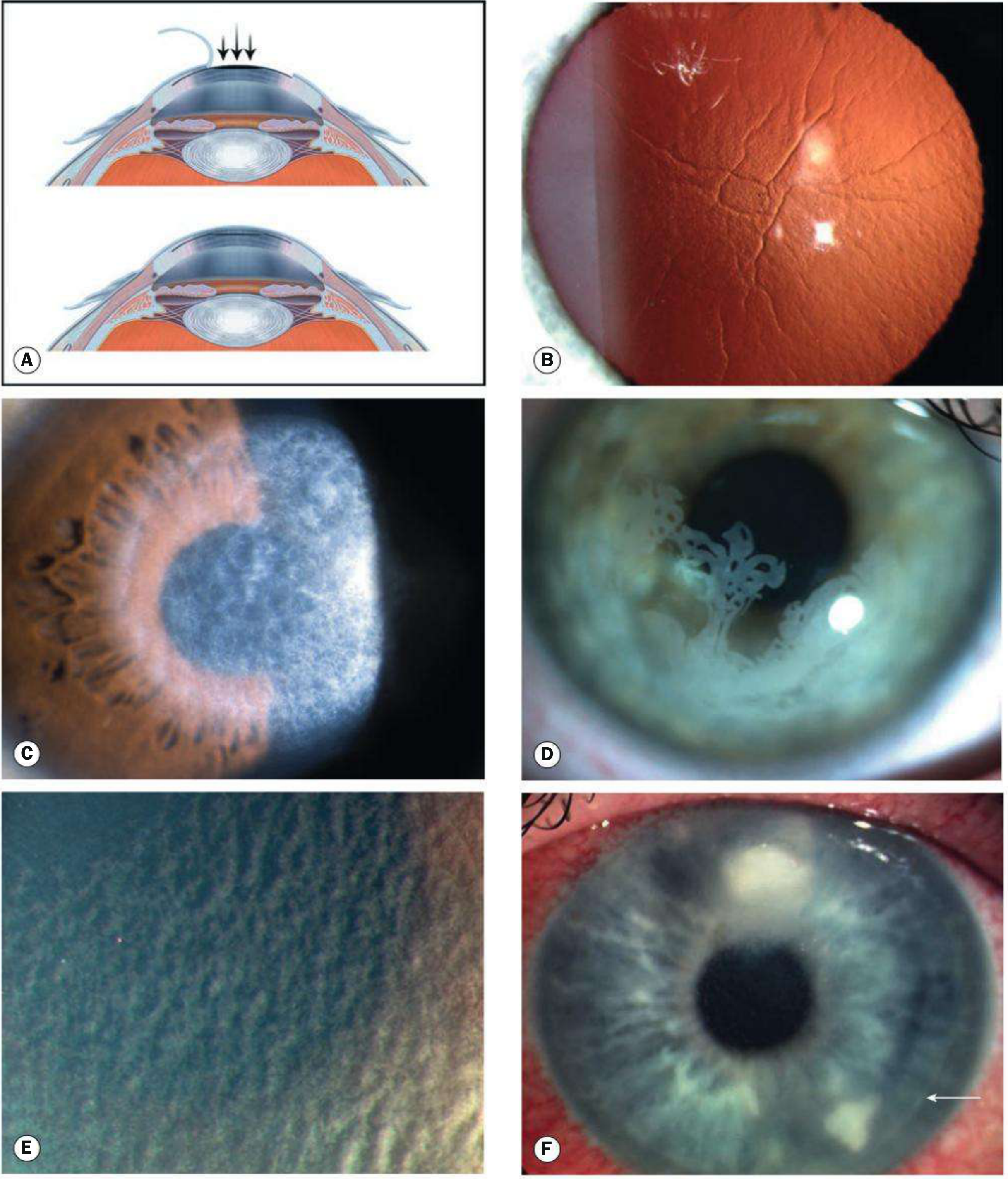
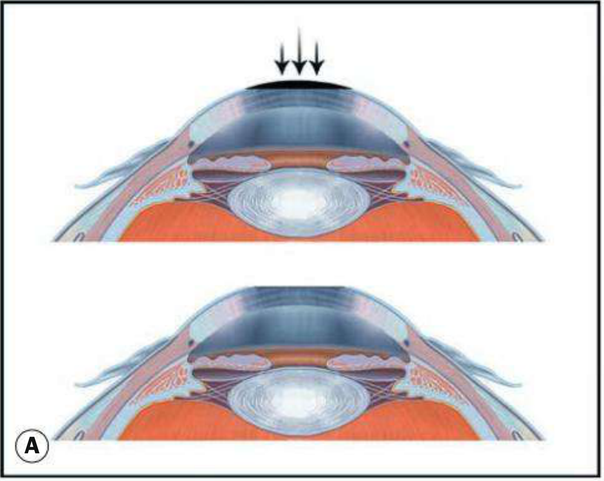
3. LASIK (Laser In Situ Keratomileusis) - The Core Topic
- Suction ring applied to globe → raises IOP substantially, stabilizes the eye, and guides the microkeratome (or centers the femtosecond laser).
- Flap created - either mechanically (microkeratome) or, now more commonly, with a femtosecond laser giving a precise uniform flap thickness of 100-120 μm.
- Flap reflected; excimer laser reshapes the exposed stromal bed.
- Flap repositioned (no sutures needed).
- Wavefront-guided LASIK: ablation pattern based on wavefront aberrometry measurements
- "Optimised" LASIK: larger optical zones/better asphericity to reduce glare
- LASIK Plus: combines with corneal cross-linking (riboflavin + UV light) in patients at risk of ectasia or with high myopic correction (>-7 D)
Complications - the exam-heavy part
- Buttonholing or amputation of the flap
- Incomplete/irregular flap creation
- Rare anterior chamber penetration (All much less likely with femtosecond flap creation)
- Flap decentration
- Wrinkling, distortion, or dislocation of the flap
- Interface debris
| Complication | Key facts |
|---|---|
| Under/overcorrection | Refractive stability takes up to 3 months |
| Dry eye/tear film instability | Commonest - up to 70%, usually self-resolving |
| Transient light sensitivity syndrome | 2-8 weeks post-op; 9% after hyperopic LASIK vs 5% myopic LASIK vs 1% SMILE; responds to topical steroids |
| Subepithelial haze | Glare, halos, starbursts at night; resolves in 3-6 months |
| Epithelial ingrowth under flap | 1-2%; treat by lifting flap, scraping cells, ± ethanol/mitomycin C |
| Diffuse lamellar keratitis (DLK, "sands of the Sahara") | Onset day 1-7; granular deposits at flap interface; treat with intensive topical antibiotic + steroid |
| Bacterial keratitis | Rare; culture-guided antibiotics |
| Epithelial hyperplasia | Causes regression; diagnosed on epithelial OCT map |
| Persistent epithelial defects | - |
| Pressure-induced stromal keratitis (PISK) | Fluid cleft at interface from elevated IOP (often steroid-induced); mimics undercorrection/regression |
| Corneal ectasia | Devastating but uncommon (0.2-0.6%). Risk factors: abnormal preop topography (forme fruste keratoconus, pellucid marginal degeneration), low preop corneal thickness, low residual stromal bed thickness, high percentage tissue altered, high myopia, young age |
4. Surface Ablation Procedures (PRK / LASEK / Epi-LASIK)
- Corrects myopia up to 6 D, astigmatism up to ~3 D, and low-moderate hyperopia
- Technique: epithelium removed first (sponge, Amoils automated brush, or alcohol) → excimer ablation of Bowman's layer + anterior stroma (30-60 sec, eye-tracking systems pause if decentred)
- Advantages over LASIK: no flap → lower risk of ectasia and late flap dislocation; better choice for thin corneas, borderline dry eye, epithelial basement membrane disease, prior corneal transplant/RK, large pupils, or high-risk occupations (contact sports)
- Disadvantages: lower correctable range, slower epithelial healing, more unpredictable postoperative discomfort
5. SMILE (Small Incision Lenticule Extraction)
- Uses a femtosecond laser (Carl Zeiss VisuMax) to cut a lens-shaped piece of stromal tissue (lenticule) within the intact cornea.
- Lenticule removed through a minimally invasive 4 mm incision.
- If removed via a LASIK-style flap instead → called ReLEx (refractive lenticule extraction).
- Results comparable to LASIK.
- Low risk of flap-related complications (no flap!)
- Biomechanical advantage (stronger residual cornea)
- Faster recovery of dry eye symptoms
- Quicker corneal reinnervation
- Minimal surface disturbance compared to surface ablation
- Only 1% incidence of transient light sensitivity syndrome (vs 5-9% for LASIK)
Quick Comparison Table (great for rapid revision)
| Feature | RK/AK | PRK/LASEK | LASIK | SMILE | Phakic IOL |
|---|---|---|---|---|---|
| Flap? | No (incisions) | No (epithelium removed) | Yes | No (lenticule via small incision) | N/A (intraocular) |
| Max correction | Historical | 6D myopia, 3D astig | 5D+ myopia (up to 4D hyperopia) | High myopia + astigmatism | Very high myopia/hyperopia |
| Ectasia risk | N/A | Lower | 0.2-0.6% | Very low | None (cornea untouched) |
| Recovery | - | Slower, more discomfort | Fast, minimal pain | Fast | Fast |
| Key complication | Diurnal fluctuation | Haze, discomfort | DLK, flap issues, ectasia | Rare | Endothelial loss, cataract, glaucoma |