
20 mark answer on craniofacial synostosis with flowchart and management
Reading File
Finding Sources
Finding Sources
Searching PubMed
craniosynostosis AND management
Searching the Web
craniosynostosis management guideline diagnosis surgery multidisciplinary care
Searching Images
craniosynostosis skull shapes sagittal coronal metopic lambdoid diagram
Reading File
Reading File
Reading File
Reading Web Page
https://pmc.ncbi.nlm.nih.gov/articles/PMC7769187
Craniofacial synostosis (Craniosynostosis)
Definition
Craniosynostosis is the premature fusion of one or more cranial sutures. It restricts skull growth perpendicular to the fused suture, while compensatory growth occurs in the remaining open sutures. This produces a characteristic abnormal head shape and, in severe cases, can cause raised intracranial pressure (ICP), visual impairment, neurodevelopmental problems, airway obstruction, and facial deformity.
The important sutures are sagittal, coronal, metopic, and lambdoid. Normal sutures permit skull expansion in response to rapid brain growth in the first two years of life. - Scott-Brown's Otorhinolaryngology Head & Neck Surgery, p. 1948
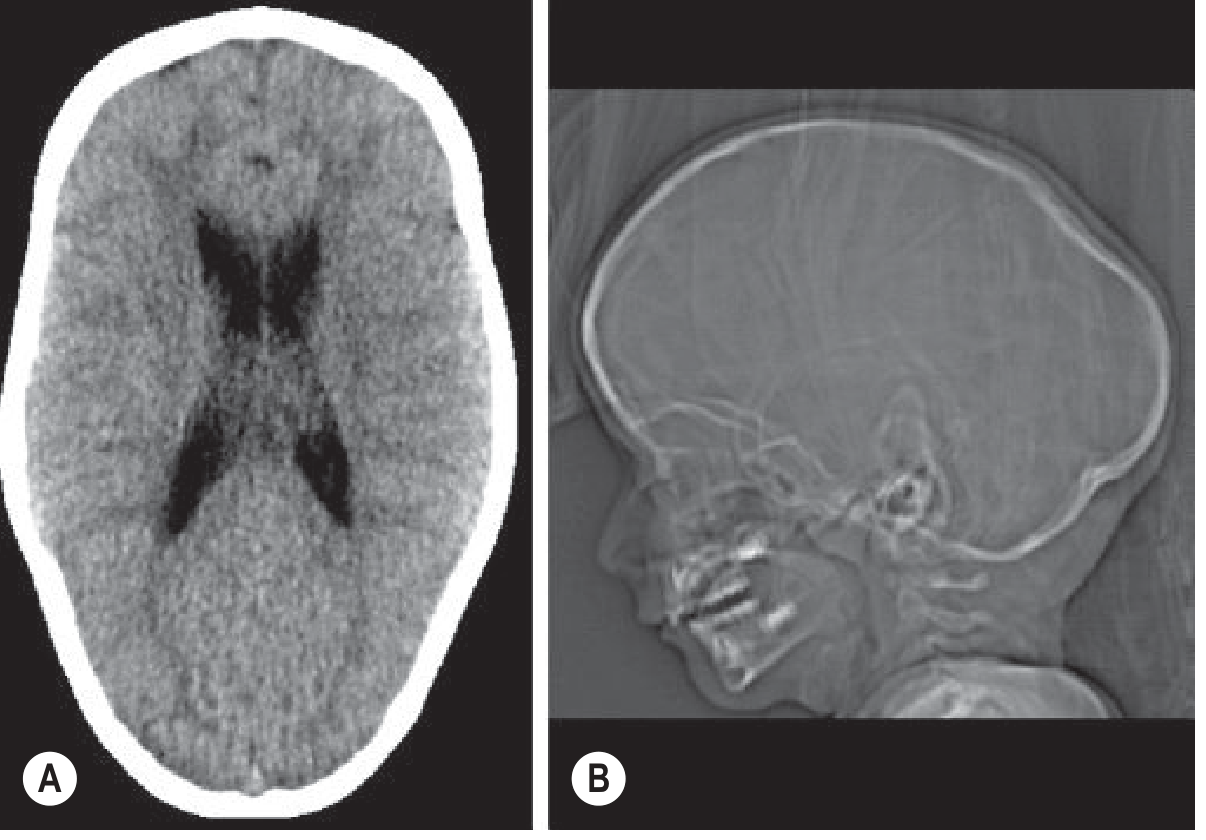
Sagittal synostosis with the typical elongated, boat-shaped skull (scaphocephaly). - Grainger & Allison's Diagnostic Radiology, p. 2001
Classification
1. Primary craniosynostosis
Due to intrinsic premature suture fusion.
A. Non-syndromic/simple
- Usually involves a single suture.
- Most common: sagittal synostosis.
B. Syndromic/complex
- Multiple sutures may be involved.
- Often associated with mutations involving fibroblast growth factor receptor pathways.
- Common syndromes:
- Crouzon syndrome
- Apert syndrome
- Pfeiffer syndrome
- Muenke syndrome
- Saethre-Chotzen syndrome
2. Secondary craniosynostosis
Occurs because of impaired brain growth or another systemic disorder, for example:
- Microcephaly
- Metabolic bone disease
- Haematological disorders
- Drug exposure
- Treated hydrocephalus or other causes of reduced brain growth
Primary and secondary forms must be distinguished because management differs. - Grainger & Allison's Diagnostic Radiology, p. 2000
Types and characteristic deformities
| Fused suture | Deformity | Typical clinical appearance |
|---|---|---|
| Sagittal | Scaphocephaly | Long, narrow, boat-shaped skull; frontal and occipital bossing |
| Metopic | Trigonocephaly | Triangular forehead, midline metopic ridge, hypotelorism |
| Unilateral coronal | Anterior plagiocephaly | Asymmetrical forehead, ipsilateral orbital elevation, facial asymmetry |
| Bilateral coronal | Brachycephaly | Short, broad skull with flattened forehead |
| Unilateral lambdoid | Posterior plagiocephaly | Posterior skull asymmetry; rare |
| Multiple sutures | Turribrachycephaly or cloverleaf skull | Severe cranial deformity, often syndromic; high risk of raised ICP |
Sagittal synostosis produces scaphocephaly; metopic synostosis produces trigonocephaly; and unilateral coronal fusion produces anterior plagiocephaly. - Bailey and Love's Short Practice of Surgery, p. 741
Clinical features
Local craniofacial findings
- Abnormal skull shape from birth or progressing in infancy
- Palpable hard ridge over a fused suture
- Absent or early closure of fontanelle
- Frontal, parietal, or occipital bossing
- Facial asymmetry
- Orbital dystopia, proptosis, hypertelorism or hypotelorism
- Midface hypoplasia in syndromic cases
Features of raised ICP
- Irritability, persistent vomiting, headache in older children
- Bulging fontanelle may occur, although it can be absent
- Papilloedema
- Visual deterioration or optic atrophy
- Developmental delay
- Sleep disturbance
Associated abnormalities in syndromic disease
- Airway obstruction and obstructive sleep apnoea
- Hearing loss and recurrent otitis media
- Hydrocephalus or Chiari malformation
- Dental malocclusion
- Limb anomalies, especially syndactyly in Apert syndrome
- Developmental and behavioural difficulties
Differential diagnosis
The major differential is deformational or positional plagiocephaly, which is much more common.
| Craniosynostosis | Positional plagiocephaly |
|---|---|
| Suture may be ridged or fused | Sutures are patent |
| Skull deformity follows a suture pattern | Flattening due to external pressure or preferred sleeping position |
| May worsen despite repositioning | Often improves with repositioning and physiotherapy |
| May need surgery | Usually managed conservatively |
| Associated orbital and facial asymmetry can occur | Usually no severe orbital deformity |
True unilateral lambdoid synostosis is uncommon and must be differentiated from positional plagiocephaly. - Grainger & Allison's Diagnostic Radiology, p. 2001
Diagnostic and management flowchart
Infant with abnormal head shape / facial asymmetry
                    │
                    ▼
Detailed history and examination
- Onset at birth, progression, sleeping position
- Head circumference and developmental assessment
- Palpate sutures/fontanelles
- Look for syndromic features, airway and eye problems
                    │
                    ▼
Is craniosynostosis clinically suspected?
          │                            │
         No                           Yes
          │                            │
          ▼                            ▼
Consider positional              Urgent referral to a
plagiocephaly, torticollis,      multidisciplinary craniofacial team
or other causes                         │
                                       ▼
                         Ophthalmology + genetics assessment
                         Hearing, airway and developmental review
                                       │
                                       ▼
                     Imaging when required for confirmation/
                     operative planning:
                     low-dose CT with 3-D reconstruction
                     ± ultrasound / MRI for brain, hydrocephalus,
                     Chiari malformation or venous anatomy
                                       │
                                       ▼
          ┌─────────────────────────────────────────────┐
          │ Single-suture, mild, no functional problem   │
          │ versus multisuture/syndromic/raised ICP      │
          └─────────────────────────────────────────────┘
                     │                         │
                     ▼                         ▼
          Individual surveillance        Early cranial vault expansion/
          or planned correction          remodelling as indicated
                     │                         │
                     └───────────────┬─────────┘
                                     ▼
                         Long-term multidisciplinary follow-up:
                         head growth, skull shape, vision/ICP,
                         development, hearing, airway, dentition
Management
1. Principles
Management should occur in a specialist multidisciplinary craniofacial centre, involving paediatric neurosurgery, craniofacial or plastic surgery, maxillofacial surgery, paediatrics, anaesthesia, ophthalmology, ENT/audiology, genetics, speech therapy, psychology, orthodontics, and specialist nursing. - Scott-Brown's Otorhinolaryngology Head & Neck Surgery, p. 1948
The aims are to:
- Allow adequate intracranial volume and prevent or treat raised ICP.
- Correct cranial and facial deformity.
- Preserve vision and manage orbital complications.
- Treat airway, hearing, dental, developmental, and psychosocial problems.
- Provide genetic diagnosis and family counselling.
Current guidance also recommends coordinated multidisciplinary care, because visual, airway, hearing, developmental, and surgical issues may coexist, as outlined in this management guideline.
2. Initial assessment and investigations
- Document head circumference, skull shape, growth, and developmental status.
- Photograph or use three-dimensional surface imaging where available.
- Ophthalmological assessment for papilloedema, optic atrophy, strabismus, and exposure keratopathy.
- ENT/audiology assessment, particularly in syndromic children.
- Assess feeding, airway obstruction, and obstructive sleep apnoea. Polysomnography is useful if sleep-disordered breathing is suspected.
- Clinical geneticist review and molecular testing in multisuture disease, syndromic features, or positive family history.
- Low-dose CT with three-dimensional reconstruction is used selectively to confirm suture fusion and plan surgery.
- MRI brain and cervicomedullary junction when hydrocephalus, Chiari malformation, intracranial anomaly, or venous abnormality is suspected.
Diagnosis begins with clinical assessment of skull shape; imaging confirms suture involvement and evaluates the skull base, orbits, hydrocephalus, and visual complications. - Grainger & Allison's Diagnostic Radiology, p. 2000
3. Surgical management
Indications
- Significant cranial deformity
- Raised ICP or restricted intracranial volume
- Progressive deformity
- Threat to visual function
- Multisuture or syndromic craniosynostosis
- Severe orbital, facial, or airway consequences
A. Endoscopic strip craniectomy
- Minimally invasive removal of the fused suture.
- Best suited to selected infants presenting early, often in the first few months of life.
- Usually followed by postoperative moulding helmet therapy.
- Commonly considered for early sagittal, metopic, and selected unilateral coronal/lambdoid cases.
B. Open cranial vault remodelling
- Open strip craniectomy with cranial vault expansion and reshaping.
- Used when presentation is later, deformity is severe, multiple sutures are involved, or an endoscopic approach is unsuitable.
- May include fronto-orbital advancement for coronal or metopic deformity.
- Usually performed during infancy, with timing tailored to the affected suture, severity, surgical technique, and centre protocol.
C. Procedures for syndromic disease
May require staged procedures:
- Posterior cranial vault expansion
- Fronto-orbital advancement
- Midface advancement, for example Le Fort III or monobloc advancement
- Management of hydrocephalus, Chiari malformation, and severe proptosis
- Tracheostomy or airway surgery in severe obstructive sleep apnoea
The objective of surgery is correction of deformity and prevention of raised ICP. - Bailey and Love's Short Practice of Surgery, p. 741
4. Non-surgical and supportive treatment
- Reassure and educate parents.
- Treat positional plagiocephaly with repositioning, physiotherapy for torticollis, and selected helmet therapy, not cranial surgery.
- Nutritional and feeding support where needed.
- Hearing aids or ventilation tubes for recurrent middle-ear disease.
- Continuous positive airway pressure or airway surgery for obstructive sleep apnoea when indicated.
- Speech, language, occupational, developmental, and psychological support.
- Orthodontic treatment and dental surveillance.
5. Follow-up
Long-term follow-up is essential, particularly in syndromic or multisuture disease.
Monitor:
- Head circumference and skull shape
- Developmental progress and school performance
- Headache, vomiting, irritability, and sleep symptoms
- Ophthalmic examination for papilloedema and optic nerve changes
- Hearing and middle-ear disease
- Airway and sleep apnoea
- Dental development and malocclusion
- Need for repeat cranial or facial surgery
Annual ophthalmological review is commonly used to detect signs of elevated ICP, particularly in children with syndromic craniosynostosis, as described in this multidisciplinary follow-up review.
Complications
- Raised ICP and visual loss
- Hydrocephalus
- Chiari malformation
- Venous sinus abnormalities
- Neurodevelopmental and learning difficulties
- Obstructive sleep apnoea
- Hearing impairment
- Persistent or recurrent cranial deformity
- Blood loss, infection, dural tear, and need for reoperation after surgery
Conclusion
Craniofacial synostosis is premature fusion of cranial sutures causing characteristic skull deformity and possible functional complications. Early recognition, differentiation from positional plagiocephaly, referral to a multidisciplinary craniofacial team, appropriately timed cranial expansion or remodelling, and long-term surveillance of vision, ICP, airway, hearing, and development are the foundations of management.
This is a shared conversation. Sign in to Orris to start your own chat.