
Management of IBD according to new guidelines of ACG or from slesingers
"inflammatory bowel disease" AND "guidelines" AND "management"
ACG guidelines IBD management 2024 2025 Crohn's ulcerative colitis
https://mediacdn.gi.org/giorg/wp-content/uploads/2025/09/170…
PMID: 40701556
IBD inflammatory bowel disease treatment algorithm step-up therapy
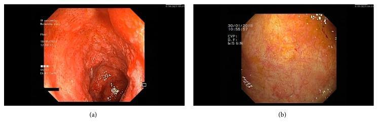
This comparison chart features two endoscopic images of the ileal mucosa, illustrating the visual progression of inflammatory bowel disease (IBD) treatment. Image (a) depicts the initial ileoscopy, showing severe mucosal inflammation characterized by intense erythema, edema, and a friable texture. Spontaneous bleeding and loss of normal vascular patterns are evident, consistent with active pan-enteritis or ulcerative colitis. In contrast, image (b) shows a follow-up ileoscopy after five months of vedolizumab therapy. This image demonstrates significant endoscopic healing, with the mucosa appearing healthy and pale pink with visible, normal vascular architecture and an absence of friability, edema, or spontaneous bleeding. These diagnostic images serve as an educational comparison of active disease versus therapeutic remission in a clinical gastroenterology context, highlighting the efficacy of biologic therapy in restoring mucosal integrity.
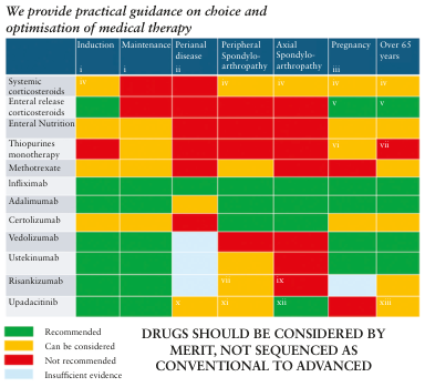
Summary : This figure provides a color-coded practical guidance matrix for the choice and optimization of medical therapy in various clinical scenarios, specifically for inflammatory bowel disease (IBD) and related conditions. It categorizes different drugs and interventions by their appropriateness (recommended, can be considered, not recommended, insufficient evidence) across treatment phases and patient groups. heat-map: Title & Axes : • No explicit title, but the guidance is for "choice and optimisation of medical therapy" in IBD. • Columns (top, left to right): Induction, Maintenance, Perianal disease, Peripheral Spondyloarthropathy, Axial Spondyloarthropathy, Pregnancy, Over 65 years. • Rows (left, top to bottom): Systemic corticosteroids, Enteral release corticosteroids, Enteral Nutrition, Thiopurines monotherapy, Methotrexate, Infliximab, Adalimumab, Certolizumab, Vedolizumab, Ustekinumab, Risankizumab, Upadacitinib. Legend & Colour Coding : • Green: Recommended • Yellow: Can be considered • Red: Not recommended • Blue: Insufficient evidence Data Matrix (Selected Examples) : • Systemic corticosteroids: Green for Induction, Red for Maintenance, Red for Perianal disease, Yellow for all other columns. • Enteral release corticosteroids: Green for Induction, Red for all other columns. • Enteral Nutrition: Green for Induction, Red for all other columns. • Thiopurines monotherapy: Yellow for Induction, Green for Maintenance, Yellow for Perianal disease, Red for all other columns. • Methotrexate: Yellow for Induction, Green for Maintenance, Yellow for Perianal disease, Red for all other columns. • Infliximab, Adalimumab, Certolizumab: Green for Induction, Maintenance, Perianal disease; Red or Yellow for other columns. • Vedolizumab, Ustekinumab, Risankizumab, Upadacitinib: Green for Induction and Maintenance, with variable recommendations for other columns. • Several cells contain Roman numerals (i–xiii), likely referencing footnotes or specific clinical considerations. Design Encodings : • Each cell is color-coded according to the recommendation level. • Roman numerals in some cells indicate special notes or exceptions. • A note at the bottom: "DRUGS SHOULD BE CONSIDERED BY MERIT, NOT SEQUENCED AS CONVENTIONAL TO ADVANCED." Analysis : • Systemic corticosteroids are recommended only for induction, not for maintenance or special populations. • Most biologics (Infliximab, Adalimumab, Vedolizumab, Ustekinumab) are recommended for both induction and maintenance, but their use in pregnancy, elderly, and spondyloarthropathy is more nuanced. • Enteral release corticosteroids and enteral nutrition are only recommended for induction. • There is insufficient evidence for some drugs in certain scenarios (blue cells). • The matrix emphasizes individualized therapy selection rather than a stepwise escalation from conventional to advanced drugs.
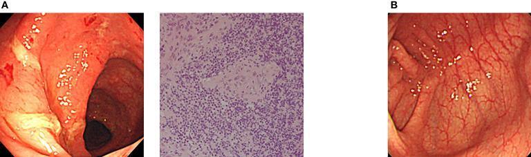
This composite clinical figure illustrates the treatment response in a patient with XIAP deficiency-associated inflammatory bowel disease (IBD). Panel A displays pre-treatment findings including a colonoscopic view and a histopathology slide. The endoscopy shows significant mucosal inflammation characterized by marked edema, friability, spontaneous hemorrhage, and whitish fibrinous ulcerations. The adjacent H&E-stained biopsy specimen reveals nonspecific active inflammation with a dense infiltrate of mononuclear cells and neutrophils, alongside architectural disruption. Panel B presents a follow-up colonoscopic image after allogeneic cord blood transplantation (CBT). It demonstrates complete mucosal healing with the restoration of normal vascular patterns, resolution of edema, and absence of ulcerations or hemorrhage. The educational focus is on the efficacy of hematopoietic stem cell transplantation as a curative option for monogenic PIDs presenting with refractory early-onset IBD, highlighting the clinical transition from severe inflammatory pathology to endoscopic remission.
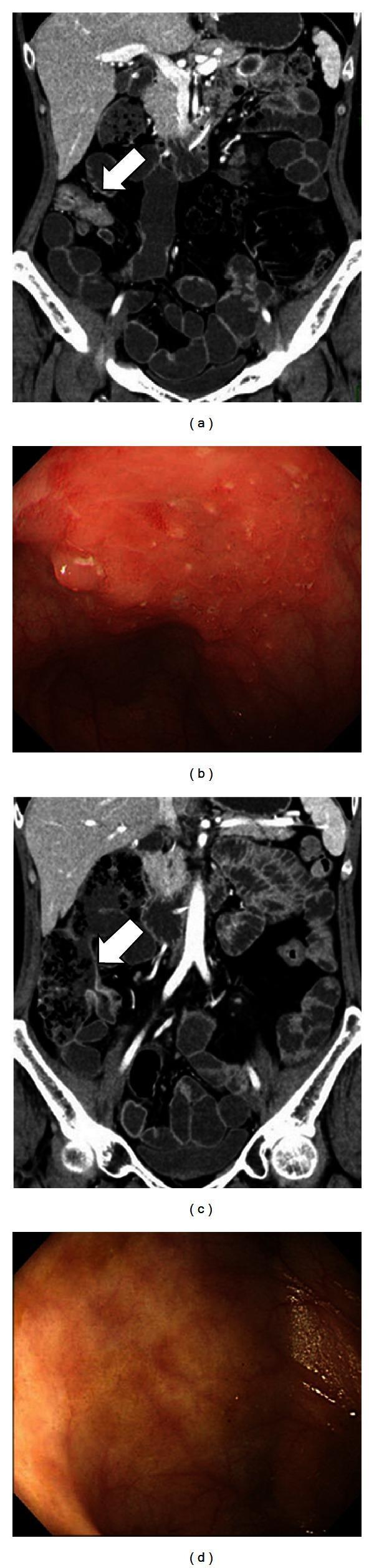
This composite clinical figure illustrates the correlation between diagnostic imaging and endoscopic findings in a patient with Inflammatory Bowel Disease (IBD) before and after biologic therapy. Panels (a) and (c) are coronal CT enteroclysis (CTE) images. Panel (a) displays an active disease state characterized by intestinal wall thickening (arrow) and increased density in the mesenteric fat (fat stranding), indicative of acute inflammation. Panel (c) shows the same region 84 days later, demonstrating therapeutic response with a reduction in wall thickness and normalization of mesenteric fat density. Panels (b) and (d) provide the corresponding endoscopic views of the ileocecal mucosa. Panel (b) reveals severe active inflammation with significant mucosal edema, erythema, and ulcerations. Panel (d) shows post-treatment resolution, featuring a smoother, non-congested mucosal surface without active ulcers or edema. This comparison serves as an educational tool for monitoring treatment efficacy in Crohn's disease using multimodality assessment, highlighting the transition from an active to an inactive inflammatory group.
Management of Inflammatory Bowel Disease (IBD)
Based on ACG 2025 Guidelines + Sleisenger & Fordtran
Overview
- Ulcerative Colitis (UC) - continuous mucosal inflammation, colon only, rectum involved in 95%
- Crohn's Disease (CD) - transmural, discontinuous ("skip lesions"), any part of GI tract
Key Paradigm Shifts in ACG 2025
- Mesalamine (5-ASA) is NO longer recommended for Crohn's disease (induction or maintenance) - strongly discouraged due to insufficient efficacy
- Strict step-up therapy abandoned for moderate-to-severe CD - patients should NOT be required to fail thiopurines/methotrexate before starting biologics
- Early advanced therapy is superior to accelerated step-up (PROFILE trial data)
- Treat-to-target strategy: goal is mucosal healing (endoscopic remission), not just symptom control
- Intestinal ultrasound (IUS) formally endorsed as non-invasive monitoring tool
- Fecal calprotectin >50-100 mg/g endorsed as practical cut-off to distinguish inflammatory from non-inflammatory disease
ULCERATIVE COLITIS - Management
Disease Severity Classification
- Mild-to-moderate: Truelove-Witts criteria - <4 stools/day, no systemic features
- Moderate-to-severe: >6 stools/day with blood, systemic features
- Acute Severe UC (ASUC): >6 bloody stools/day + systemic toxicity (fever, tachycardia, anemia, ESR >30)
Mild-to-Moderate UC
| Extent | Induction | Maintenance |
|---|---|---|
| Proctitis | Topical mesalamine suppository 1g/day (preferred) or topical steroid | Topical 5-ASA |
| Left-sided (proctosigmoiditis) | Oral 5-ASA ≥2g/day + topical mesalamine enema | Oral 5-ASA ≥1.5g/day |
| Extensive colitis | Oral 5-ASA ≥2g/day | Oral 5-ASA ≥1.5g/day |
- If intolerant/non-responsive to 5-ASA: Budesonide MMX 9 mg/day for induction (Strong recommendation)
- Once-daily vs more frequent 5-ASA dosing: equal efficacy - choose based on patient preference to optimize adherence
- Steroids (systemic, budesonide MMX, or topical) are NOT recommended for maintenance of remission
Moderate-to-Severe UC - Biologic and Small Molecule Therapy
| Drug Class | Agent | Recommendation |
|---|---|---|
| Anti-TNF | Infliximab | Strong (high evidence) - also for ASUC |
| Anti-TNF | Adalimumab, golimumab | Strong recommendation |
| Anti-integrin | Vedolizumab | Strong recommendation |
| Anti-IL12/23 | Ustekinumab | Strong recommendation |
| Anti-IL23p19 | Guselkumab, mirikizumab, risankizumab | Strong recommendation |
| JAK inhibitors | Upadacitinib, tofacitinib | Strong recommendation |
- When infliximab is used for induction in moderate-to-severe UC, combination with a thiopurine (azathioprine) is recommended (Strong, moderate quality) to reduce immunogenicity
- 5-ASA should NOT be added for additional clinical efficacy when biologics/JAK inhibitors are being used (Conditional recommendation)
Acute Severe Ulcerative Colitis (ASUC)
- IV corticosteroids (methylprednisolone 60mg/day or hydrocortisone 300-400mg/day) - first-line
- If no response in 3-5 days: Cyclosporine IV (2-4 mg/kg/day) OR infliximab (5 mg/kg)
- Cyclosporine responders: Bridge to thiopurine or vedolizumab for maintenance
- Surgery: Subtotal colectomy with end ileostomy for refractory ASUC or perforation
CROHN'S DISEASE - Management
Mild-to-Moderate CD (ACG 2025)
| Situation | Recommendation |
|---|---|
| Ileocecal CD | Budesonide 9 mg/day for induction (Strong) |
| Any site | Strongly against mesalamine for induction or maintenance |
| Colonic CD (mild only) | Sulfasalazine can be considered |
| Low risk of progression | Diet-based strategies with careful monitoring acceptable |
| Maintenance | Budesonide not recommended for maintenance |
Moderate-to-Severe CD - Biologic and Small Molecule Therapy
- Infliximab (IV/subcutaneous), adalimumab, vedolizumab, ustekinumab, risankizumab, mirikizumab, guselkumab
- Risankizumab is recommended over ustekinumab in this setting
- Upadacitinib (JAK1 inhibitor) recommended for induction and maintenance in moderate-to-severe CD with prior anti-TNF exposure
- Infliximab 5 mg/kg at weeks 0, 2, 6 (induction), then every 8 weeks (maintenance)
- ACCENT I trial: infliximab maintenance superior to episodic therapy for mucosal healing and fistula closure
- Adalimumab 160mg → 80mg → 40mg every 2 weeks; CHARM trial showed 36% remission vs 12% placebo at 56 weeks
- Certolizumab approved for CD (not UC); useful in pregnancy (poor placental transfer)
- Gut-selective (anti-α4β7 integrin) - acts only on gut, not systemic immune suppression
- GEMINI trials: ~47% clinical response at week 6 vs 26% placebo in UC; durable at week 52
- VARSITY trial (UC): vedolizumab 31% remission vs adalimumab 22% at week 52 - vedolizumab superior
- IV induction dose 6 mg/kg → then SC 90 mg every 8 weeks for maintenance
- UNITI trials: effective in anti-TNF refractory CD
- More selective than ustekinumab (targets IL-23 only, not IL-12)
- Preferred over ustekinumab post-anti-TNF failure per ACG 2025
Immunomodulators (Thiopurines, Methotrexate)
| Drug | Role |
|---|---|
| Azathioprine (2-2.5 mg/kg/day) | Maintenance, combination with infliximab to prevent antibody formation |
| 6-Mercaptopurine (1-1.5 mg/kg/day) | Same as azathioprine |
| Methotrexate (25 mg SC/IM weekly) | Induction and maintenance in CD; not recommended in UC per ACG 2025 |
Perianal/Fistulizing CD
- Examination under anesthesia (EUA) + seton placement (surgical drainage first)
- Infliximab - only anti-TNF with Level 1 evidence for fistula closure (ACCENT II trial: 36% vs 19% fistula closure)
- Combination anti-TNF + thiopurine for fistulizing disease
- Adalimumab, certolizumab - also effective
- Ustekinumab - emerging data
Special Situations (Sleisenger & Fordtran)
IBD in Pregnancy
- Safe: 5-ASA, sulfasalazine (+ folic acid), certolizumab (safest biologic - low placental transfer), glucocorticoids if needed
- Use with caution: Other anti-TNFs - generally continued if benefit > risk; stop infliximab/adalimumab at 24-26 weeks if disease in remission
- Avoid: Methotrexate (teratogenic), thiopurines in 1st trimester if possible
- Active IBD during pregnancy is more harmful than most medications
IBD in the Elderly (>65)
- Higher infection risk with biologics and thiopurines
- Prefer vedolizumab (gut-selective, lower systemic infection risk) as first-line biologic
- JAK inhibitors associated with higher cardiovascular and VTE risk - use with caution
IBD and Infection (Clostridioides difficile)
- C. diff worsens IBD course dramatically
- Treat C. diff first; consider fecal microbiota transplant (FMT) for recurrent C. diff in IBD
Cancer Surveillance
- Colonoscopy screening starts 8 years after UC onset (or CD colitis onset)
- Chromoendoscopy with targeted biopsy preferred over random biopsies
- Dysplasia management: low-grade dysplasia - can consider colectomy or intensive surveillance; high-grade dysplasia - colectomy recommended
Treatment Algorithm Summary
Monitoring (Treat-to-Target)
| Target | Tool |
|---|---|
| Mucosal healing | Colonoscopy |
| Transmural healing | CT/MR enterography, intestinal ultrasound (IUS) |
| Biochemical remission | CRP, fecal calprotectin <150 mg/g |
| Clinical remission | CDAI <150 (CD), Mayo score 0-1 (UC) |
Sources
- ACG 2025 UC Guideline Update (Rubin DT et al., Am J Gastroenterol 2025, PMID 40701556)
- ACG 2025 Crohn's Disease Guideline Update - June 2025
- AGA Living Guideline: Pharmacological Management of Moderate-to-Severe UC (Singh S et al., Gastroenterology 2024, PMID 39572132)
- Sleisenger and Fordtran's Gastrointestinal and Liver Disease, 11th Ed., Chapter 116 (pp. 2331-2409)