
Explain action of aldosterone
Reading File
Now let me search the library for aldosterone content.
Finding Sources
Finding Sources
Good findings. Let me now read the key textbook sections in parallel.
Reading File
Reading File
Reading File
Excellent content retrieved. Let me now read the detailed Ganong's mechanism section and the Brenner kidney section on non-epithelial actions and SGK1.
Reading File
Reading File
Reading File
I now have very rich content from multiple authoritative textbooks. Let me also fetch the key diagram image and read the Ganong section on regulation of aldosterone.
Loading Image
Reading File
I now have comprehensive, authoritative content from Ganong's and Brenner & Rector's Kidney to give a thorough answer. Let me compose the complete response.
Action of Aldosterone
Aldosterone is the principal mineralocorticoid secreted by the zona glomerulosa of the adrenal cortex. It plays a central role in sodium, potassium, water, and acid-base homeostasis.
1. Source and Basic Properties
- Synthesized exclusively in the zona glomerulosa via the enzyme aldosterone synthase (CYP11B2), which performs sequential 11β-hydroxylation, 18-hydroxylation, and 18-methyl oxidation of deoxycorticosterone.
- Secreted at 50-200 µg/day; plasma levels ~0.006 µg/dL (much lower than cortisol).
- Half-life ~20 min; poorly protein-bound.
(Brenner and Rector's The Kidney, p. 490)
2. Primary Target: The Aldosterone-Sensitive Distal Nephron (ASDN)
Aldosterone acts on the principal cells (P cells) of the:
- Distal convoluted tubule (DCT)
- Connecting tubule (CNT)
- Cortical collecting duct (CCD)
These segments express the mineralocorticoid receptor (MR) and the protective enzyme 11β-hydroxysteroid dehydrogenase type 2 (11β-HSD2).
3. Genomic (Classical) Mechanism of Action
Aldosterone is a steroid hormone and acts primarily through a nuclear receptor mechanism:
- Aldosterone diffuses into the principal cell and binds to the cytoplasmic mineralocorticoid receptor (MR).
- The aldosterone-MR complex translocates to the nucleus.
- It binds to mineralocorticoid response elements (MREs) on DNA.
- This alters transcription of target mRNAs, leading to synthesis of aldosterone-induced proteins (AIPs).
The key AIPs and their effects:
| Protein | Effect |
|---|---|
| SGK1 (serum- and glucocorticoid-regulated kinase 1) | Phosphorylates Nedd4-2, preventing it from ubiquitinating and removing ENaC from the membrane → increased apical ENaC expression |
| ENaC subunits (α, β, γ) | Aldosterone increases transcription of all three ENaC subunits; α-ENaC induction is rate-limiting (relieves a bottleneck in ENaC assembly and trafficking) |
| Na⁺-K⁺-ATPase (α₁ and β₁ subunits) | Increased expression on the basolateral membrane drives the Na⁺ gradient |
| CHIF (channel-inducing factor) | Further stimulates ENaC trafficking to the apical membrane |
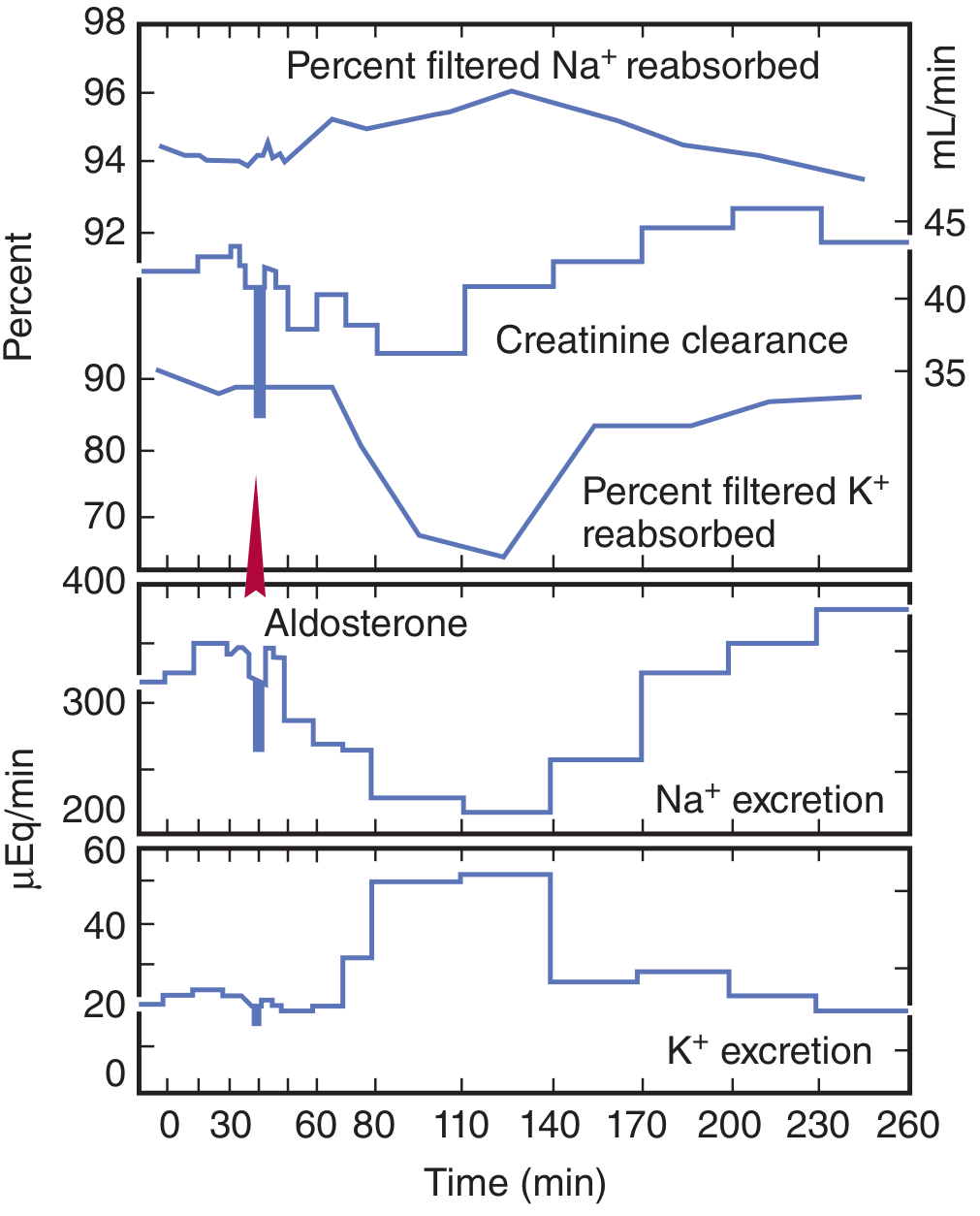
The SGK1 gene is an early response gene - its mRNA rises within 15 min and protein within 30 min of aldosterone stimulation. This is why the peak effect on Na⁺ transport takes 10-30 minutes to develop and continues to build over hours.
(Ganong's Review of Medical Physiology, p. 358; Brenner and Rector's The Kidney, p. 502)
4. Net Renal Effect
Na⁺ reabsorption ↑ → K⁺ and H⁺ secretion ↑ → ECF volume expansion
- Apical ENaC reabsorbs Na⁺ from the tubular lumen into the principal cell.
- Basolateral Na⁺-K⁺-ATPase pumps Na⁺ into the interstitium (3 Na⁺ out, 2 K⁺ in).
- The electronegativity created in the tubular lumen drives K⁺ secretion via ROMK channels and H⁺ secretion via H⁺-ATPase on intercalated cells.
- Water follows Na⁺ passively → ECF volume expansion.
- Aldosterone also induces SLC26A4 (apical Cl⁻/HCO₃⁻ exchanger) on intercalated cells, contributing to acid-base regulation.
The graph below from Ganong's shows the time-course of these effects after a single aldosterone dose:
5. Non-Genomic (Rapid) Mechanism
In addition to its slow genomic action, aldosterone also binds to cell membrane receptors and rapidly (within minutes) increases the activity of membrane Na⁺-K⁺ exchangers through a non-genomic pathway. The second messenger is likely IP₃. However, this rapid effect is considered minor compared to the dominant genomic mechanism.
(Ganong's Review of Medical Physiology, p. 358)
6. Role of 11β-HSD2 (Specificity Guard)
The mineralocorticoid receptor (MR) has a higher affinity for cortisol than for aldosterone in vitro, and plasma cortisol is ~100-fold more abundant. To prevent cortisol from occupying MR and mimicking aldosterone:
- 11β-HSD2 in mineralocorticoid-sensitive tissues converts cortisol → cortisone (inactive at MR) and corticosterone → its 11-oxy derivative (also inactive).
- Aldosterone is left untouched by 11β-HSD2.
Clinical relevance: Inhibition of 11β-HSD2 (e.g., by licorice's glycyrrhetinic acid, or genetic deficiency) leads to Apparent Mineralocorticoid Excess (AME) - cortisol floods the MR causing hypertension and hypokalemia, with low plasma aldosterone and low renin (Clinical Box 19-3 - Ganong's).
7. Actions Beyond the Kidney
| Site | Effect |
|---|---|
| Sweat glands, salivary glands, colon | Increased Na⁺ reabsorption and K⁺ secretion (same ENaC-mediated mechanism) |
| Vascular wall | Vasoconstriction in response to volume depletion (expresses 11β-HSD2, so aldosterone-selective) |
| Nucleus tractus solitarius (brain) | Stimulates salt appetite; also expresses 11β-HSD2 |
| Taste buds | ENaC-mediated Na⁺ taste transduction |
(Brenner and Rector's The Kidney, p. 507)
8. Regulation of Aldosterone Secretion
Aldosterone secretion is primarily regulated by:
- Angiotensin II (RAAS activation in response to volume depletion/low BP) - most important
- Plasma K⁺ - direct stimulation of zona glomerulosa
- ACTH - minor role (short-term stimulation; important for the "aldosterone synthesis" pathway through StAR protein)
Summary Diagram of Mechanism
Volume depletion / ↑K⁺
        ↓
Angiotensin II / ACTH
        ↓
Adrenal Zona Glomerulosa
        ↓
   ALDOSTERONE secreted
        ↓
Enters principal cell → binds cytoplasmic MR
        ↓
Aldosterone-MR complex → nucleus → binds MREs
        ↓
↑ Transcription of SGK1, ENaC subunits, Na⁺-K⁺-ATPase
        ↓
↑ Apical ENaC → Na⁺ reabsorption ↑
↑ Basolateral Na⁺-K⁺-ATPase → driving force ↑
↑ K⁺ secretion (ROMK) + H⁺ secretion (H⁺-ATPase)
        ↓
ECF volume expansion, ↓K⁺, metabolic alkalosis
Key clinical conditions related to aldosterone action:
- Primary hyperaldosteronism (Conn's syndrome): Hypertension, hypokalemia, metabolic alkalosis, low renin
- Hypoaldosteronism / Addison's disease: Na⁺ wasting, hyperkalemia, metabolic acidosis
- Pseudo-hypoaldosteronism: MR mutation - unresponsive to aldosterone
- Drugs: Spironolactone and eplerenone competitively antagonize the MR; amiloride/triamterene block ENaC directly
This is a shared conversation. Sign in to Orris to start your own chat.