
I will give you subject surgery topics of mbbs explain me that topics and make neat clean notes pointwise also add clinical correlation and diagrams for neet pg inicet …. Okay can you do it?
- Pointwise structured content (etiology, clinical features, investigations, management)
- High-yield clinical correlations and exam pearls
- Relevant diagrams/images where useful
Create a PDF with diagrams and clinical correlations for quick reference
Which MBBS Surgery topic should I create the NEET PG/INICET quick-reference PDF for?
- Pointwise structured content (etiology, clinical features, investigations, management)
- High-yield clinical correlations and exam pearls
- Relevant diagrams/images where useful
Wound healing tissue repair and scar
phases of wound healing diagram
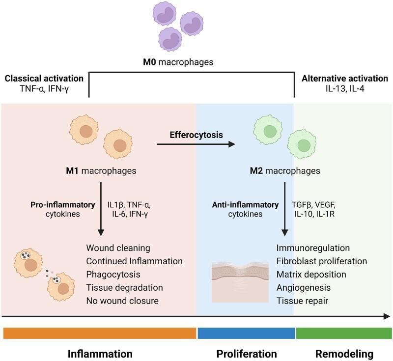
A pathophysiology diagram illustrating macrophage polarization during the stages of wound healing. The process begins with M0 macrophages (undifferentiated) which undergo 'Classical activation' via TNF-α and IFN-γ to become M1 macrophages. This pro-inflammatory phenotype predominates during the 'Inflammation' phase, secreting cytokines (IL-1β, TNF-α, IL-6, IFN-γ) and performing phagocytosis, tissue degradation, and wound cleaning. A transition via efferocytosis leads to 'Alternative activation' stimulated by IL-13 and IL-4, resulting in M2 macrophages. The M2 phenotype characterizes the 'Proliferation' and 'Remodeling' phases, producing anti-inflammatory and growth factors (TGFβ, VEGF, IL-10, IL-1R). These actions promote immunoregulation, fibroblast proliferation, matrix deposition, and angiogenesis, ultimately leading to successful tissue repair. The diagram uses color-coded backgrounds (orange for inflammation, blue for proliferation, green for remodeling) to link cellular phenotypes to clinical wound progression.
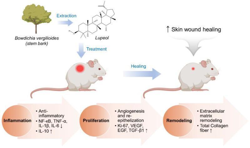
A medical infographic and pathophysiology diagram illustrating the therapeutic effect of Lupeol on the skin wound healing process. The diagram utilizes a rat model to visualize transition through three clinical phases: Inflammation, Proliferation, and Remodeling. Lupeol, shown as a chemical structure extracted from Bowdichia virgilioides, is applied as a treatment to a cutaneous wound. During the Inflammation phase, Lupeol modulates molecular pathways by decreasing pro-inflammatory markers (NF-κB, TNF-α, IL-1β, and IL-6) and increasing the anti-inflammatory cytokine IL-10. In the Proliferation phase, it promotes angiogenesis and re-epithelization through the upregulation of Ki-67, VEGF, EGF, and TGF-β1. The final Remodeling phase is characterized by increased total collagen fiber production and extracellular matrix remodeling, resulting in visible wound closure. The illustration demonstrates the multi-target mechanism of action of Lupeol in accelerating cutaneous repair by modulating growth factor expression and cytokine levels.
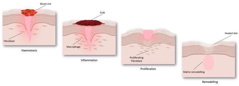
An anatomical pathophysiology diagram illustrating the four sequential stages of cutaneous wound healing: haemostasis, inflammation, proliferation, and remodeling. The illustration depicts a cross-section of the skin layers (epidermis and dermis). During 'Haemostasis', a red blood clot forms on the surface above a funnel-shaped injury, with fibroblasts visible in the surrounding dermis. In the 'Inflammation' stage, a dark-colored scab replaces the initial clot, and macrophages are identified in the underlying tissue. The 'Proliferation' stage demonstrates the formation of pinkish granulation tissue filling the wound gap, accompanied by 'proliferating fibroblasts' migrating into the site. Finally, the 'Remodeling' stage shows 'healed skin' with a restored epidermal surface and an underlying zone of 'matrix remodeling' replacing the previous injury site. The diagram serves as an educational summary of tissue repair mechanisms and cellular involvement at different phases of recovery.
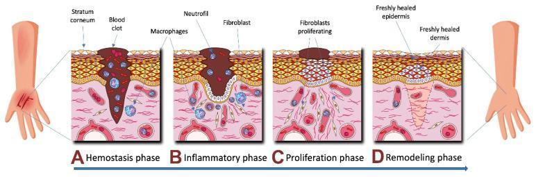
A medical pathophysiology diagram illustrating the four sequential phases of human skin wound healing: Hemostasis (A), Inflammation (B), Proliferation (C), and Remodeling (D). The cross-sectional views depict the stratum corneum, epidermis, and dermis. In the Hemostasis phase (A), a large blood clot fills the deep wound tissue with early macrophage presence. The Inflammatory phase (B) shows a persisting clot with an influx of neutrophils, macrophages, and early fibroblasts. The Proliferation phase (C) illustrates significant fibroblast activity and cellular multiplication beneath a shrinking scab as the epidermal gap begins to close. The final Remodeling phase (D) depicts a freshly healed epidermis and dermis with organized tissue structure and the absence of the initial clot and inflammatory cells. This illustration is an educational resource for understanding dermatological repair mechanisms and cellular recruitment during tissue injury.
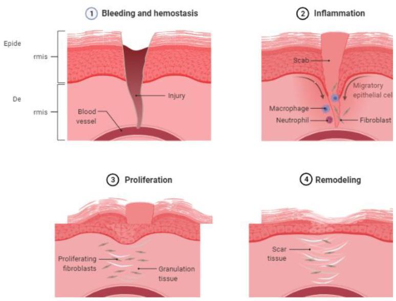
Anatomical diagram illustrating the four sequential phases of cutaneous wound healing: (1) Bleeding and Hemostasis, (2) Inflammation, (3) Proliferation, and (4) Remodeling. The cross-sectional illustrations represent the epidermis and dermis layers. Phase 1 shows a full-thickness injury penetrating through to the underlying blood vessel, depicting acute hemorrhage. Phase 2 (Inflammation) displays the formation of a surface scab, with arrows indicating the movement of migratory epithelial cells, alongside an infiltration of macrophages, neutrophils, and fibroblasts. Phase 3 (Proliferation) demonstrates the development of granulation tissue, characterized by proliferating fibroblasts and the reconstruction of the epidermal layer beneath the residual scab. Phase 4 (Remodeling) illustrates the final stage where the epidermis is fully restored, and the dermis contains organized scar tissue in place of the original injury site. This educational infographic provides a visual timeline of cellular responses and tissue structural changes during the natural course of human skin repair, useful for medical students and clinical practitioners studying pathophysiology.
keloid vs hypertrophic scar clinical photo
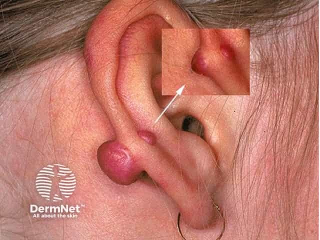
hypertrophic scar burn
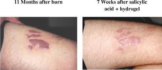
This clinical photograph represents a comparison of a hypertrophic burn scar on the medial thigh of a 42-year-old male before and after treatment. The left panel, labeled '11 Months after burn', shows a well-demarcated, elevated, and intensely erythematous (reddish-purple) scar with irregular borders, characteristic of hypertrophic scarring following a deep partial-thickness or full-thickness burn. The right panel, labeled '7 Weeks after salicylic acid + hydrogel', demonstrates the clinical progression after topical intervention. There is a visible reduction in erythema, with the scar tissue appearing lighter and more pinkish. The lesion also shows a noticeable decrease in vertical height (elevation) and thickness compared to the baseline image. The surrounding skin appears less inflamed, and the overall morphology of the scar is more flattened and less prominent. This comparison illustrates the therapeutic efficacy of combining salicylic acid and hydrogel in managing the physical and vascular characteristics of chronic hypertrophic scars in dermatology and burn surgery.
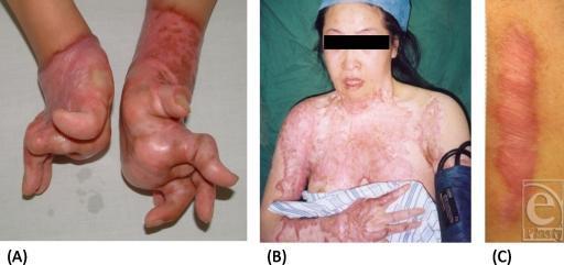
A clinical photograph comparison consisting of three panels (A, B, and C) illustrating different morphological presentations of cutaneous scarring. Panel (A) displays severe burn-related scar contractures of the bilateral hands and wrists. The skin is hyperpigmented, shiny, and thickened, causing fixed deformities and significant functional impairment of the fingers. Panel (B) shows an extensive hypertrophic scar involving a large total body surface area (TBSA), specifically the anterior chest, neck, and right upper extremity. The tissue exhibits a mottled, erythematous, and irregular texture, characteristic of post-burn sequelae. Panel (C) provides a close-up of a linear hypertrophic scar, demonstrating a raised, rope-like, and erythematous appearance against normal skin. This educational visual contrasts localized, linear scars with complex, functionally debilitating scarring, highlighting the limitations of standard scar assessment scales (e.g., VSS, POSAS) in evaluating large-scale or contracture-prone lesions. The content is relevant to dermatology, plastic surgery, and burn rehabilitation.
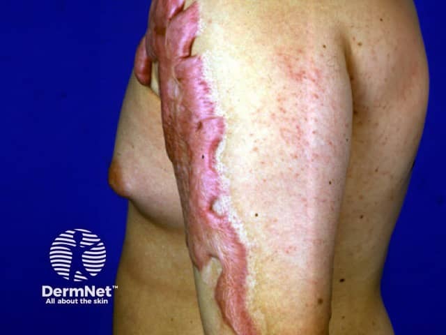
Clinical photography of the left upper extremity showing a long, well-demarcated hypertrophic scar following a burn injury. The scar runs longitudinally along the lateral/posterior aspect of the arm from mid-upper arm to proximal forearm. The scar tissue is raised, firm, and erythematous-pink with a shiny, contracted surface and irregular parallel banding consistent with post-burn scar formation. Surrounding skin shows residual erythema and hypopigmentation with patchy telangiectasia and minor textural changes. No active ulceration is evident. The scar margin is relatively sharp with limited pliability; subtle contour irregularities indicate localized contracture potential that may restrict flexion or extension at the elbow depending on involvement. The image demonstrates typical scar morphology: dense collagenous bundles, altered dermal architecture, and abnormal pigmentation. This type of lesion is common after partial-thickness to full-thickness burns and can contribute to restricted range of motion and cosmetic concerns. Clinically, such scars are evaluated for severity using scales like Vancouver Scar Scale or Patient and Observer Scar Assessment; management options include silicone gel sheeting, pressure therapy, intralesional corticosteroids, laser therapy, and physical therapy. The image is intended for dermatology, burn surgery, and rehabilitative medicine education, illustrating scar characterization, differential diagnosis (hypertrophic vs keloid scar), and therapeutic planning.
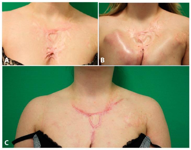
This clinical photograph series illustrates the surgical reconstruction of hypertrophic scarring on the chest and décolleté area of an 18-year-old female patient following burn injuries. Panel A displays the initial presentation of extensive, erythematous hypertrophic scar tissue characterized by thick, raised, branching cords and central tethering. Panel B shows the intermediate stage following tissue expander implantation; a significant subcutaneous volume prominence is visible on the right chest wall, stretching the overlying skin to gain healthy surface area for subsequent reconstruction. Panel C demonstrates the six-month postoperative outcome following scar resection and closure. The final image shows a flattened, significantly reduced linear scar following the natural contours of the décolleté, with improved skin texture and normalized chest wall contour compared to the preoperative state. The series serves as an educational example of plastic surgery techniques for managing complex post-burn contractures using tissue expansion and secondary resection.
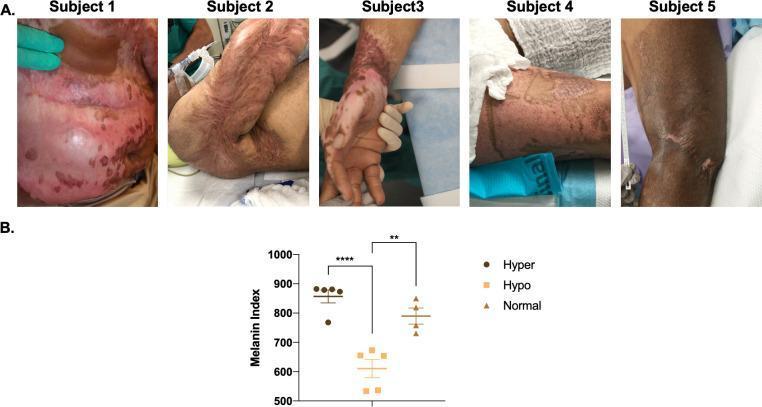
This composite educational image illustrates post-burn hypertrophic scarring (HTS) and associated dyschromia. Panel A contains five clinical photographs (Subjects 1–5) showing heterogeneous skin pigmentation on the torso and extremities. The photographs demonstrate a range of clinical findings, including hyperpigmented (darkened) and hypopigmented (depigmented) patches, mottled skin patterns, and raised, thickened scar tissue characteristic of hypertrophic remodeling. Panel B presents a dot plot quantifying the Melanin Index across three clinical phenotypes: hyperpigmented scar, hypopigmented scar, and normal skin (n=5). The data shows that hyperpigmented areas have the highest melanin content (mean ~875), followed by normal skin (mean ~800), and hypopigmented areas (mean ~625). Statistical significance is indicated, with p<0.01 between hyper- and hypopigmented groups and p<0.0001 between hyperpigmented and normal skin. This material serves to correlate clinical visual manifestations of post-burn dyschromia with objective non-invasive skin probe measurements, highlighting the physiological variability in melanin distribution following severe cutaneous injury.
mkdir -p /tmp/workspace/wound-healing-notes/assets /tmp/workspace/wound-healing-notes/output && ls -la /tmp/workspace/
~/wound-healing-notes/src/build_pdf.py
# -*- coding: utf-8 -*-
from reportlab.lib.pagesizes import A4
from reportlab.lib.units import cm
from reportlab.lib import colors
from reportlab.lib.styles import getSampleStyleSheet, ParagraphStyle
from reportlab.platypus import (SimpleDocTemplate, Paragraph, Spacer, Image, Table,
TableStyle, ListFlowable, ListItem, PageBreak, HRFlowable)
from reportlab.lib.enums import TA_CENTER, TA_LEFT
import os
ASSETS = "/tmp/workspace/wound-healing-notes/assets"
OUT = "/tmp/workspace/wound-healing-notes/output/Wound_Healing_Tissue_Repair_Scar_NEETPG_Notes.pdf"
styles = getSampleStyleSheet()
title_style = ParagraphStyle('TitleX', parent=styles['Title'], fontSize=20, textColor=colors.HexColor('#0B3954'), spaceAfter=6)
subtitle_style = ParagraphStyle('Subtitle', parent=styles['Normal'], fontSize=11, textColor=colors.HexColor('#3D5A80'), alignment=TA_CENTER, spaceAfter=14)
h1 = ParagraphStyle('H1', parent=styles['Heading1'], fontSize=15, textColor=colors.white, backColor=colors.HexColor('#0B3954'), spaceBefore=14, spaceAfter=8, leftIndent=6, borderPadding=(6,6,6,6))
h2 = ParagraphStyle('H2', parent=styles['Heading2'], fontSize=12.5, textColor=colors.HexColor('#0B3954'), spaceBefore=10, spaceAfter=4)
body = ParagraphStyle('Body', parent=styles['Normal'], fontSize=9.7, leading=13.5, spaceAfter=3)
bullet = ParagraphStyle('Bullet', parent=body, leftIndent=10, bulletIndent=0, spaceAfter=2)
subbullet = ParagraphStyle('SubBullet', parent=body, leftIndent=22, spaceAfter=2, fontSize=9.3)
caption = ParagraphStyle('Caption', parent=styles['Normal'], fontSize=8, textColor=colors.HexColor('#555555'), alignment=TA_CENTER, spaceAfter=10, spaceBefore=2)
pearl_title = ParagraphStyle('PearlTitle', parent=styles['Normal'], fontSize=10, textColor=colors.white, fontName='Helvetica-Bold')
pearl_body = ParagraphStyle('PearlBody', parent=styles['Normal'], fontSize=9.3, leading=13, textColor=colors.HexColor('#1a1a1a'))
story = []
def H1(text):
story.append(Paragraph(text, h1))
def H2(text):
story.append(Paragraph(text, h2))
def P(text):
story.append(Paragraph(text, body))
def BL(items):
story.append(ListFlowable([ListItem(Paragraph(i, bullet), bulletColor=colors.HexColor('#0B3954')) for i in items],
bulletType='bullet', start='circle', leftIndent=12))
story.append(Spacer(1,4))
def SUBBL(items):
story.append(ListFlowable([ListItem(Paragraph(i, subbullet), bulletColor=colors.HexColor('#3D5A80')) for i in items],
bulletType='bullet', start='-', leftIndent=22))
story.append(Spacer(1,4))
def PEARL(title_text, points):
rows = [[Paragraph(title_text, pearl_title)]]
for pt in points:
rows.append([Paragraph("• " + pt, pearl_body)])
t = Table(rows, colWidths=[16.5*cm])
t.setStyle(TableStyle([
('BACKGROUND', (0,0), (0,0), colors.HexColor('#EE6C4D')),
('BACKGROUND', (0,1), (-1,-1), colors.HexColor('#FFF3EE')),
('BOX', (0,0), (-1,-1), 0.8, colors.HexColor('#EE6C4D')),
('LEFTPADDING', (0,0), (-1,-1), 8),
('RIGHTPADDING', (0,0), (-1,-1), 8),
('TOPPADDING', (0,0), (-1,0), 5),
('BOTTOMPADDING', (0,0), (-1,0), 5),
('TOPPADDING', (0,1), (-1,-1), 3),
('BOTTOMPADDING', (0,1), (-1,-1), 3),
]))
story.append(Spacer(1,4))
story.append(t)
story.append(Spacer(1,8))
def IMG(path, cap, width=11*cm):
if os.path.exists(path):
try:
img = Image(path, width=width, height=width*0.68)
img.hAlign = 'CENTER'
story.append(Spacer(1,4))
story.append(img)
story.append(Paragraph(cap, caption))
except Exception as e:
story.append(Paragraph(f"[Image unavailable: {cap}]", caption))
else:
story.append(Paragraph(f"[Image unavailable: {cap}]", caption))
def TBL(data, colWidths=None):
t = Table(data, colWidths=colWidths, repeatRows=1)
t.setStyle(TableStyle([
('BACKGROUND', (0,0), (-1,0), colors.HexColor('#0B3954')),
('TEXTCOLOR', (0,0), (-1,0), colors.white),
('FONTNAME', (0,0), (-1,0), 'Helvetica-Bold'),
('FONTSIZE', (0,0), (-1,-1), 8.8),
('GRID', (0,0), (-1,-1), 0.6, colors.HexColor('#AAAAAA')),
('VALIGN', (0,0), (-1,-1), 'TOP'),
('ROWBACKGROUNDS', (0,1), (-1,-1), [colors.white, colors.HexColor('#EEF3F7')]),
('LEFTPADDING', (0,0), (-1,-1), 5),
('RIGHTPADDING', (0,0), (-1,-1), 5),
('TOPPADDING', (0,0), (-1,-1), 4),
('BOTTOMPADDING', (0,0), (-1,-1), 4),
]))
story.append(t)
story.append(Spacer(1,8))
# ================= TITLE PAGE =================
story.append(Spacer(1, 2*cm))
story.append(Paragraph("WOUND HEALING, TISSUE REPAIR & SCAR", title_style))
story.append(Paragraph("General Surgery | MBBS Quick Reference Notes for NEET PG / INICET", subtitle_style))
story.append(HRFlowable(width="100%", thickness=1.2, color=colors.HexColor('#0B3954')))
story.append(Spacer(1, 0.3*cm))
P("This note covers the phases of wound healing, types of healing intention, biochemistry of repair, "
"factors affecting healing, and abnormal scarring (keloid vs hypertrophic scar) - high-yield for surgery vivas, "
"theory exams, and NEET PG / INICET single-best-answer questions.")
story.append(Spacer(1, 0.4*cm))
# ================= SECTION 1: DEFINITIONS =================
H1("1. Basic Definitions")
BL([
"<b>Healing</b> - restoration of tissue continuity and function after injury; used interchangeably with <b>repair</b>.",
"<b>Regeneration</b> - replacement of lost tissue by cells identical to the original (e.g., liver, epidermis, GI mucosa) - occurs only in tissues with dividing (labile/stable) cells and intact connective tissue scaffold.",
"<b>Repair by scarring</b> - occurs when tissue cannot regenerate or when the connective tissue framework is damaged; lost tissue is replaced by <b>fibrocollagenous scar</b> (type I collagen), which is structurally strong but not functionally identical.",
"<b>Fibrosis</b> - term used when scar-type collagen deposition occurs in an organ (lung, liver, kidney, heart) after chronic injury.",
])
IMG(f"{ASSETS}/robbins_regeneration_vs_scar.png",
"Fig 1. Mechanisms of tissue repair - regeneration (mild injury, epithelium only) vs scar formation (severe injury, connective tissue damaged). Robbins Pathologic Basis of Disease.")
# ================= SECTION 2: TYPES OF WOUND HEALING =================
H1("2. Types of Wound Healing (by Intention)")
data = [
["Type", "Description", "Example"],
["Primary intention\n(Healing by first intention)", "Clean, incised wound; edges apposed with sutures/staples/glue; minimal scarring, fast", "Surgical incision closed primarily"],
["Secondary intention", "Wound left open; heals by granulation tissue formation + wound contraction + epithelialization from margins; larger scar", "Infected/contaminated wound, abscess cavity, pressure sore"],
["Tertiary intention\n(Delayed primary closure)", "Wound initially left open (to allow debridement/observation for infection), then surgically closed after 3-5 days once clean", "Contaminated traumatic wound, dirty surgical wound"],
]
TBL(data, colWidths=[4.3*cm, 8.2*cm, 4*cm])
PEARL("CLINICAL CORRELATION", [
"Incisions placed along <b>Langer's lines (relaxed skin tension lines)</b> heal with thinner, cosmetically better scars - key principle in elective surgical planning and plastic surgery.",
"Delayed primary closure (tertiary intention) is used in <b>contaminated/dirty wounds</b> to reduce infection risk while still achieving a primary-type scar.",
])
# ================= SECTION 3: PHASES =================
H1("3. Phases of Wound Healing")
IMG(f"{ASSETS}/phases_cross_section.png",
"Fig 2. The four sequential phases of cutaneous wound healing: haemostasis, inflammation, proliferation and remodeling.")
data = [
["Phase", "Time", "Key Events / Cells", "Key Mediators"],
["1. Hemostasis", "Seconds - minutes (Day 0)", "Vasoconstriction -> platelet plug -> coagulation cascade -> fibrin clot (provisional matrix)", "Thromboxane A2, ADP, PDGF, TGF-beta (released from platelet alpha-granules)"],
["2. Inflammation", "Day 0 - Day 3-5", "<b>Neutrophils</b> first (peak 24-48h, debride bacteria/debris) -> <b>Macrophages</b> (Day 2-3, KEY orchestrator cell - phagocytosis + release growth factors)", "IL-1, TNF-alpha, PDGF, TGF-beta, VEGF, FGF"],
["3. Proliferation", "Day 3 - Day 21", "Fibroblast migration/proliferation -> <b>granulation tissue</b> (new capillaries + fibroblasts); angiogenesis; re-epithelialization from wound margins; wound contraction by <b>myofibroblasts</b>", "TGF-beta, FGF, VEGF, EGF, PDGF"],
["4. Maturation / Remodeling", "Day 21 - up to 1-2 years", "Type III collagen replaced by stronger <b>Type I collagen</b>; collagen cross-linking; scar remodeling; decreasing vascularity and cellularity; wound contracts and pales", "MMPs (collagenases) balance new synthesis vs breakdown"],
]
TBL(data, colWidths=[3.3*cm, 2.7*cm, 6.7*cm, 3.8*cm])
PEARL("HIGH-YIELD NEET PG FACTS", [
"<b>Macrophage</b> is the single most important cell of wound healing - orchestrates the transition from inflammation to proliferation; its absence causes the most severe defect in healing.",
"<b>Granulation tissue</b> = capillary buds + fibroblasts + inflammatory cells; clinically appears beefy-red, granular, and bleeds easily on touch.",
"<b>Collagen switch</b>: Type III (early, thin, laid by fibroblasts) → Type I (mature, thick, cross-linked) - defective switch/ratio is seen in <b>keloids</b> and in <b>Ehlers-Danlos syndrome</b>.",
"<b>Wound tensile strength</b>: ~20% of normal at 3 weeks; reaches only ~70-80% of original skin strength by 3 months and <b>never reaches 100%</b>.",
"<b>Myofibroblasts</b> (contain alpha-smooth muscle actin) mediate wound contraction; excessive/abnormal contraction over joints causes a <b>contracture</b> (e.g., post-burn contractures).",
"Suture removal timing is guided by tensile strength gain: face 5-7 days, scalp 7-10 days, trunk/limbs 10-14 days, palms/soles 14 days (approximate, exam-relevant ranges).",
])
# ================= SECTION 4: GROWTH FACTORS =================
H1("4. Key Growth Factors in Wound Healing")
data = [
["Growth Factor", "Source", "Main Action"],
["PDGF (Platelet-derived GF)", "Platelets, macrophages", "Chemotaxis for neutrophils, macrophages, fibroblasts; stimulates fibroblast proliferation"],
["TGF-beta", "Platelets, macrophages, fibroblasts", "Stimulates collagen synthesis & fibrosis; master regulator of scarring (overactivity -> keloid/hypertrophic scar)"],
["VEGF", "Macrophages, keratinocytes", "Angiogenesis - new capillary formation in granulation tissue"],
["EGF / TGF-alpha", "Platelets, macrophages, keratinocytes", "Stimulates keratinocyte migration & re-epithelialization"],
["FGF (basic)", "Macrophages, fibroblasts", "Angiogenesis, fibroblast proliferation, epithelialization"],
]
TBL(data, colWidths=[4.5*cm, 4.5*cm, 7*cm])
# ================= SECTION 5: FACTORS AFFECTING =================
H1("5. Factors Affecting Wound Healing")
H2("Local Factors")
BL([
"Infection (most common cause of delayed healing) and foreign body / necrotic tissue",
"Poor blood supply / ischemia (e.g., peripheral arterial disease, tight sutures, hematoma)",
"Wound tension, movement, or repeated trauma at the site",
"Radiation therapy at the wound site (causes endarteritis obliterans, fibrosis)",
])
H2("Systemic Factors")
BL([
"<b>Malnutrition</b> - protein deficiency impairs collagen synthesis",
"<b>Vitamin C deficiency (scurvy)</b> - impaired hydroxylation of proline/lysine -> defective collagen cross-linking -> poor healing, gum bleeding, old scars break down",
"<b>Zinc deficiency</b> - impaired epithelialization and fibroblast proliferation",
"<b>Diabetes mellitus</b> - impaired healing via microangiopathy, neuropathy, and increased infection risk",
"<b>Corticosteroids</b> - inhibit the inflammatory phase (decrease macrophage function & collagen synthesis); effect partially reversed by <b>Vitamin A</b>",
"Smoking (nicotine-induced vasoconstriction, tissue hypoxia), old age, jaundice/uremia, anemia, obesity, immunosuppression, chemotherapy",
])
PEARL("CLASSIC EXAM ASSOCIATIONS", [
"<b>Scurvy</b> (Vitamin C deficiency) -> defective collagen hydroxylation -> wound dehiscence, gum disease, perifollicular hemorrhages.",
"<b>Steroids delay healing</b>; give <b>Vitamin A</b> to counteract steroid-induced impairment of wound healing (classic INICET/NEET PG one-liner).",
"<b>Ehlers-Danlos syndrome</b> and <b>Osteogenesis imperfecta</b> = heritable collagen disorders presenting with fragile skin, poor wound healing, and abnormal scarring.",
])
story.append(PageBreak())
# ================= SECTION 6: ABNORMAL HEALING =================
H1("6. Abnormal / Excessive Healing")
H2("Deficient Scar Formation")
BL([
"<b>Wound dehiscence</b> - separation of wound edges, often 5-8 days post-op, associated with infection, raised intra-abdominal pressure, poor nutrition, steroid use.",
"<b>Incisional hernia</b> - late complication of a weak/dehisced abdominal wound scar.",
"<b>Chronic non-healing ulcer</b> - persistent inflammatory phase (e.g., venous ulcer, diabetic foot ulcer, pressure sore).",
"<b>Marjolin's ulcer</b> - squamous cell carcinoma arising in a chronic scar, burn scar, or long-standing sinus/ulcer - classically painless and slow-growing due to poor scar lymphatics; suspect with a non-healing ulcer edge/floor change in an old scar.",
])
H2("Excessive Scar Formation - Keloid vs Hypertrophic Scar")
IMG(f"{ASSETS}/keloid.png", "Fig 3. Extensive keloids showing growth beyond the margins of the original wound.")
IMG(f"{ASSETS}/hypertrophic_scar.jpg", "Fig 4. Hypertrophic scar following a burn injury - raised, erythematous, but confined to the original wound margins.")
data = [
["Feature", "Keloid", "Hypertrophic Scar"],
["Extension", "Extends <b>beyond</b> original wound margins, clawlike projections", "Confined <b>within</b> original wound margins"],
["Onset", "May appear months after injury, keeps growing", "Appears within weeks, may regress spontaneously over months"],
["Common sites", "Earlobe, deltoid, presternal region, face", "Any site of high tension, especially flexor surfaces & burns"],
["Race/Genetics", "More common in darker skin types; familial tendency", "No strong racial predilection"],
["Histology", "Thick, hyalinized, whorled collagen bundles; scanty elastic tissue", "Nodular fibroblasts/myofibroblasts, less hyalinization"],
["Regression", "Rarely regresses; recurs after simple excision", "Often regresses/flattens spontaneously with time"],
["Treatment", "Intralesional triamcinolone +/- 5-FU, silicone sheeting, pressure, excision + adjuvant radiotherapy (high recurrence with excision alone)", "Silicone gel sheeting, pressure garments, intralesional steroid, Z-plasty for tension release, laser"],
]
TBL(data, colWidths=[3*cm, 6.6*cm, 6.4*cm])
PEARL("MUST-KNOW ONE-LINERS", [
"Keloid = scar tissue that grows BEYOND the wound boundary; Hypertrophic scar stays WITHIN the boundary - single most tested differentiator.",
"Keloids are managed primarily <b>non-surgically</b> first (steroid injection) because excision alone has high recurrence; adjuvant radiotherapy reduces recurrence after excision.",
"<b>Z-plasty</b> releases scar tension and lengthens a contracted scar at the expense of width - useful for hypertrophic scar contractures across joints/web spaces.",
"A rapidly growing, ulcerating, or bleeding nodule within an old keloid or burn scar should raise suspicion for malignant change (Marjolin's ulcer / rarely dermatofibrosarcoma-like change) - biopsy atypical lesions.",
])
# ================= SECTION 7: SPECIAL TOPIC =================
H1("7. Special High-Yield Points")
BL([
"<b>Fetal wound healing</b> is scarless up to mid-gestation - due to high hyaluronic acid, minimal inflammatory response, and different collagen deposition pattern (favors regeneration research).",
"<b>Hyperbaric oxygen therapy</b> and negative pressure wound therapy (VAC) are adjuncts used for chronic, ischemic, or diabetic wounds to promote angiogenesis and granulation.",
"Wound classification by contamination (for antibiotic/closure decisions): <b>Clean -> Clean-contaminated -> Contaminated -> Dirty/Infected</b> - determines primary vs delayed closure and prophylactic antibiotic use.",
"Bone healing differs from soft tissue: goes through hematoma -> soft (fibrocartilaginous) callus -> hard (bony) callus -> remodeling; requires adequate blood supply and immobilization.",
])
story.append(Spacer(1, 0.6*cm))
story.append(HRFlowable(width="100%", thickness=0.8, color=colors.HexColor('#0B3954')))
story.append(Paragraph("Compiled for quick revision - Sources: Bailey & Love's Short Practice of Surgery (28e), Robbins & Cotran Pathologic Basis of Disease, Schwartz's Principles of Surgery (11e), Andrews' Diseases of the Skin, Fitzpatrick's Dermatology.", caption))
doc = SimpleDocTemplate(OUT, pagesize=A4, topMargin=1.4*cm, bottomMargin=1.4*cm, leftMargin=1.5*cm, rightMargin=1.5*cm,
title="Wound Healing, Tissue Repair and Scar - NEET PG Notes")
doc.build(story)
print("PDF built at:", OUT)
cd /tmp/workspace/wound-healing-notes/assets && \ curl -sL -o robbins_regeneration_vs_scar.png "https://cdn.orris.care/cdss_images/bf02fb0bd95d1bac53eac5cbd060dac52ffebe95cc6ccdab7e6fe1ca48ca4382.png" && \ curl -sL -o phases_cross_section.png "https://cdn.orris.care/cdss_images/pmc_clinical_VQA_657b0019b41f392704c6eb42cd19190b0b2ed37790e44ec5ceb2d4d2c1da6a97.jpg" && \ curl -sL -o keloid.png "https://cdn.orris.care/cdss_images/d9b75e105fb92e91b92604f9a97a8a736eddaa89abcb4b171770b3e084e5d7eb.png" && \ curl -sL -o hypertrophic_scar.jpg "https://cdn.orris.care/cdss_images/DermNetNZ_1760030514668_0d44d7ba-2bbb-4aa2-b457-849a916a614c.jpg" && \ ls -la
cd /tmp/workspace/wound-healing-notes/src && python3 build_pdf.py
cd /tmp/workspace/wound-healing-notes/output && ls -la && python3 -c " from pypdf import PdfReader r = PdfReader('Wound_Healing_Tissue_Repair_Scar_NEETPG_Notes.pdf') print('pages:', len(r.pages)) "
Wound Healing Tissue Repair Scar NEETPG Notes
PDF Document · PDF
- Basic definitions - healing vs regeneration vs repair by scarring vs fibrosis, with the Robbins diagram of regeneration vs scar formation
- Types of wound healing - primary, secondary, tertiary (delayed primary) intention, table + clinical correlation on Langer's lines
- Phases of wound healing - hemostasis → inflammation → proliferation → maturation/remodeling, with time frames, key cells (neutrophils, macrophages, fibroblasts, myofibroblasts), mediators, and a labeled cross-section diagram
- Growth factors table - PDGF, TGF-beta, VEGF, EGF, FGF and their roles
- Factors affecting healing - local (infection, ischemia, radiation) and systemic (vitamin C/zinc deficiency, diabetes, steroids) with classic exam associations (scurvy, steroid + Vitamin A reversal, Ehlers-Danlos/osteogenesis imperfecta)
- Abnormal healing - dehiscence, incisional hernia, chronic ulcers, Marjolin's ulcer, and a full keloid vs hypertrophic scar comparison table with real clinical photos of both
- Special high-yield points - fetal scarless healing, hyperbaric oxygen/VAC therapy, wound contamination classification, bone healing sequence