
Hemothorax mbbs final year university exam answer 20 marks surgery theory exam
Reading File
Now let me search the medical library for comprehensive hemothorax content in parallel.
Finding Sources
Finding Sources
Excellent sources found. Let me read the most comprehensive surgery and respiratory medicine sections in parallel.
Reading File
Reading File
Excellent content. Let me read more of the Murray & Nadel section and also the Tintinalli section for management details.
Reading File
Reading File
Let me read the Murray & Nadel key points and management section, plus the Current Surgical Therapy retained hemothorax and complications.
Reading File
Reading File
I now have very comprehensive content from multiple authoritative surgical and emergency medicine textbooks. Let me fetch the key chest X-ray image showing massive hemothorax for inclusion.
Loading Image
Loading Image
I now have comprehensive material from multiple authoritative surgery textbooks. Let me compile the full 20-mark university exam answer.
HEMOTHORAX
Surgery Theory Exam - 20 Marks Answer
DEFINITION
Hemothorax is defined as the accumulation of blood within the pleural space. It is confirmed when the hematocrit of the pleural fluid is greater than 50% of the patient's peripheral blood hematocrit. Due to the significant color change of pleural fluid even with small amounts of blood, laboratory analysis of the fluid is essential to confirm the diagnosis.
The pleural space can accumulate up to 3 litres of blood. Trauma is the most common cause, accounting for nearly 300,000 cases per year in the United States.
ETIOLOGY / CAUSES
Hemothorax is broadly classified into three types:
1. Traumatic Hemothorax (Most Common)
- Blunt trauma: Motor vehicle collisions, falls - injures intercostal vessels, lung parenchyma, diaphragm, great vessels, or mediastinal structures
- Penetrating trauma: Gunshot wounds, stab wounds - 83% of penetrating thoracic injuries are associated with hemopneumothorax
- Rib fractures: Strong correlation - patients with >5 rib fractures have ≥48% likelihood of concomitant hemothorax
- Sources of bleeding:
- Chest wall: Intercostal arteries, internal mammary artery (arterial - more severe, requires intervention)
- Lung parenchyma: Most common; low pulmonary arterial pressure + lung thromboplastin limit bleeding (usually stops with drainage alone)
- Major vessels: Pulmonary artery/vein, aorta (life-threatening)
- Mediastinum, diaphragm
2. Non-Traumatic (Spontaneous) Hemothorax
- Spontaneous pneumothorax: Most common non-traumatic cause; hemothorax develops in 2-7% of cases, from torn pleural adhesions or rupture of vascularized bullae
- Vascular pathology: Aortic dissection/rupture (usually left-sided), pulmonary arteriovenous malformations (associated with hereditary hemorrhagic telangiectasia)
- Neoplasms: Malignant pleural tumors, metastatic disease
- Coagulopathy / anticoagulant therapy
- Spontaneous: From pleural endometriosis (catamenial hemothorax)
3. Iatrogenic Hemothorax
- Central venous catheter insertion
- Thoracentesis, pleural biopsy
- Post-cardiac surgery
- Pacemaker placement
PATHOPHYSIOLOGY
Blood enters the pleural space and initially has full coagulation ability. Three mechanisms limit bleeding in lung parenchymal injuries:
- Compressing effect of blood accumulation in the pleural space
- High concentration of lung thromboplastin promoting clot formation
- Low pulmonary arterial pressure (compared to systemic circulation)
Bleeding from systemic vessels (intercostal, internal mammary arteries) does NOT benefit from these low-pressure mechanisms and almost always requires surgical intervention.
As blood accumulates:
- Respiratory compromise: Lung is compressed and collapsed, causing ventilation-perfusion mismatch and hypoxia
- Hemodynamic compromise: Significant blood loss leads to hypovolemic shock
- Massive hemothorax (>1500 mL) can cause tension physiology - mediastinal shift, decreased venous return, reduced cardiac output (similar to tension pneumothorax)
CLASSIFICATION
| Type | Volume |
|---|---|
| Small | <300 mL |
| Moderate | 300 - 1500 mL |
| Massive | >1500 mL |
Massive hemothorax (>1500 mL or >1/3 hemithorax opacification) is a surgical emergency.
CLINICAL FEATURES
Symptoms
- Chest pain (pleuritic or dull)
- Dyspnea, breathlessness
- Signs of hemorrhagic shock (tachycardia, hypotension, pallor) depending on volume lost
Signs
- Asymmetric chest expansion on the affected side
- Decreased/absent breath sounds on the affected side
- Stony dullness to percussion on the affected side (distinguishes from pneumothorax)
- Tracheal deviation to the opposite side in massive hemothorax with tension component
- Hypotension, tachycardia - from both blood loss and tension physiology
Key clinical distinction: Hemothorax = dull percussion + absent breath sounds; Tension pneumothorax = hyper-resonant percussion + absent breath sounds + tracheal deviation
INVESTIGATIONS / DIAGNOSIS
Diagnosis is based on high index of suspicion, physical examination, and thoracic imaging.
1. Chest X-Ray (CXR) - First-Line Investigation
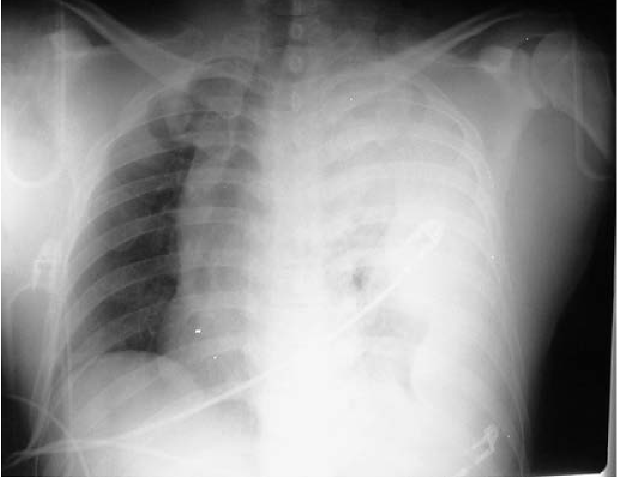
CXR: Large left-sided tension hemothorax with complete opacification and tracheal deviation - Current Surgical Therapy 14e
Findings on CXR:
- Blunting of costophrenic angle - requires ~200 mL of fluid (upright PA view)
- Blunting of cardiophrenic angle
- Obliteration of hemidiaphragm - requires ~500-650 mL
- Homogeneous opacity of hemithorax (white-out)
- Mediastinal shift away from the effusion in large collections
- Supine CXR: May only show a diffuse haziness; >1000 mL of blood may be missed due to posterior layering
- Lateral decubitus CXR: Most sensitive plain film; can detect as little as 10 mL
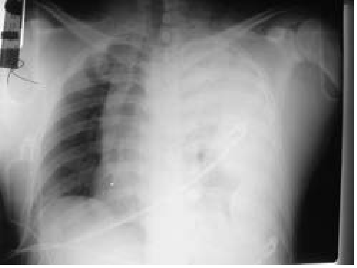
CXR: Massive hemothorax with tracheal deviation - Current Surgical Therapy 14e
2. Ultrasound (FAST / eFAST)
- Point-of-care ultrasound can rapidly detect pleural fluid in the trauma bay
- Extended FAST (eFAST): Sensitivity 77%, negative predictive value close to 100%
- Pleural fluid appears as anechoic collection superior to the diaphragm
- Can be used in the critically ill patient at bedside without radiation
3. CT Scan (Gold Standard)
- CT thorax is the gold standard for detection of hemothorax
- Detects exact location, volume, and associated injuries (pneumothorax, rib fractures, aortic injury)
- Shows "hematocrit effect" - fluid-fluid level between denser blood cells inferiorly and serum superiorly
- Identifies active contrast extravasation (active hemorrhage)
- Ideal for stable patients; NOT appropriate for hemodynamically unstable patients
4. Diagnostic Thoracocentesis / Pleural Fluid Analysis
- Confirms diagnosis: pleural fluid hematocrit >50% of peripheral blood hematocrit
- pH, LDH, cytology as needed
- Not usually required in trauma setting where diagnosis is clinical
MANAGEMENT
Initial Resuscitation (ATLS Protocol)
All patients should be managed with:
- Airway: Secure airway, supplemental oxygen
- Two large-bore IV access: Aggressive IV fluid resuscitation
- Blood transfusion as required (O-negative in emergencies)
- Monitoring: BP, pulse, SpO2, urine output
Definitive Treatment Algorithm
Step 1: Is the patient hemodynamically unstable?
UNSTABLE → Immediate tube thoracostomy (chest drain) without waiting for imaging
STABLE → Imaging (eFAST/CXR/CT) → Plan treatment based on volume and ongoing bleeding
1. Observation (Small Hemothorax, <300 mL, Asymptomatic)
- Supportive: Supplemental oxygen, analgesia, incentive spirometry, early ambulation
- Serial CXR monitoring for progression
- No drainage required if asymptomatic and no ongoing bleeding
2. Tube Thoracostomy (Chest Drain) - MAINSTAY OF TREATMENT
Indications: Hemothorax >200-300 mL, symptomatic patient, hemodynamic instability with suspected hemothorax
Procedure:
- Position: Supine with ipsilateral arm extended overhead
- Site: 4th or 5th intercostal space, midaxillary line (triangle of safety)
- Under local anesthesia (lignocaine)
- Large-bore tube (28-32 Fr) is preferred for hemothorax to prevent clot blockage
- Connect to underwater seal drain / Pleur-Evac at -20 cmH2O suction
- Secure with sutures
Outcomes with chest drain:
- In ~85% of cases, bleeding stops as the lung re-expands (low pulmonary vascular pressure allows tamponade)
- Initial output >1500 mL OR ongoing output >200 mL/hour for 4 hours = indication for thoracotomy
Autotransfusion: Shed blood should be collected in a sterile fashion for autotransfusion, particularly valuable in massive hemothorax.
3. Emergency/Urgent Thoracotomy
Indications for Thoracotomy:
| Indication | Threshold |
|---|---|
| Initial chest drain output | >1500 mL (massive hemothorax) |
| Ongoing hemorrhage | >200 mL/hour for 4 consecutive hours |
| Hemodynamic instability despite resuscitation | - |
| Massive continuous air leak | Suggests major airway or lung injury |
| Penetrating cardiac injury | Immediately |
| Thoracic great vessel injury | Immediately |
Emergency Room Thoracotomy (ERT): Performed in the ER for patients in cardiac arrest or peri-arrest following penetrating thoracic trauma - direct cardiac massage, control of aorta/pulmonary hilum.
Operative approach: Anterolateral thoracotomy (quick access), posterolateral thoracotomy (best exposure for elective cases)
4. Video-Assisted Thoracoscopic Surgery (VATS)
Indications:
- Retained hemothorax (most common indication)
- Empyema from infected retained hemothorax
- Fibrothorax / trapped lung
- Continuous air leak not resolving with chest drain
- Clotted hemothorax within 7-10 days (before fibrous peel organizes)
Advantages over open thoracotomy: Less pain, smaller incision, shorter hospital stay, lower morbidity.
COMPLICATIONS
1. Retained Hemothorax (Most Common Complication)
- Occurs in approximately 10-15% of hemothoraces treated by tube thoracostomy
- Defined as persistent pleural opacity after adequate chest tube placement
- Clotted blood unable to drain through tube
- Risk: Empyema, fibrothorax
- Treatment: Early VATS evacuation (ideally within 3-5 days, before day 10)
- Intrapleural fibrinolytics (tPA/DNase) can be used for smaller retained collections
2. Empyema
- Infected retained hemothorax
- Organisms: S. aureus, gram-negative rods
- Presents with fever, leukocytosis, chest pain
- Treatment: Tube drainage + antibiotics ± VATS decortication
3. Fibrothorax
- Organized fibrous peel encasing the lung, preventing re-expansion
- Leads to restrictive lung disease
- Treatment: Surgical decortication
4. Hypovolemic Shock
- Due to massive blood loss
- Can be compounded by tension hemothorax physiology
5. Tension Hemothorax
- Massive hemothorax causing mediastinal shift, kinking of great veins, decreased cardiac output
- Presents with hypotension + absent breath sounds + tracheal deviation
- Immediate chest tube decompression
SPECIAL SITUATIONS
Hemopneumothorax
- Associated pneumothorax present in 2.4% of blunt trauma and 83% of penetrating thoracic injury cases
- Managed with same chest drain; tube positioned posteriorly for blood, superiorly for air
Delayed Hemothorax
- Can occur hours to days after apparently minor trauma
- Risk factors: Anticoagulant use, rib fractures
- Requires vigilance with serial imaging in high-risk patients
Non-Operative Management of Occult Hemothorax
- Small hemothorax incidentally found on CT in a stable patient without respiratory compromise
- Can be observed with serial imaging
- Failure rate under observation: ~6% (higher if on positive pressure ventilation)
KEY POINTS SUMMARY (for exam recall)
| Feature | Detail |
|---|---|
| Definition | Blood in pleural space; pleural hematocrit >50% of blood hematocrit |
| Most common cause | Trauma (blunt > penetrating) |
| Most common bleeding source | Lung parenchyma |
| Mainstay of treatment | Intercostal chest drain (tube thoracostomy) |
| Bleeding stops with drain alone | ~85% of cases |
| Indication for thoracotomy | >1500 mL initial, or >200 mL/hr x 4 hrs |
| Most common complication | Retained hemothorax |
| Complication of retained hemothorax | Empyema → fibrothorax |
| VATS indication | Retained hemothorax, empyema, fibrothorax |
| Gold standard imaging | CT thorax |
| Diagnosis confirmed by | Pleural hematocrit >50% of blood hematocrit |
FLOWCHART: MANAGEMENT OF HEMOTHORAX
Suspected Hemothorax (chest trauma / hemodynamic instability)
              |
    ┌─────────┴──────────┐
Unstable                Stable
    |                    |
Immediate ICD       Imaging (CXR/eFAST/CT)
    |                    |
  ┌─┴──┐           ┌────┴────┐
Stop  Cont.      Small     Large
bleed  bleed    (<300 mL)  (>300 mL)
  |     |          |          |
Obs. Thoracotomy  Observe    ICD + drain
         |          |          |
      (>1500 mL  Serial    Stops (~85%)
       initial   CXR       |  Continues
       or >200            Obs.   |
       mL/hr x 4h)           Thoracotomy
                              (>1500 mL or
                               >200 mL/hr x4h)
              Retained? → VATS
Sources: Current Surgical Therapy 14e (pp. 1311-1312) | Murray & Nadel's Textbook of Respiratory Medicine, 2-Volume Set (Ch. 113) | Tintinalli's Emergency Medicine: A Comprehensive Study (Ch. 261) | Current Surgical Therapy 14e (Retained Hemothorax)
This is a shared conversation. Sign in to Orris to start your own chat.