
Chickenguniya
"chikungunya"[MeSH Terms] AND "vaccine"
chikungunya vaccine Ixchiq Vimkunya current status 2026
chikungunya rash or joint swelling
Chikungunya
- It is caused by a single-stranded RNA virus in the Alphavirus genus (Togaviridae).
- It is spread mainly by Aedes aegypti and Aedes albopictus mosquitoes. Humans are the main host during epidemics.
- Rare routes are bloodborne exposure and mother-to-baby transmission. The baby can be infected in utero, mostly in the second trimester, or at delivery if the mother is viremic. No cases have been reported through breastfeeding.
- One infection likely gives protection against future infection. (Red Book 2021, p. 449-450)
- The incubation period is 1 to 12 days, usually 2 to 3. (Tintinalli's Emergency Medicine)
- Most infected people develop symptoms. Onset is abrupt, with high fever (usually above 39°C) and polyarthralgia. The fever lasts several days to a week and can be biphasic.
- Joint pain is often severe and disabling. It is usually bilateral and symmetric, mostly in the hands and feet.
- Other features are headache, myalgia, conjunctivitis, nausea and vomiting, and a maculopapular rash. The rash can itch and usually involves the trunk and limbs, sometimes the palms, soles and face.
- Lab findings include lymphopenia, thrombocytopenia, raised creatinine and raised transaminases.
- The name comes from Kimakonde, meaning "to become contorted," which describes the stooped posture caused by joint pain. (Red Book 2021, p. 449)
- Rare but notable complications are meningoencephalitis, myelitis, Guillain-Barré syndrome and cranial nerve palsies. Others are uveitis, retinitis, myocarditis, hepatitis, nephritis, bullous skin lesions and hemorrhage.
- Neonates infected around birth, adults over 65, and people with hypertension, diabetes, or cardiovascular or kidney disease are at higher risk of severe disease.
- Chronic arthralgia, arthritis and tenosynovitis can last months to years. Risk factors are age over 50, arthritis in the acute phase, and a severe or prolonged initial illness. Mortality is rare.
- Diagnosis is by RT-PCR in the first week or so, then by IgM or neutralizing antibodies. (Tintinalli's and Red Book)
- Zika and dengue circulate in the same regions and look similar, so test for them too in travelers returning from the tropics.
- Treatment is supportive: rest, fluids and acetaminophen.
- Avoid NSAIDs and aspirin until dengue has been excluded.
- Persistent arthritis may need NSAIDs, or rheumatology input for DMARDs.
- Prevention relies on mosquito avoidance and vector control.
Vaccines (recent changes)
- Ixchiq (Valneva, live-attenuated):
- The US FDA suspended its license in August 2025 because of serious adverse reactions.
- In February 2026 the UK MHRA contraindicated it for adults over 60 and for people with hypertension, cardiovascular disease, diabetes or chronic kidney disease.
- On 20 October 2026 the MHRA withdrew its UK license. It cited chikungunya-like illness and rare neurological events, including encephalitis, some leading to hospitalization or death. (Reported by The Pharmaceutical Journal.)
- Vimkunya (Bavarian Nordic, non-replicating virus-like particle):
- It is now the only licensed UK option, approved for ages 12 and over.
- In trials, 98% of people aged 12 to 64 developed neutralizing antibodies at day 22. This fell to 86% at 6 months.
- A 2026 systematic review (PMID 41564838) compares the immunogenicity and safety of the two vaccines. I haven't read its full text.
Chickenguniya pathology
chikungunya virus
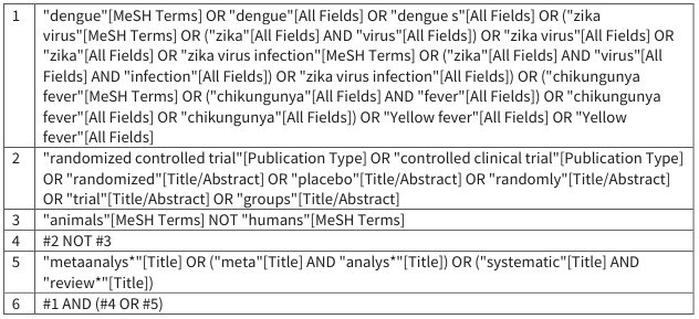
<table><tr><td>1</td><td>"dengue"[MeSH Terms] OR "dengue"[All Fields] OR "dengue s"[All Fields] OR ("zika virus"[MeSH Terms] OR ("zika"[All Fields] AND "virus"[All Fields]) OR "zika virus"[All Fields] OR "zika"[All Fields] OR "zika virus infection"[MeSH Terms] OR ("zika"[All Fields] AND "virus"[All Fields] AND "infection"[All Fields]) OR "zika virus infection"[All Fields]) OR ("chikungunya fever"[MeSH Terms] OR ("chikungunya"[All Fields] AND "fever"[All Fields]) OR "chikungunya fever"[All Fields] OR "chikungunya"[All Fields]) OR "Yellow fever"[All Fields] OR "Yellow fever"[All Fields]</td></tr><tr><td>2</td><td>"randomized controlled trial"[Publication Type] OR "controlled clinical trial"[Publication Type] OR "randomized"[Title/Abstract] OR "placebo"[Title/Abstract] OR "randomly"[Title/Abstract] OR "trial"[Title/Abstract] OR "groups"[Title/Abstract]</td></tr><tr><td>3</td><td>"animals"[MeSH Terms] NOT "humans"[MeSH Terms]</td></tr><tr><td>4</td><td>#2 NOT #3</td></tr><tr><td>5</td><td>"metaanalys*"[Title] OR ("meta"[Title] AND "analys*"[Title]) OR ("systematic"[Title] AND "review*"[Title])</td></tr><tr><td>6</td><td>#1 AND (#4 OR #5)</td></tr></table>
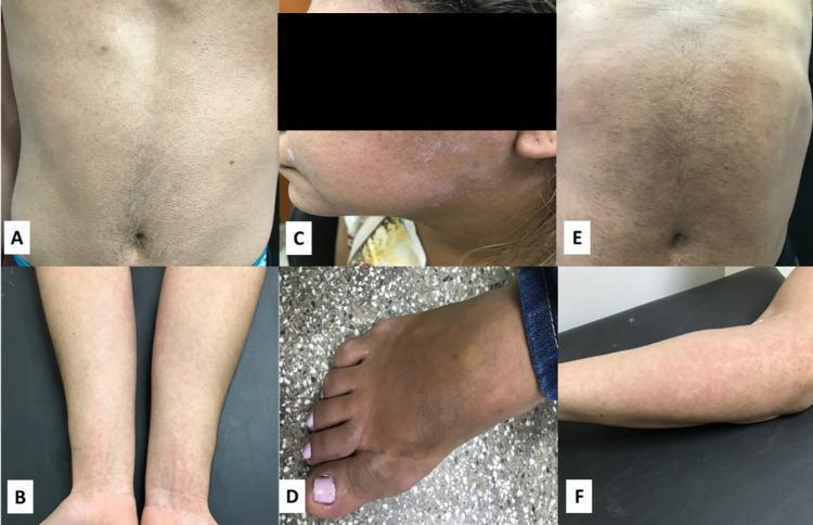
A series of clinical photographs (panels A-F) demonstrating dermatological manifestations of common arboviral infections including Zika virus (ZIKV), Chikungunya virus (CHIKV), and Dengue virus (DENV). Panels A and B illustrate a ZIKV-positive patient with a widespread maculopapular rash involving the thorax and upper limbs, characterized by small, erythematous, non-confluent spots. Panel C displays fine desquamation and peeling on the facial skin of a CHIKV-positive patient, while panel D shows localized peripheral edema in the lower limb (foot) of the same patient. Panels E and F represent a DENV-positive case, showing a diffuse, erythematous rash across the abdomen, thorax, and upper limbs. The images highlight key clinical findings used in the diagnosis of acute arboviral diseases: the classic maculopapular eruption of Zika and Dengue, and the specific secondary skin changes (peeling) and inflammatory edema often associated with Chikungunya. This collection serves as an educational reference for distinguishing clinical presentations of tropical viral exanthems.
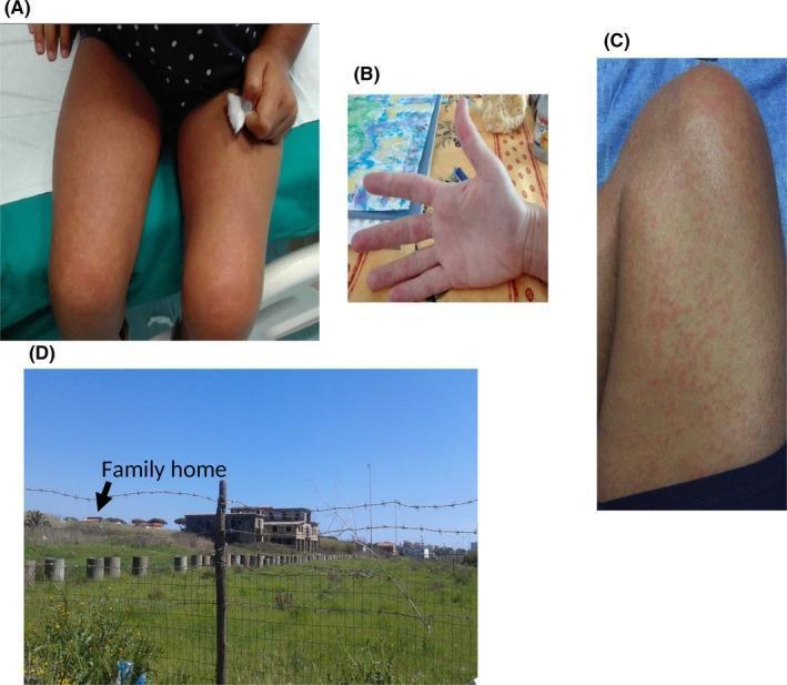
A composite image consisting of three clinical photographs (A, B, C) and one environmental photograph (D). Images A, B, and C demonstrate the dermatological manifestations of a family cluster infected with Chikungunya virus. Panels A and C show widespread, blanchable, erythematous maculopapular rashes on the inferior limbs (thighs and knees) of a child and an adult, characterized by diffuse reddish patches and small raised lesions. Panel B depicts similar maculopapular eruptions on the palmar surface and digits of the hand. These cutaneous findings are classic for the post-febrile phase of arboviral infections. Panel D provides epidemiological context, showing an abandoned building near the family's residence with numerous containers capable of holding stagnant water. This highlights an environmental risk factor for the breeding of Aedes mosquitoes, the primary vector for Chikungunya transmission. The collection serves as an educational tool for identifying clinical signs of Chikungunya and understanding the role of environmental surveillance in public health.
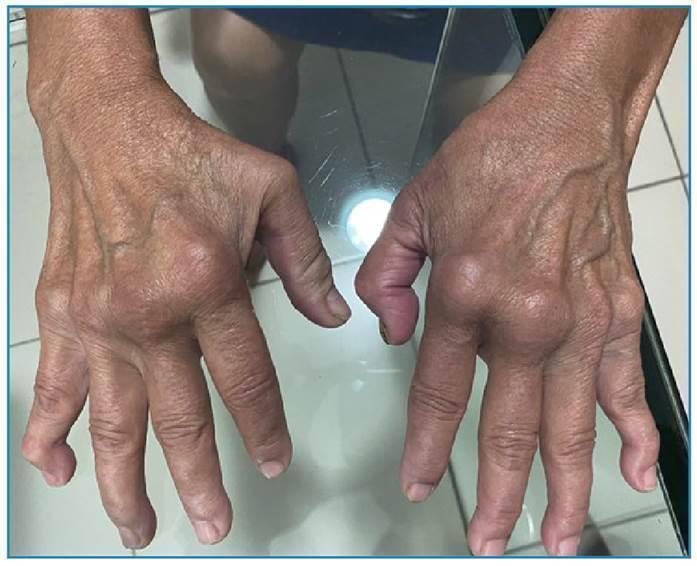
Clinical photograph of both hands in a dorsal view, demonstrating chronic, severe polyarticular joint deformities following chikungunya virus infection. Key findings include prominent, symmetrical swelling and enlargement of the proximal interphalangeal (PIP) joints, particularly affecting the index and middle fingers bilaterally. Notable distal interphalangeal (DIP) joint involvement is observed, including a fixed flexion deformity of the left index finger. There is evidence of muscle wasting and thinning of the overlying skin, which appears translucent and wrinkled with visible superficial dorsal veins. The clinical presentation mimics chronic rheumatoid arthritis (RA), highlighting the long-term sequelae of chronic chikungunya arthritis (CCA). The educational focus is on the long-term musculoskeletal impact of arboviral infections and the differential diagnosis of inflammatory polyarthritis.
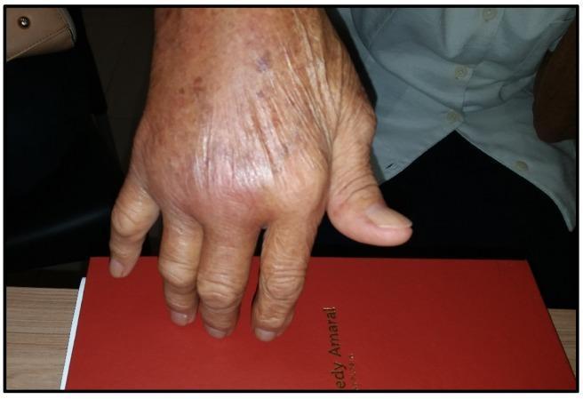
Clinical photograph of the dorsal aspect of an 82-year-old female's hand, demonstrating chronic polyarthritis as a sequela of Chikungunya virus infection (CHIKV). The image shows significant fusiform swelling and soft tissue inflammation predominantly localized to the metacarpophalangeal (MCP) joints and the wrist. Erythema is visible over the affected joints, suggesting active synovitis. There is a notable loss of normal anatomical contour and a slight ulnar deviation of the fingers, mimicking the clinical presentation of rheumatoid arthritis (RA). The overlying skin exhibits age-related changes including wrinkles, thinning, and actinic purpura/solar lentigines. This visual serves as a primary example of Chronic Chikungunya Arthritis (CCA) and its potential to act as an 'RA mimic' in geriatric populations, highlighting the long-term musculoskeletal morbidity associated with arboviral infections.
"chikungunya virus"[MeSH Terms] AND "pathogenesis"
Chikungunya pathology
1. Virus
- CHIKV is an enveloped, positive-strand RNA alphavirus of about 12 kb, in the Togaviridae family. Aedes aegypti and Ae. albopictus transmit it. (Firestein & Kelley's Rheumatology, p. 2556)
- Other alphaviruses that cause arthritis are O'nyong-nyong, Ross River, Sindbis, Mayaro and Semliki Forest virus.
2. Entry, cell targets and viremia
- Receptor: The entry receptor is mxra8 (matrix remodeling associated 8, also called limitrin). It binds a surface-exposed domain of the viral envelope protein. (Firestein)
- Main target: Fibroblasts are the main cells that CHIKV infects and replicates in. This includes human synovial and skin fibroblasts. (Firestein)
- Other targets: The virus also infects endothelial and epithelial cells, hepatocytes, neurons, astrocytes, muscle satellite cells and osteoblasts. (Firestein)
- Partial resistance: Resting fibroblasts and a CD141+/CD90+ subset of synovial fibroblasts resist infection to some degree.
- Viremia: Viremia is brief, usually 5 to 7 days. Type I interferon and antibodies mainly control it. (Firestein)
- Early disease: Most early manifestations come from the high viremia. It drives an intense inflammatory response, with immune cells infiltrating the joints and surrounding tissues. (Fitzpatrick's Dermatology, p. 3170)
- Joint pain: Direct infection of osteoblasts may help explain the arthralgia and arthritis. (Fitzpatrick's)
3. Joint and musculoskeletal pathology
- Acute stage: Synovitis and tenosynovitis develop, with infiltration of monocytes and macrophages.
- Chronic stage:
- Viral antigen and RNA persist in macrophages for up to 90 days. In one patient, viral RNA and protein were found in synovial macrophages 18 months after infection. (Fitzpatrick's, p. 3170)
- One study found no detectable viral persistence in synovial fluid at 22 months. The persistence mechanism is therefore not settled.
- Imaging: MRI findings include joint effusion, bone erosion, marrow edema, synovial thickening, tendonitis and tenosynovitis. Ultrasound often shows tenosynovitis or enthesopathy rather than true synovitis. (Firestein)
- Chronic arthritis: Some patients progress to erosive arthritis 1 to 2 years after infection. RF and anti-CCP are usually negative, so the picture looks like RA but is not classic seropositive RA. About one third of patients with joint pain in one study met ACR criteria for RA. (Firestein)
- Chronic pain: The pain is probably partly neuropathic as well as nociceptive. Patients often respond poorly to ordinary analgesics. (Firestein)
- Predictors of non-recovery: These are age over 45, severe initial joint pain, and underlying osteoarthritis. (Firestein)
4. Skin pathology
- Morbilliform rash: It affects 40 to 50% of patients, or more in some series, and may leave islands of sparing. Later hyperpigmentation is common. (Andrews' Diseases of the Skin; Fitzpatrick's)
- Bullous disease in infants:
- It occurs mostly in infants under 1 year. Blisters are flaccid, Nikolsky sign is positive, and the picture can resemble toxic epidermal necrolysis, with up to 80% of body surface area denuded.
- Biopsy: It shows an intraepidermal blister with free-floating acantholytic cells.
- Virus in the blister: Viral titers in blister fluid exceed those in blood. (Andrews')
- Other skin findings: Aphthous-like ulcers, acral lymphedema, and neuropathic acral changes (Raynaud-like, erythromelalgia) as late sequelae.
5. Other organs
- Nervous system: Meningoencephalitis, myelitis, Guillain-Barré syndrome and cranial nerve palsies.
- Other organs: Myocarditis, hepatitis, nephritis, uveitis and retinitis. (Red Book 2021)
- Lab changes: Lymphopenia, thrombocytopenia, and raised creatinine and transaminases.
- Perinatal infection: Vertical transmission can cause severe neonatal disease.
Images
Limits of this answer
- The library textbooks I found don't describe tissue histology of the synovium or other organs beyond the skin biopsy above.
- Recent PubMed searches on pathogenesis returned only bibliometric and drug reviews, with nothing that contradicts the textbook account.
- I can go deeper on the immunopathology of chronic arthritis (cytokines, T cells, and how it differs from RA) if you want.