
CHOLELITHIASIS classification, colour, Types, diagnosis, sing, pathology, diagnosis, treatment
cholelithiasis gallstones types cholesterol pigment
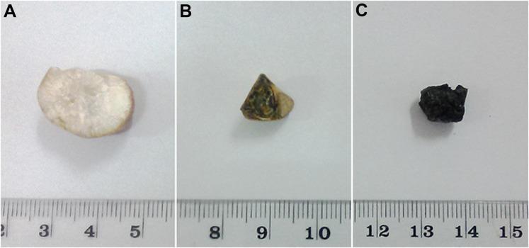
A clinical photograph displaying the cross-sectional morphology of three distinct types of human gallstones (cholelithiasis), presented with a centimeter scale for size reference. Stone A represents a pure cholesterol gallstone, characterized by a pale white color and a radially crystalline internal structure extending from the center to the periphery. Stone B depicts a mixed cholesterol gallstone, featuring a heterogeneous internal appearance with alternating light (cholesterol) and dark (pigment) concentric, crescentric layers. Stone C illustrates a pigment gallstone, showing a uniformly dark, black coloration throughout its cross-section with an irregular, rough surface texture. The image serves as an educational reference for distinguishing gallstone subtypes based on macroscopic internal and external characteristics, including color distribution, layering patterns, and crystalline arrangement.
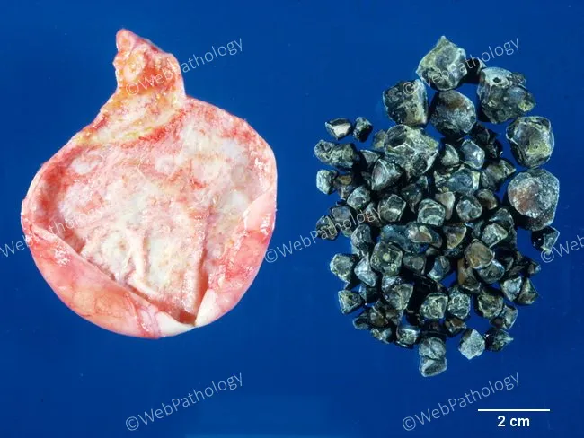
This gross pathology photograph documents massive cholelithiasis with densely packed pigment stones within the gallbladder. The left panel shows a partially opened gallbladder specimen with a pale mucosal surface and residual bile, while the right cluster consists of numerous dark stones laid out for visualization. The stones are black to dark green in color, with a mulberry-like, irregular morphology and spiculated or molded contours. They are small in size, typically 0.2–0.5 cm, but a minority can reach larger dimensions; in this case, a very heavy burden is present with approximately 130 stones. The stones are hard and resistant to crushing, contrasting with brown pigment or cholesterol stones which are usually softer. The overall appearance is characteristic of black pigment gallstones (calcium bilirubinate) formed in the setting of chronic biliary infection or hemolysis. This stone type is often radiopaque and may be associated with pigment deposition in the gallbladder mucosa and chronic cholecystitis. Clinically, pigment stones accompany biliary stasis and inflammatory changes and increase risk for gallstone-related complications such as biliary colic, cholecystitis, or pancreatitis. This specimen provides a valuable teaching example for stone morphology, composition, and differential diagnosis between pigment and cholesterol stones, and illustrates the extreme stone burden possible in pigment stone disease.
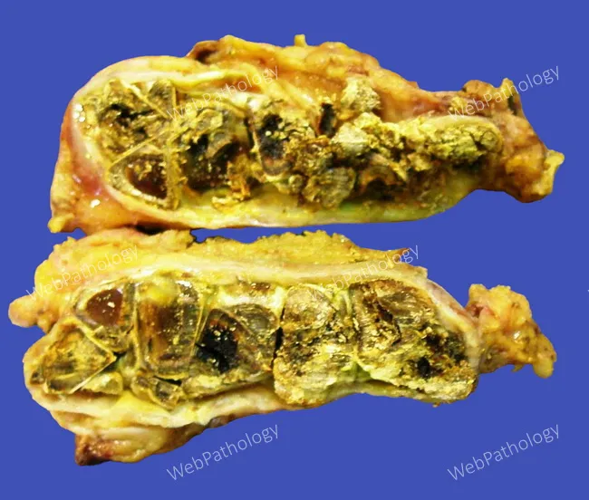
Gross pathology photograph of brown pigment gallstones (choleliths) retrieved from the gallbladder, featuring a cross sectional view of laminated surfaces. These pigment stones are composed of calcium salts of unconjugated bilirubin (monomeric calcium bilirubinate) embedded in a glycoprotein matrix, with interleaved calcium salts of fatty acids such as palmitate and stearate. Relative to black pigment stones, brown pigment stones contain a higher proportion of cholesterol and frequently incorporate small quantities of bile salts, phospholipids, and bacterial residues. The cut surface is stratified (lamellated), with bilirubinate-rich layers alternating with calcium palmitate-rich layers, reflecting alternating deposition of pigment and fatty acid calcium salts during growth. The overall appearance on the specimen can closely resemble fecal matter, a feature highlighted in this photograph. Image courtesy of Dr. Ibrahim Zardawi, used with permission. Clinically, brown pigment stones are associated with biliary infection and stasis, and are more common in settings of cholangitis or enteric bacterial activity producing beta-glucuronidase that promotes pigment precipitation. These stones are typically radiolucent and may be mixed with cholesterol components. They contribute to cholelithiasis risk and may require cholecystectomy. Potential diagnostic uses include educational reference for gallstone composition, histologic correlation, and differential diagnosis with black pigment stones and cholesterol stones.
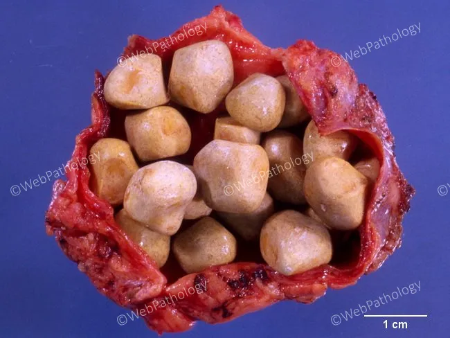
This is a macroscopic gross pathology photograph of a cholecystectomy specimen demonstrating cholelithiasis confined to the gallbladder. The lumen is densely packed with multiple cholesterol stones, disc-like to rounded, with smooth to slightly faceted surfaces and a pale yellow to tan coloration. Individual calculi measure approximately 0.5–1.5 cm in diameter, as inferred from the included 1 cm scale bar. The surrounding gallbladder wall is partially exposed with a red, friable serosa and mucosa not fully visible due to tissue handling. The stones appear radiolucent on plain radiographs in life; here they are visible only as macroscopic calculus clusters. The specimen illustrates classic features of cholesterol stones: low pigment, cholesterol-supersaturated bile and hypomotility contributing to stone formation. The image is optimized to highlight gross morphology suitable for educational and diagnostic reference in surgical pathology, hepatobiliary disease, and gastroenterology. Clinically, cholelithiasis is common in adults and may present with biliary colic or cholecystitis; cholesterol stones constitute the majority of gallstones. The image provides visual confirmation of stone burden within the gallbladder and can aid differential with pigment stones or mixed stones and in teaching radiology-pathology correlations. This image illustrates macroscopic appearance for teaching gross pathology and correlating with surgical pathology findings globally.
gallbladder ultrasound cholelithiasis diagnosis acoustic shadow
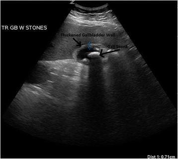
This diagnostic ultrasound image in the transverse plane demonstrates a gallbladder with classic sonographic findings of acute cholecystitis and cholelithiasis. A large, prominent hyperechoic gallstone is visible within the lumen of the gallbladder, exhibiting a characteristic posterior acoustic shadow that extends inferiorly, indicating the dense nature of the calculus. The gallbladder wall is clearly labeled and appears significantly thickened (measuring 0.71 cm as per the distal measurement marker), which is a primary diagnostic criterion for inflammation. Additionally, there is evidence of pericholecystic edema, visualized as a hypoechoic, hazy area surrounding the outer wall of the gallbladder. These features—cholelithiasis, wall thickening, and pericholecystic fluid/edema—collectively support a diagnosis of acute cholecystitis. The image serves as an educational tool for identifying the sonographic hallmarks of biliary disease in a clinical setting.
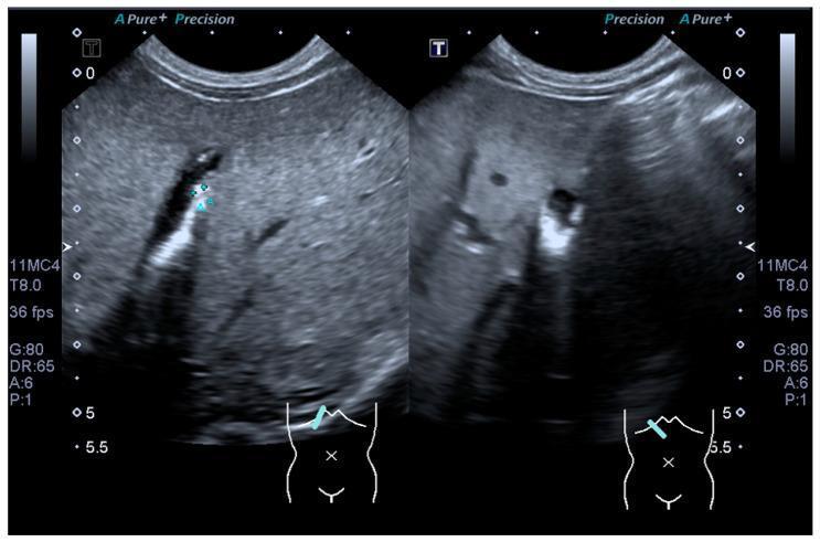
This diagnostic ultrasound image displays a dual-view scan of the gallbladder within the upper right quadrant. The primary finding consists of multiple hyperechoic foci located within the dependent portion of the gallbladder lumen. These structures exhibit high echogenicity relative to the surrounding anechoic bile. Distinctive posterior acoustic shadowing is visible deep to these foci, where the ultrasound signal is attenuated, a classic hallmark of cholelithiasis (gallstones) or dense biliary sludge. The surrounding liver parenchyma appears homogeneous with normal echotexture. These images are captured using a curvilinear probe, as indicated by the sector-shaped field of view and the anatomical orientation marker showing placement in the subcostal region. The presence of these findings is clinically significant for diagnosing gallbladder pathology and distinguishing between biliary sludge and formed calculi based on the intensity of the acoustic shadow.
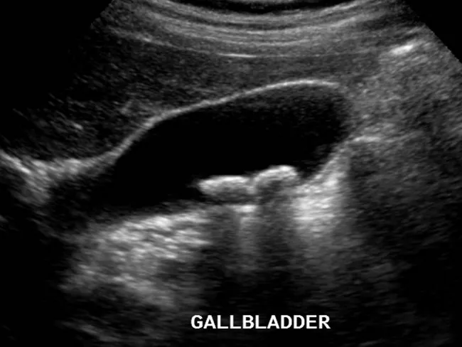
Ultrasound examination of the gallbladder using grayscale B-mode ultrasonography (transabdominal approach) demonstrates two echogenic foci within the gallbladder lumen. Both features are dependent on the gallbladder, implying intraluminal calculi rather than extrinsic objects. Each lesion casts a posterior acoustic shadow, a hallmark of gallstones, and their appearance distinguishes calcified cholesterol stones from gallbladder polyps, which typically lack shadowing. The stones measure over 2 millimeters in diameter, consistent with cholelithiasis and enabling reliable detection by ultrasound. The gallbladder wall appears without significant thickening, and there is no overt pericholecystic fluid evident on this image, arguing against acute cholecystitis at the time of imaging. The study highlights the classic sonographic criteria for gallstone disease: echogenic mobile calculi with clean shadowing, gravity-dependent location, and recognition of potential sludge in the gallbladder lumen. This imaging feature set supports clinical decisions regarding symptomatic biliary colic, risk stratification for complications, and surgical planning such as cholecystectomy. In differential considerations, gallbladder polyps or sludge can mimic stones; however, shadowing and mobility aid differentiation. Overall, ultrasound remains the initial modality of choice for suspected biliary pathology due to safety, accessibility, and high diagnostic yield. Correlative clinical data and laboratory tests further refine management decisions. Guiding patient-specific treatment options.
CHOLELITHIASIS (Gallstones) - Comprehensive Review
Definition
Classification / Types
1. Cholesterol Stones (most common in the West)
- Contain >50% crystalline cholesterol monohydrate
- Account for 80% of gallstones in the USA and Europe
- The remaining 20% is an admixture of calcium salts, bile acids, bile pigments, and phospholipids
- Arise exclusively in the gallbladder
2. Pigment Stones
| Feature | Black Pigment Stones | Brown Pigment Stones |
|---|---|---|
| Location | Sterile gallbladder bile | Infected bile ducts |
| Composition | Oxidized polymers of calcium bilirubinate + calcium carbonate/phosphate + mucin glycoprotein | Calcium bilirubinate + calcium palmitate + calcium stearate + cholesterol |
| Cause | Chronic hemolysis, cirrhosis | Bile stasis + bacterial infection (beta-glucuronidase) |
| Associations | Hereditary spherocytosis, sickle cell disease, mechanical heart valves | Clonorchis sinensis, Ascaris, biliary stents, biliary strictures |
3. Mixed Stones
- Contain 50-99% pure cholesterol plus admixture of other components
- Often faceted due to tight opposition to adjacent stones
Color
| Stone Type | Color |
|---|---|
| Pure cholesterol stones | Pale yellow |
| Mixed cholesterol stones | Gray-white to black (with increasing calcium) |
| Black pigment stones | Jet black |
| Brown pigment stones | Brown (resembles fecal matter on cross-section) |
Pathogenesis
Cholesterol Stone Formation (4 key steps):
- Supersaturation of bile with cholesterol - when cholesterol concentrations exceed the solubilizing capacity of bile salts and lecithin micelles
- Hypomotility of the gallbladder - stasis allows crystal aggregation
- Accelerated cholesterol crystal nucleation - formation of solid cholesterol monohydrate crystals
- Hypersecretion of mucus - traps nucleated crystals, leading to macroscopic stone growth
Cholesterol is rendered soluble in bile by forming micelles with bile salts and lecithins. When bile is supersaturated (e.g., due to obesity, oral contraceptives, ileal disease), unstable phospholipid vesicles form, and cholesterol crystals nucleate.
Pigment Stone Formation:
- Elevated unconjugated bilirubin in bile is the key step
- Bacterial beta-glucuronidases (E. coli, Ascaris, Clonorchis) hydrolyze bilirubin glucuronides → unconjugated bilirubin precipitates as calcium bilirubinate
- In hemolytic anemias: increased secretion of conjugated bilirubin → ~1% deconjugates → pigment stone formation
- Hereditary factor: ABCG8 gene variant is associated with increased cholesterol gallstone risk
Risk Factors ("4 Fs" + more)
The Classic Mnemonic: 4 Fs
- Female
- Fat (obesity)
- Forty (age >40)
- Fertile (pregnancy/estrogen)
For Cholesterol Stones:
- Northern European, North/South American, Native American (Pima, Hopi, Navajo - prevalence up to 75%)
- Female sex hormones, oral contraceptives, pregnancy
- Obesity and metabolic syndrome
- Rapid weight loss (incidence 30% after bariatric surgery)
- Gallbladder stasis, TPN, prolonged fasting
- Hyperlipidemia, inborn bile acid metabolism disorders
- Hereditary (ABCG8 mutations)
For Pigment Stones:
- Chronic hemolytic anemias (sickle cell, spherocytosis, thalassemia)
- Biliary infection/parasites
- Ileal disease (Crohn's disease), ileal resection, cystic fibrosis
- Cirrhosis
- Rural populations in Asia
Morphology (Gross Pathology)
| Feature | Cholesterol Stone | Black Pigment Stone | Brown Pigment Stone |
|---|---|---|---|
| Shape | Round/ovoid, faceted | Small, irregular, spiculated | Lamellated (layered cross-section) |
| Surface | Hard, finely granular | Mulberry-like, rough | Stratified layers |
| Cross-section | Glistening radiating crystalline palisade | Uniformly dark | Alternating bilirubinate/calcium layers |
| Radiology | Radiolucent (10-20% radiopaque if calcified) | Often radiopaque | Radiolucent |
Clinical Signs & Symptoms
Symptomatic Presentation:
- Biliary "colic" - constant (not truly colicky), severe, excruciating pain
- Location: Right upper quadrant (RUQ) or epigastric, may radiate to right shoulder or back
- Precipitated by fatty meals (gallbladder contraction forces stone against outlet)
- Associated nausea, vomiting
- Pain often starts at night and wakes the patient
- Flat tolerance and food intolerance (especially fatty foods)
Key Clinical Signs:
- Murphy's sign: RUQ tenderness exacerbated during inspiration on deep palpation in right subcostal region - pathognomonic of acute cholecystitis
- Courvoisier's sign: Palpable, non-tender gallbladder in the presence of jaundice - NOT due to gallstones; suggests periampullary malignancy
- Palpable mass (omentum walling off inflamed gallbladder)
Complications (Summary Box from Bailey & Love):
- Acute cholecystitis
- Chronic cholecystitis
- Empyema
- Mucocele
- Perforation
- Biliary obstruction (obstructive jaundice)
- Acute cholangitis
- Acute pancreatitis
- Gallstone ileus (large stone erodes into duodenum → intestinal obstruction)
- Bouveret syndrome (stone lodges at duodenal bulb)
- Mirizzi syndrome (stone compresses CBD from outside)
- Increased risk of gallbladder carcinoma
Small stones ("gravel") are more dangerous than large ones - they are more likely to enter the cystic or common bile duct and cause obstruction.
Diagnosis
Imaging
1. Ultrasound (USG) - Investigation of Choice
- Sensitivity and specificity >90% for cholelithiasis
- Shows: hyperechoic foci with posterior acoustic shadow within gallbladder lumen
- Gravity-dependent movement of stones (distinguishes from polyps)
- Also shows: gallbladder wall thickening, pericholecystic fluid (in cholecystitis)
- Safe in pregnancy
2. Plain X-ray (AXR)
- Only 10-20% of cholesterol stones are radiopaque
- Black pigment stones are often radiopaque
- Brown pigment stones are radiolucent
- Mercedes-Benz sign: radiating lucencies in cholesterol stones
3. CT Scan
- Useful for complications (pancreatitis, perforation, fistula)
- Better at detecting CBD stones and biliary complications
4. MRCP (Magnetic Resonance Cholangiopancreatography)
- Gold standard for CBD stones / choledocholithiasis
- Non-invasive; no radiation
5. ERCP (Endoscopic Retrograde Cholangiopancreatography)
- Both diagnostic and therapeutic for CBD stones
- Allows sphincterotomy and stone extraction
6. HIDA Scan (Hepatobiliary Iminodiacetic Acid)
- Functional imaging; useful when USG equivocal
- Assesses gallbladder ejection fraction
Laboratory Investigations
| Test | Finding |
|---|---|
| CBC | Leukocytosis in cholecystitis/cholangitis |
| LFTs (ALP, bilirubin, transaminases) | Elevated in CBD obstruction/cholangitis |
| Serum amylase/lipase | Elevated if associated pancreatitis |
| CRP | Elevated in acute cholecystitis |
Tokyo Guidelines Diagnostic Criteria (Acute Cholecystitis):
- A - Local signs: Murphy's sign, RUQ mass/pain/tenderness
- B - Systemic signs: Fever, elevated CRP, elevated WBC
- C - Imaging: Gallstones + wall thickening/pericholecystic fluid on USG
Treatment
1. Asymptomatic Cholelithiasis
- Expectant (watchful waiting) - evidence from GREPCO studies supports this
- Cumulative probability of biliary colic: 11.9% at 2 years, 25.8% at 10 years; complications only 3% at 10 years
- Prophylactic cholecystectomy NOT routinely indicated
2. Symptomatic Cholelithiasis
Surgical - Gold Standard: Laparoscopic Cholecystectomy
- Treatment of choice for symptomatic cholelithiasis
- Short hospital stay (~1 day), low complication rate
- Should NOT be delayed (recurrence rate of symptoms: 92% if initially in 1st trimester, 64% in 2nd, 44% in 3rd)
- Open cholecystectomy reserved for complicated cases (dense adhesions, perforation, malignancy)
Medical Management (Dissolution Therapy)
- Ursodeoxycholic acid (Ursodiol): 8-10 mg/kg/day
- Effective only for small (<5mm), radiolucent cholesterol stones
- Incomplete stone clearance; high recurrence after stopping
- Used prophylactically (300-400 mg BID) after bariatric surgery to prevent stone formation (reduces incidence from 30% to 4%)
- Treatment duration: 6-24 months
Endoscopic
- ERCP + sphincterotomy: For choledocholithiasis (CBD stones)
- Pre-operative ERCP followed by laparoscopic cholecystectomy is standard if CBD stones suspected
Other
- Extracorporeal Shock Wave Lithotripsy (ESWL): Fragmentation of stones; rarely used now
- Contact dissolution: Direct instillation of methyl tert-butyl ether (MTBE); historical
Special Situations:
- Laparoscopic cholecystectomy is preferred over open surgery
- Safest in 1st or 2nd trimester
- Laparoscopic approach: reduced fetal mortality (OR 0.28-0.63), maternal complications, and shorter hospital stay vs open
- Gallstone pancreatitis in pregnancy: maternal mortality 15%, fetal mortality 60% - requires surgery
- Synchronous cholecystectomy recommended if symptomatic cholelithiasis present
- Ursodiol 400 mg BID for 6 months post-op if rapid weight loss expected
- IV fluids, analgesics, antibiotics (covering gram-negatives and anaerobes)
- Early laparoscopic cholecystectomy (within 72 hours) is preferred over delayed approach
Summary: Key Mnemonics
| Mnemonic | Content |
|---|---|
| 4 Fs | Female, Fat, Forty, Fertile |
| Cholesterol stone "4 factors" | Supersaturation, Hypomotility, Nucleation, Mucus trapping |
| USG findings | Hyperechoic + Posterior acoustic shadow + Gravity-dependent |
| Murphy's sign | Acute cholecystitis (NOT cholelithiasis per se) |
| Courvoisier's sign | Jaundice + palpable gallbladder = NOT stones (think malignancy) |