
Oligodendroglioma imaging features
I'll read the medical library skill first, then search for oligodendroglioma imaging features.
Reading File
Now I'll run parallel searches across the library and PubMed simultaneously.
Finding Sources
Searching PubMed
oligodendroglioma MRI imaging features
The Grainger & Allison radiology textbook has the most relevant imaging section. Let me read it along with other key sources in parallel.
Reading File
Reading File
Reading File
I have rich imaging content. Let me now read the remaining Grainger & Allison radiology content and fetch the key imaging figures.
Reading File
Loading Image
I now have comprehensive information from multiple authoritative sources. Here is the full answer:
Oligodendroglioma: Imaging Features
Molecular definition (WHO 2021): Oligodendroglioma is defined by IDH mutation + 1p/19q codeletion. This molecular signature takes diagnostic priority over microscopic appearances alone.
Location
- Almost exclusively cerebral hemispheres
- Frontal lobe is the most common site (followed by temporal lobe)
- Typically involves both subcortical white matter and cortex (cortico-subcortical involvement is characteristic)
- Patients often present with years of antecedent seizures before diagnosis
CT Features
- Calcification is the hallmark: present in up to 90% of oligodendrogliomas on CT
- Can be central, peripheral, or ribbon-like (gyriform/cortical pattern is characteristic)
- Coarse, dense calcification is more common than in other low-grade gliomas
- Large, partially solid tumour involving cortex
- Cysts may be present
- Mild to moderate enhancement in at least 20% of WHO grade II tumours; does not automatically imply anaplasia
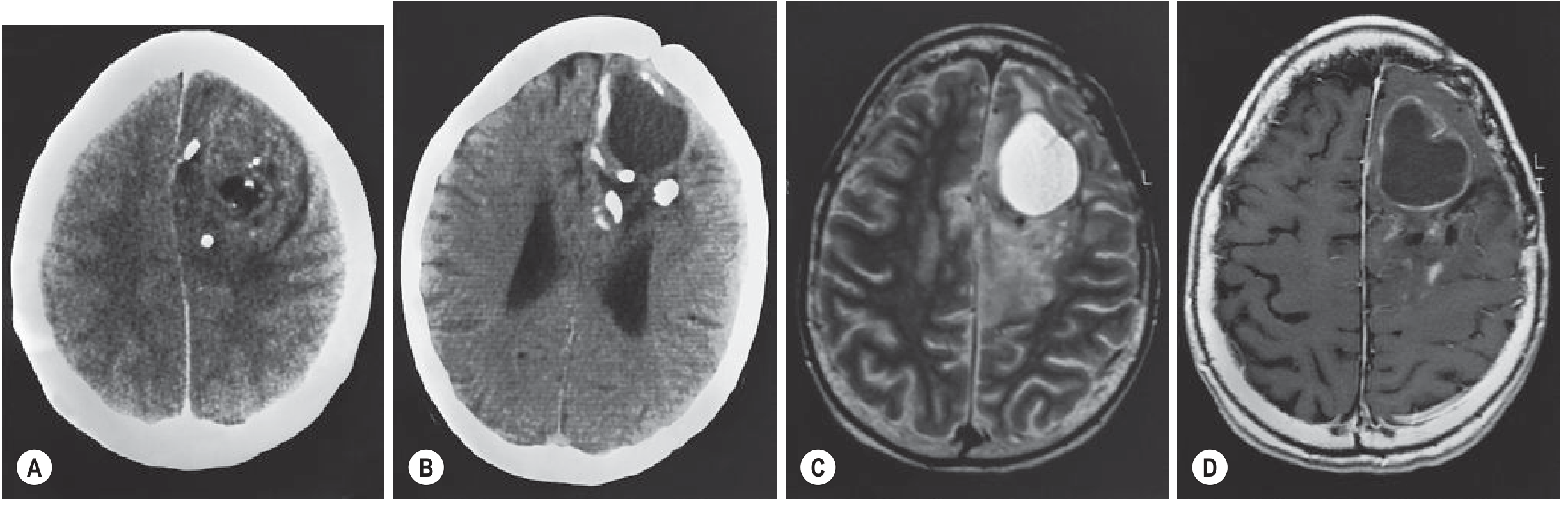
Fig. 55.7 (Grainger & Allison) - WHO Grade II Oligodendroglioma shown below: CT (A) shows large left frontal tumour with cortical involvement, predominantly solid with irregular enhancement, coarse calcification, and cysts. Follow-up CT (B), T2 MRI (C), and T1 post-contrast MRI (D) demonstrate progressive cyst formation and calcification.
MRI Features
| Sequence | Appearance |
|---|---|
| T2/FLAIR | Hyperintense (diffusely infiltrative) |
| T1 | Hypointense |
| T1 post-contrast | Mild-moderate enhancement in ~20-50% of grade II; more common in grade III |
| T2*/SWI | Marked signal loss ("blooming") due to calcification or haemorrhage |
| DWI | Typically no restricted diffusion (unlike higher-grade gliomas) |
Key MRI points:
- Calcification on MRI: Typically T2 hypointense and T1 hyperintense; causes marked signal dropout on T2*/SWI (susceptibility-weighted) sequences
- No FLAIR-mismatch sign - the FLAIR-mismatch sign (T2 bright but FLAIR suppressed solid core) is specific for IDHmut astrocytoma (1p19q-intact), NOT oligodendroglioma - its absence helps differentiate
- Posterior extent and infiltration are best seen on MRI rather than CT
- Calcification appears as non-specific low signal areas on MRI and is much less apparent than on CT
Advanced MRI Features
Perfusion MRI (DSC/PWI)
- Low-grade oligodendrogliomas commonly show moderately elevated rCBV - this can overlap with IDH wild-type (higher grade) features and may prompt early tissue diagnosis
- Both WHO II and III oligodendrogliomas express proangiogenic mitogens and contain regions of increased vascular density with finely branching capillaries ("chicken wire" pattern)
MR Spectroscopy (MRS)
- Increased myo-inositol/glycine, glutamine, and glutamate in low-grade tumours
- Presence of lipid/lactate peaks suggests anaplastic (grade III) transformation
- Where perfusion is non-specifically elevated, MRS can help differentiate anaplastic oligodendroglioma
Enhancement and Grading
- New enhancement or accelerated growth in a known IDHmut 1p/19q-codeleted glioma raises concern for anaplastic transformation (WHO grade III)
- Few, if any, progressive IDHmut 1p/19qdel gliomas ever fulfil histological criteria for glioblastoma (unlike IDHwt astrocytoma)
Imaging vs. Astrocytoma
| Feature | Oligodendroglioma | IDHmut Astrocytoma |
|---|---|---|
| Calcification | Very common (up to 90% on CT) | Less common |
| Enhancement | More common (~20-50% grade II) | Less common in grade II |
| FLAIR mismatch | Absent | Present (~50%) - specific |
| rCBV (perfusion) | Moderately elevated | Usually lower in grade II |
| Gyriform calcification | Characteristic | Rare |
Key Teaching Points
- Calcification on CT is the imaging hallmark - gyriform/cortical pattern is characteristic but not pathognomonic
- On MRI, calcification "blooms" on SWI/T2* - the best sequence to detect it
- Enhancement does not equal high grade, but new/progressive enhancement in a known oligodendroglioma warrants repeat biopsy
- Absence of FLAIR-mismatch sign helps suggest oligodendroglioma over IDHmut astrocytoma when molecular data is unavailable
- Despite elevated perfusion suggesting aggressiveness, prognosis is significantly better than astrocytoma (mean survival ~8-10+ years for grade II)
Sources: Grainger & Allison's Diagnostic Radiology, p. 1407-1408 | Robbins & Kumar Basic Pathology, p. 859-860 | Bradley and Daroff's Neurology in Clinical Practice
This is a shared conversation. Sign in to Orris to start your own chat.