
Acid base balance in the body and it's regulation in detail
Reading File
Reading File
Reading File
Finding Sources
Finding Sources
Finding Sources
Searching PubMed
"Acid-Base Equilibrium"[MeSH Terms] AND regulation
Searching the Web
Merck Manual acid-base balance regulation buffers respiratory renal compensation
Finding Sources
Finding Sources
Reading Web Page
https://www.merckmanuals.com/professional/nephrology/acid-ba…
Reading File
Reading File
Searching Images
acid base balance renal regulation bicarbonate phosphate ammonium buffer diagram
Finding Sources
Reading File
Reading File
Reading File
Acid-base balance: definition and normal values
Acid-base balance is the maintenance of hydrogen ion concentration, ([H^+]), within a narrow range so enzymes, ion channels, proteins, and cellular metabolism function normally.
- pH = -log₁₀[H⁺]
- Normal arterial blood pH: 7.35-7.45 (approximately 7.4)
- Acidemia: pH < 7.35
- Alkalemia: pH > 7.45
- Usual arterial values:
- PaCO₂: 35-45 mmHg
- HCO₃⁻: 22-26 mEq/L
Even small pH changes matter because the pH scale is logarithmic. A fall in pH means a rise in free hydrogen ions. Costanzo notes that normal arterial pH is about 7.37-7.42 and intracellular pH is slightly lower, about 7.2. - Costanzo Physiology, 7th ed., p. 317
The central principle: bicarbonate to carbon dioxide ratio
The main extracellular buffer is the bicarbonate-carbonic acid system:
[
CO_2 + H_2O \leftrightarrow H_2CO_3 \leftrightarrow H^+ + HCO_3^-
]
Carbonic anhydrase, especially abundant in red cells and renal tubular cells, speeds this reaction.
The relationship is expressed by the Henderson-Hasselbalch equation:
[
\text{pH} = 6.1 + \log\left(\frac{[HCO_3^-]}{0.03 \times P_{CO_2}}\right)
]
At normal values:
[
\text{pH} = 6.1 + \log\left(\frac{24}{0.03 \times 40}\right) \approx 7.40
]
So pH is governed by a ratio:
[
\frac{\text{Kidney-controlled } HCO_3^-}{\text{Lung-controlled } P_{CO_2}}
]
-
More HCO₃⁻ raises pH.
-
More CO₂ lowers pH.
-
The lungs regulate the denominator, PaCO₂.
-
The kidneys regulate the numerator, plasma HCO₃⁻.
-
Guyton and Hall Textbook of Medical Physiology, pp. 409-410
Sources of acid in the body
1. Volatile acid: carbon dioxide
Aerobic cellular metabolism continuously produces large quantities of CO₂. CO₂ acts as an acid source because it forms carbonic acid in water. It is called volatile acid because the lungs can excrete it.
[
CO_2 + H_2O \rightarrow H_2CO_3 \rightarrow H^+ + HCO_3^-
]
Approximately 13,000-20,000 mmol of CO₂ may be produced daily. It is eliminated mainly through ventilation. - Costanzo Physiology, 7th ed., p. 317
2. Fixed or nonvolatile acids
These cannot be removed by the lungs and must ultimately be excreted by the kidneys. They include:
- Sulfuric acid from metabolism of sulfur-containing amino acids such as methionine and cysteine
- Phosphoric acid from phospholipids and dietary phosphate
- Lactic acid in hypoxia, shock, seizures, or strenuous exercise
- Ketoacids in fasting, starvation, alcoholic ketoacidosis, and diabetic ketoacidosis
- Toxic acids, for example formic acid from methanol and oxalic/glycolic acid from ethylene glycol
The routine fixed-acid burden is roughly 50 mmol/day, but rises substantially in disease. - Costanzo Physiology, 7th ed., p. 317
Three lines of defense against pH change
| Mechanism | Main action | Onset | Capacity / role |
|---|---|---|---|
| Chemical buffers | Bind or release H⁺ | Seconds | Immediate but cannot remove acid |
| Respiratory regulation | Alters CO₂ excretion | Minutes | Rapid control of volatile acid |
| Renal regulation | Excretes H⁺, regenerates HCO₃⁻ | Hours to days | Slowest but most powerful long-term control |
A useful sequence is:
Buffers minimize the initial change. Lungs remove CO₂. Kidneys remove fixed acid and restore bicarbonate.
1. Chemical buffer systems
A buffer is a weak acid plus its conjugate base. It resists a sudden pH change by accepting or donating hydrogen ions.
[
HA \leftrightarrow H^+ + A^-
]
- If acid is added: (A^-) binds H⁺ to form (HA)
- If base is added: (HA) releases H⁺, reducing the rise in pH
Buffers act immediately but do not eliminate hydrogen ions from the body. They temporarily contain the disturbance until lung and kidney responses occur.
A. Bicarbonate buffer system
[
H^+ + HCO_3^- \leftrightarrow H_2CO_3 \leftrightarrow CO_2 + H_2O
]
This is the most important extracellular buffer because both components are independently controlled:
- Lungs remove CO₂.
- Kidneys conserve, generate, or excrete HCO₃⁻.
Example: after addition of HCl,
[
HCl + NaHCO_3 \rightarrow NaCl + H_2CO_3 \rightarrow CO_2 + H_2O
]
The CO₂ can then be exhaled.
B. Protein buffers
Proteins contain negatively charged groups that accept H⁺ and acidic groups that can donate H⁺.
[
Pr^- + H^+ \leftrightarrow HPr
]
Proteins are important intracellular buffers. Hemoglobin is especially important in red blood cells.
In systemic capillaries, deoxygenated hemoglobin binds H⁺ generated from CO₂ hydration. In pulmonary capillaries, oxygenation of hemoglobin promotes H⁺ release, which combines with HCO₃⁻ to form CO₂ for exhalation.
C. Phosphate buffer system
[
HPO_4^{2-} + H^+ \leftrightarrow H_2PO_4^-
]
Phosphate is relatively less important in plasma because its concentration is low. It is much more important:
- Intracellularly
- In renal tubular fluid, where it buffers secreted H⁺ and permits urinary acid excretion
D. Bone buffering
Bone contains carbonate and phosphate salts that can buffer chronic acid loads. This contributes to buffering in prolonged acidosis, but sustained acid buffering by bone can contribute to mineral loss and bone disease.
A current Merck Manual overview likewise describes immediate intra- and extracellular buffering and notes the role of bone, particularly with acid loads.
2. Respiratory regulation of acid-base balance
The lungs regulate arterial CO₂ through alveolar ventilation.
[
P_{aCO_2} \propto \frac{\dot V_{CO_2}}{\dot V_A}
]
Where:
- (\dot V_{CO_2}) = CO₂ production
- (\dot V_A) = alveolar ventilation
Thus:
- Increased ventilation lowers PaCO₂.
- Decreased ventilation raises PaCO₂.
Because CO₂ behaves as an acid:
- Hyperventilation causes lower PaCO₂ and tends to raise pH.
- Hypoventilation causes higher PaCO₂ and tends to lower pH.
Control of ventilation
A fall in pH, rise in PaCO₂, or both stimulates respiratory drive through chemoreceptors.
Central chemoreceptors
Located in the medulla. They respond mainly to increased CO₂, which diffuses into cerebrospinal fluid and generates H⁺.
Peripheral chemoreceptors
Located in the carotid and aortic bodies. They respond to:
- Decreased arterial pH
- Increased PaCO₂
- Marked hypoxemia
Respiratory compensation
Metabolic acidosis
Primary defect: ↓ HCO₃⁻
The compensatory response is hyperventilation:
[
\downarrow HCO_3^- \rightarrow \downarrow pH \rightarrow \uparrow ventilation \rightarrow \downarrow P_{aCO_2}
]
This is seen clinically as deep, rapid Kussmaul breathing in severe diabetic ketoacidosis.
Expected compensation can be estimated using Winter's formula:
[
Expected\ P_{CO_2} = 1.5 \times [HCO_3^-] + 8 \pm 2
]
Metabolic alkalosis
Primary defect: ↑ HCO₃⁻
Compensation is hypoventilation, causing CO₂ retention. This is limited because marked hypoventilation would produce dangerous hypoxemia.
Respiratory compensation begins within minutes and is substantial but never fully corrects the primary disorder. A pH that has crossed to the opposite side of normal suggests a mixed acid-base disorder, not simple compensation. The Merck Manual discussion emphasizes that metabolic disorders prompt respiratory compensation and respiratory disorders prompt renal compensation.
3. Renal regulation of acid-base balance
The kidneys provide the final long-term defense. Their three major tasks are:
- Reabsorb nearly all filtered bicarbonate
- Secrete hydrogen ions
- Generate new bicarbonate while excreting net acid
The kidney response begins over hours and reaches maximum effectiveness over days.
A. Reabsorption of filtered bicarbonate
The kidneys filter a large amount of HCO₃⁻ daily. Under normal conditions, nearly all is reclaimed, mainly in the proximal tubule.
Proximal-tubule mechanism
- Tubular cells secrete H⁺ into the lumen, mainly through the Na⁺-H⁺ exchanger.
- Secreted H⁺ combines with filtered HCO₃⁻:
[
H^+ + HCO_3^- \rightarrow H_2CO_3
]
- Luminal carbonic anhydrase converts carbonic acid to CO₂ and water:
[
H_2CO_3 \rightarrow CO_2 + H_2O
]
- CO₂ enters the tubular cell.
- Intracellular carbonic anhydrase reforms H⁺ and HCO₃⁻.
- H⁺ is secreted again; HCO₃⁻ returns to blood.
This process reclaims filtered bicarbonate. It does not produce net acid excretion or new bicarbonate.
B. Hydrogen-ion secretion
H⁺ is secreted in:
- Proximal tubule: predominantly through Na⁺-H⁺ exchange
- Thick ascending limb
- Distal tubule and collecting duct: mainly through H⁺-ATPase and H⁺-K⁺-ATPase in alpha-intercalated cells
The collecting duct can generate a very low urine pH, usually to a minimum around 4.5. Free H⁺ excretion alone is small because urine cannot contain enough free H⁺ to dispose of the daily acid burden. Hence urinary buffers are essential.
C. Excretion as titratable acid
Secreted H⁺ binds filtered buffers, mainly phosphate:
[
HPO_4^{2-} + H^+ \rightarrow H_2PO_4^-
]
The hydrogen ion is then excreted as (H_2PO_4^-). This is called titratable acid because it can be measured by titrating urine back to plasma pH.
Each H⁺ excreted in this form adds one new HCO₃⁻ to blood.
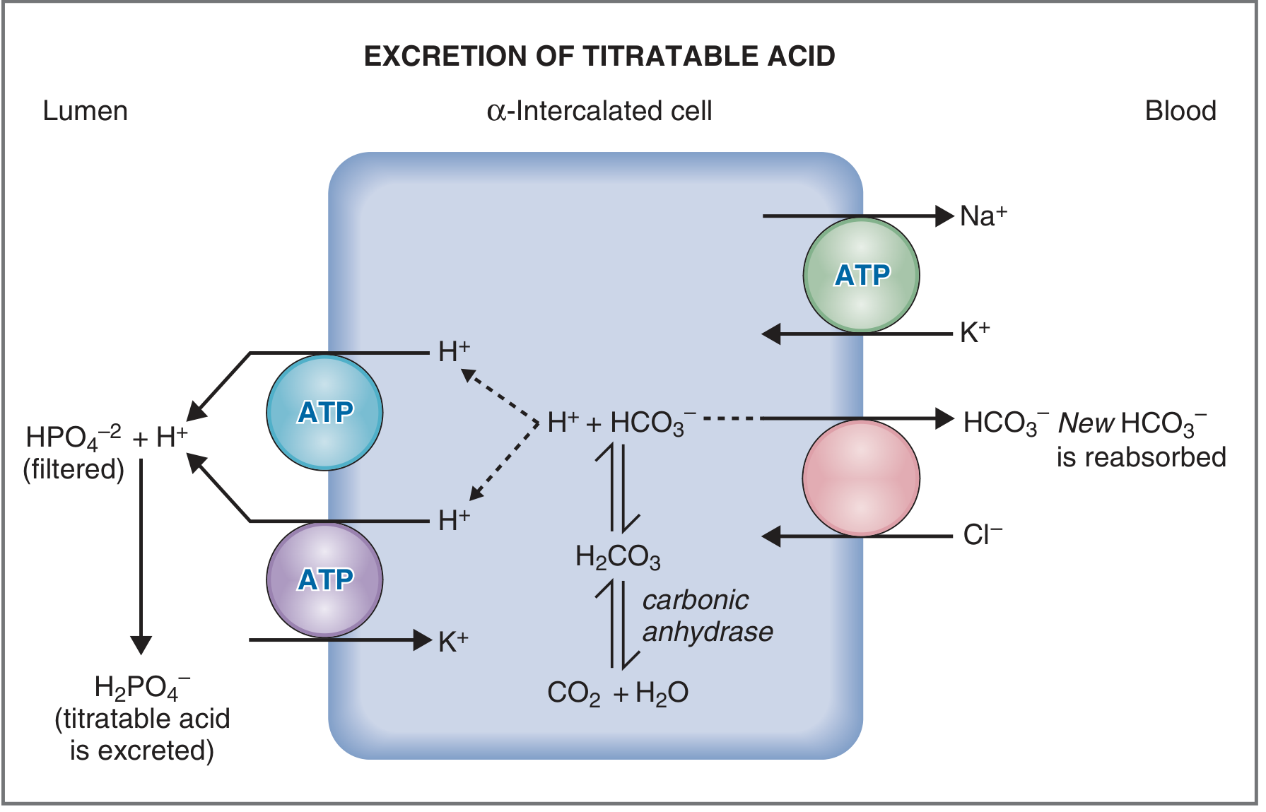
Renal tubular secretion of H⁺ and generation of bicarbonate during titratable acid excretion. - Costanzo Physiology, 7th ed., p. 325
D. Excretion as ammonium: the major adaptive mechanism in acidosis
Ammonium excretion is particularly important when acid production is high.
In proximal tubular cells:
[
Glutamine \rightarrow NH_4^+ + HCO_3^-
]
More precisely, metabolism of glutamine produces ammonium and new bicarbonate. The bicarbonate moves into the bloodstream; ammonium is secreted into tubular fluid.
In the collecting duct:
[
NH_3 + H^+ \rightarrow NH_4^+
]
NH₃ diffuses into the lumen, binds secreted H⁺, and becomes NH₄⁺. Because NH₄⁺ crosses membranes poorly, it becomes trapped in tubular fluid and is excreted. This is called diffusion trapping.
In acidosis
- ↑ Glutamine metabolism
- ↑ NH₃/NH₄⁺ production
- ↑ H⁺ secretion
- ↑ NH₄⁺ excretion
- ↑ New HCO₃⁻ added to blood
In alkalosis
- ↓ Ammonium production and excretion
- ↓ H⁺ secretion
- More bicarbonate may be excreted
Net acid excretion is:
[
\text{Net acid excretion} = NH_4^+ + \text{titratable acid} - urinary\ HCO_3^-
]
The Merck Manual renal-regulation summary describes ammonia as especially important because renal tubular cells can increase ammonia production in response to an acid load.
Renal cellular responses
Alpha-intercalated cells
These cells are active during acidosis.
- Secrete H⁺ into tubular lumen via H⁺-ATPase and H⁺-K⁺-ATPase
- Move HCO₃⁻ into blood
- Produce acidic urine
- Help restore plasma bicarbonate
Beta-intercalated cells
These are more active during alkalosis.
- Secrete HCO₃⁻ into urine via the apical Cl⁻-HCO₃⁻ exchanger, pendrin
- Return H⁺ to blood
- Help eliminate excess base
Factors affecting renal acid excretion
Potassium
Potassium and hydrogen balance are closely linked.
- Hypokalemia promotes H⁺ secretion, HCO₃⁻ reabsorption, ammoniagenesis, and metabolic alkalosis.
- Hyperkalemia suppresses ammonium production and can contribute to metabolic acidosis, particularly type 4 renal tubular acidosis.
Aldosterone
Aldosterone increases distal Na⁺ reabsorption and supports H⁺ secretion by alpha-intercalated cells.
- Excess aldosterone can promote metabolic alkalosis.
- Aldosterone deficiency or resistance can impair H⁺ excretion and promote metabolic acidosis.
Extracellular-fluid volume and chloride
Volume contraction activates angiotensin II and enhances proximal Na⁺-H⁺ exchange, increasing bicarbonate reabsorption. This helps explain contraction alkalosis, such as after vomiting or loop/thiazide diuretic use.
Chloride depletion also limits bicarbonate excretion and helps maintain metabolic alkalosis. - Costanzo Physiology, 7th ed., pp. 324-325
Primary acid-base disorders and compensation
| Primary disorder | Primary change | Direction of pH | Expected compensation |
|---|---|---|---|
| Metabolic acidosis | ↓ HCO₃⁻ | ↓ | Hyperventilation, ↓ PaCO₂ |
| Metabolic alkalosis | ↑ HCO₃⁻ | ↑ | Hypoventilation, ↑ PaCO₂ |
| Respiratory acidosis | ↑ PaCO₂ | ↓ | Renal H⁺ excretion and ↑ HCO₃⁻ |
| Respiratory alkalosis | ↓ PaCO₂ | ↑ | Renal HCO₃⁻ excretion and ↓ HCO₃⁻ |
Important principle
Compensation reduces the pH disturbance but does not correct the underlying cause. Complete normalization usually requires correction of the primary disease process.
Summary flowchart
Acid or base disturbance
        ↓
Immediate: chemical buffering
        ↓
Minutes: respiratory response alters PaCO₂
        ↓
Hours to days: renal response alters HCO₃⁻ and net acid excretion
        ↓
pH moves toward normal
Acidosis:
↑ H⁺ → buffers bind H⁺ → lungs increase ventilation → kidneys retain/generate HCO₃⁻
     → kidneys excrete H⁺ as phosphate and NH₄⁺

Alkalosis:
↓ H⁺ → buffers release H⁺ → lungs reduce ventilation within hypoxemia limits
     → kidneys excrete HCO₃⁻ and reduce H⁺/NH₄⁺ excretion
Key takeaways
- Body pH is controlled chiefly by the HCO₃⁻ to PaCO₂ ratio.
- Buffers act in seconds, but only temporarily.
- Lungs control volatile acid, CO₂, in minutes.
- Kidneys control bicarbonate and fixed-acid removal over hours to days.
- Renal ammonium excretion is the principal adaptive pathway for sustained acid loading.
- Compensation does not overcorrect a simple disorder. An inappropriate compensation suggests an additional mixed acid-base disorder.
Each acid base disorder in detail
Reading File
Finding Sources
Finding Sources
Finding Sources
Searching PubMed
"Acid-Base Equilibrium"[MeSH Terms] AND "acid-base disorders"
Searching the Web
Merck Manual Professional acid base disorders metabolic acidosis alkalosis respiratory acidosis alkalosis
Finding Sources
Finding Sources
Finding Sources
Reading Web Page
https://www.merckmanuals.com/professional/nephrology/acid-ba…
Reading File
Finding Sources
Acid-base disorders
There are four primary acid-base disorders:
- Metabolic acidosis
- Metabolic alkalosis
- Respiratory acidosis
- Respiratory alkalosis
They are classified by the primary abnormality:
- Metabolic disorders: primary change in serum bicarbonate, (HCO_3^-)
- Respiratory disorders: primary change in arterial carbon dioxide, (PaCO_2)
[
\text{pH} = 6.1 + \log\left(\frac{HCO_3^-}{0.03 \times PaCO_2}\right)
]
Thus, pH falls if bicarbonate falls or CO₂ rises; pH rises if bicarbonate rises or CO₂ falls.
Normal approximate values:
| Variable | Normal value |
|---|---|
| Arterial pH | 7.35-7.45 |
| (PaCO_2) | 35-45 mmHg |
| (HCO_3^-) | 22-26 mEq/L |
A simple disorder has one primary abnormality and expected compensation. If compensation is outside the expected range, suspect a mixed acid-base disorder. - Costanzo Physiology, 7th ed., pp. 330-336
1. Metabolic acidosis
Definition
Metabolic acidosis is a primary reduction in plasma bicarbonate concentration, causing a fall in pH.
[
\downarrow HCO_3^- \rightarrow \downarrow pH
]
Typical arterial blood gas pattern
| Variable | Finding |
|---|---|
| pH | Low |
| (HCO_3^-) | Low, primary change |
| (PaCO_2) | Low, due to respiratory compensation |
Mechanisms
Metabolic acidosis occurs due to one of four mechanisms:
- Increased endogenous acid production
- Ingestion of acid or toxin
- Loss of bicarbonate
- Failure of renal acid excretion
Respiratory compensation
A low pH stimulates peripheral chemoreceptors, especially carotid bodies, producing hyperventilation. This lowers PaCO₂.
[
\downarrow HCO_3^- \rightarrow \downarrow pH \rightarrow \uparrow ventilation \rightarrow \downarrow PaCO_2
]
Severe acidosis may cause Kussmaul respiration: deep, labored, rapid breathing, classically seen in diabetic ketoacidosis.
Winter's formula
Use this to determine whether respiratory compensation is appropriate:
[
Expected\ PaCO_2 = 1.5 \times [HCO_3^-] + 8 \pm 2
]
Example: if (HCO_3^- = 12) mEq/L:
[
Expected\ PaCO_2 = 1.5(12) + 8 = 26 \pm 2
]
- Actual PaCO₂ higher than predicted: additional respiratory acidosis
- Actual PaCO₂ lower than predicted: additional respiratory alkalosis
Anion gap
The anion gap identifies whether unmeasured acids are present.
[
AG = Na^+ - (Cl^- + HCO_3^-)
]
A common reference range is about 8-12 mEq/L when potassium is omitted, but interpret according to the laboratory reference interval.
Because albumin is a major unmeasured anion, a low albumin can conceal a raised anion gap:
[
Corrected\ AG = Measured\ AG + 2.5 \times (4 - albumin\ in\ g/dL)
]
A. High-anion-gap metabolic acidosis
Here, bicarbonate is consumed buffering an added acid, while the acid's unmeasured anion remains in plasma.
[
\downarrow HCO_3^- + \uparrow unmeasured\ anions = \uparrow AG
]
Common causes: GOLD MARK
| Cause | Examples |
|---|---|
| Glycols | Ethylene glycol, propylene glycol |
| Oxoproline | Chronic acetaminophen use, usually in susceptible patients |
| L-lactate | Shock, hypoxemia, sepsis, seizures, severe liver dysfunction |
| D-lactate | Short-bowel syndrome |
| Methanol | Formic acid accumulation |
| Aspirin | Salicylate poisoning |
| Renal failure | Retention of sulfate, phosphate, and other acids |
| Ketoacidosis | Diabetic, alcoholic, starvation ketoacidosis |
Major clinical examples
Diabetic ketoacidosis
Insulin deficiency causes lipolysis and hepatic ketone production, especially beta-hydroxybutyrate and acetoacetate.
Typical findings:
- Hyperglycemia
- Ketones
- High anion gap acidosis
- Dehydration
- Kussmaul breathing
- Total-body potassium deficit, even when initial serum potassium is normal or high
Lactic acidosis
Occurs when lactate production exceeds clearance.
Common settings:
- Sepsis or septic shock
- Hypovolemia or hemorrhage
- Cardiogenic shock
- Severe hypoxemia
- Seizures
- Mesenteric ischemia
- Severe liver dysfunction
- Some drugs and toxins
Renal failure
Reduced nephron mass impairs ammonium production, titratable-acid excretion, and regeneration of bicarbonate. Fixed acids accumulate. - Costanzo Physiology, 7th ed., pp. 328-331
B. Normal-anion-gap metabolic acidosis
Also called hyperchloremic metabolic acidosis.
Here, bicarbonate is lost and replaced electrically by chloride:
[
\downarrow HCO_3^- \quad \text{with} \quad \uparrow Cl^-
]
The anion gap stays normal.
Causes
| Mechanism | Examples |
|---|---|
| Gastrointestinal bicarbonate loss | Diarrhea, ileostomy, pancreatic or biliary drainage, intestinal fistula |
| Renal bicarbonate loss | Proximal renal tubular acidosis, carbonic anhydrase inhibitors |
| Reduced distal H⁺ secretion | Distal renal tubular acidosis |
| Low aldosterone effect | Type 4 renal tubular acidosis, adrenal insufficiency |
| Chloride administration | Large-volume 0.9% saline administration |
| Urinary diversion | Ureterosigmoidostomy |
Urine anion gap
In normal renal response to metabolic acidosis, the kidney excretes ammonium chloride, so urinary chloride rises.
[
Urine\ anion\ gap = (Urine\ Na^+ + Urine\ K^+) - Urine\ Cl^-
]
- Negative urine anion gap: high urinary NH₄⁺ excretion, suggesting an extrarenal cause such as diarrhea
- Positive urine anion gap: low urinary NH₄⁺ excretion, suggesting impaired renal acid excretion, such as renal tubular acidosis
Symptoms and signs
Symptoms depend on severity and cause:
- Deep rapid breathing
- Fatigue, weakness
- Nausea and vomiting
- Confusion, drowsiness, coma in severe cases
- Reduced cardiac contractility and hypotension in severe acidemia
- Hyperkalemia may occur, especially with mineral acidosis or renal failure
Management principles
Treatment is directed at the cause:
- Restore perfusion and oxygenation in shock or lactic acidosis
- Insulin, fluids, electrolyte monitoring, and potassium management in DKA
- Treat toxins urgently, with antidotes and dialysis where indicated
- Stop bicarbonate-losing medications or treat diarrhea
- Address renal failure or renal tubular acidosis
Bicarbonate is not routine for all acidoses. It is considered selectively in severe acidemia or specific bicarbonate-loss states, with careful monitoring.
2. Metabolic alkalosis
Definition
Metabolic alkalosis is a primary elevation in plasma bicarbonate, causing increased pH.
[
\uparrow HCO_3^- \rightarrow \uparrow pH
]
Typical blood gas pattern
| Variable | Finding |
|---|---|
| pH | High |
| (HCO_3^-) | High, primary change |
| (PaCO_2) | High, compensatory |
Mechanisms
Metabolic alkalosis results from:
- Loss of hydrogen ions
- Gain of bicarbonate
- Renal retention of bicarbonate
For metabolic alkalosis to persist, the kidney must be unable or signaled not to excrete excess bicarbonate. Common maintaining factors are:
- Volume depletion
- Chloride depletion
- Hypokalemia
- Reduced kidney function
- Mineralocorticoid excess
Respiratory compensation
The compensatory response is hypoventilation, causing CO₂ retention:
[
\uparrow HCO_3^- \rightarrow \uparrow pH \rightarrow \downarrow ventilation \rightarrow \uparrow PaCO_2
]
Expected response:
[
Expected\ PaCO_2 \approx 40 + 0.6\text{ to }0.75 \times (HCO_3^- - 24)
]
Respiratory compensation is limited because excessive hypoventilation causes hypoxemia. A compensatory PaCO₂ generally should not rise above about 55 mmHg. - Merck diagnostic guidance
Causes
A. Chloride-responsive metabolic alkalosis
Usually caused by volume depletion and chloride loss. Urine chloride is typically low:
[
Urine\ Cl^- < 20\ mEq/L
]
Common causes:
- Vomiting
- Nasogastric suction
- Loop diuretics
- Thiazide diuretics
- Post-hypercapnic alkalosis
- Chloride-losing diarrhea, rarely
- Cystic fibrosis with marked sweat chloride loss
Why vomiting causes alkalosis
Loss of gastric hydrochloric acid causes direct H⁺ and Cl⁻ loss:
[
Loss\ of\ H^+ \rightarrow \uparrow HCO_3^-
]
Volume and chloride depletion activate renin-angiotensin-aldosterone mechanisms, enhancing proximal sodium-bicarbonate reabsorption and maintaining alkalosis.
B. Chloride-unresponsive metabolic alkalosis
Urine chloride is usually high:
[
Urine\ Cl^- > 20\ mEq/L
]
Main causes:
- Primary hyperaldosteronism
- Cushing syndrome or ectopic mineralocorticoid effect
- Severe hypokalemia
- Bartter syndrome
- Gitelman syndrome
- Excess alkali intake in reduced renal function
- Licorice ingestion, causing apparent mineralocorticoid excess
Potassium relationship
Hypokalemia promotes:
- H⁺ movement into cells
- Increased renal H⁺ secretion
- Increased bicarbonate reabsorption
- Increased ammonium production
Therefore, hypokalemia both accompanies and perpetuates metabolic alkalosis.
Symptoms and signs
Mild cases may be asymptomatic. Severe alkalemia can cause:
- Weakness
- Headache
- Confusion or lethargy
- Muscle cramps
- Paresthesia
- Tetany
- Seizures
- Cardiac arrhythmias
Alkalemia increases protein binding of calcium and lowers ionized calcium, which contributes to tetany and carpopedal spasm. - Costanzo Physiology, 7th ed., p. 336
Management principles
- Treat vomiting, gastric suction, or diuretic excess
- Correct extracellular-volume depletion with chloride-containing fluid when appropriate
- Correct potassium and magnesium deficits
- Treat mineralocorticoid excess when present
- Consider acetazolamide in selected patients, especially when volume expansion is undesirable
- Severe, resistant alkalemia requires urgent specialist management
A urine chloride below 20 mEq/L generally suggests a chloride-responsive cause, whereas a higher value supports chloride-unresponsive alkalosis, assuming kidney function is adequate. - Merck metabolic alkalosis review
3. Respiratory acidosis
Definition
Respiratory acidosis is a primary increase in PaCO₂ due to inadequate alveolar ventilation.
[
\uparrow PaCO_2 \rightarrow \uparrow H_2CO_3 \rightarrow \uparrow H^+ \rightarrow \downarrow pH
]
Typical blood gas pattern
| Variable | Finding |
|---|---|
| pH | Low |
| (PaCO_2) | High, primary change |
| (HCO_3^-) | High if compensation occurs |
Main mechanism: hypoventilation
[
\downarrow alveolar\ ventilation \rightarrow \uparrow PaCO_2
]
Any condition that reduces effective ventilation can cause respiratory acidosis.
Causes
A. Decreased respiratory drive
- Opioid intoxication
- Sedatives, anesthetics, barbiturates
- Brainstem stroke, tumor, trauma, or infection
- Central sleep apnea
- Obesity hypoventilation syndrome
B. Neuromuscular failure
- Guillain-Barré syndrome
- Myasthenia gravis
- Motor neuron disease
- Spinal cord injury
- Severe hypokalemia or hypophosphatemia
- Neuromuscular blocking drugs
C. Airway obstruction
- Severe COPD exacerbation
- Severe asthma with fatigue or impending respiratory failure
- Upper airway obstruction
- Obstructive sleep apnea
D. Impaired gas exchange or increased work of breathing
-
Severe pneumonia
-
Pulmonary edema
-
Acute respiratory distress syndrome
-
Severe obesity or chest-wall restriction
-
Costanzo Physiology, 7th ed., pp. 333-334
Acute versus chronic respiratory acidosis
The distinction matters because the kidneys require time to retain bicarbonate and excrete acid.
Acute respiratory acidosis
Occurs over minutes to hours, for example opioid overdose or sudden airway obstruction.
- Intracellular buffering is the main initial response.
- Bicarbonate rises only slightly.
[
\text{For every 10 mmHg rise in } PaCO_2,\ HCO_3^- \text{ rises by about 1-2 mEq/L}
]
Example: PaCO₂ rises from 40 to 60 mmHg.
Expected acute bicarbonate:
[
HCO_3^- \approx 24 + 2\text{ to }4 = 26\text{ to }28
]
Chronic respiratory acidosis
Occurs over days, for example stable COPD with chronic CO₂ retention.
- Kidneys increase H⁺ secretion.
- Ammonium and titratable-acid excretion increase.
- New bicarbonate is generated and retained.
[
\text{For every 10 mmHg rise in } PaCO_2,\ HCO_3^- \text{ rises by about 3-4 mEq/L}
]
Example: PaCO₂ rises from 40 to 60 mmHg.
Expected chronic bicarbonate:
[
HCO_3^- \approx 24 + 6\text{ to }8 = 30\text{ to }32
]
The pH is lower in acute respiratory acidosis than in chronic respiratory acidosis at the same PaCO₂ because chronic renal compensation is more effective.
Symptoms and signs
Severity depends on how rapidly CO₂ rises.
- Headache
- Drowsiness
- Confusion
- Asterixis
- Warm, flushed skin
- Dyspnea
- Reduced level of consciousness
- CO₂ narcosis and coma in severe cases
Chronic hypercapnia may be tolerated better than an abrupt CO₂ rise.
Management principles
- Support airway, breathing, and oxygenation
- Reverse or stop the underlying cause where possible
- Naloxone for opioid-induced hypoventilation, when appropriate
- Treat COPD or asthma exacerbation
- Manage pneumonia, pulmonary edema, or neuromuscular weakness
- Use noninvasive ventilation or invasive ventilation when indicated
In a chronic CO₂ retainer, abrupt overventilation can rapidly lower PaCO₂ while bicarbonate remains elevated, producing post-hypercapnic metabolic alkalosis. Correction must be monitored clinically and with serial blood gases.
4. Respiratory alkalosis
Definition
Respiratory alkalosis is a primary reduction in PaCO₂ caused by excessive ventilation.
[
\downarrow PaCO_2 \rightarrow \downarrow H^+ \rightarrow \uparrow pH
]
Typical blood gas pattern
| Variable | Finding |
|---|---|
| pH | High |
| (PaCO_2) | Low, primary change |
| (HCO_3^-) | Low if compensation occurs |
Causes
A. Hypoxemia-driven hyperventilation
- Pulmonary embolism
- Pneumonia
- Pulmonary edema
- Asthma exacerbation
- High altitude
- Severe anemia, rarely
- Right-to-left shunt
B. Increased respiratory drive
- Pain
- Anxiety or panic
- Fever
- Sepsis
- Pregnancy, due partly to progesterone
- Liver disease
- CNS disorders: stroke, trauma, tumor, meningitis, encephalitis
- Salicylate toxicity, especially early
C. Iatrogenic causes
- Excessive mechanical ventilation
Never assume hyperventilation is due to anxiety until potentially serious causes, especially hypoxemia, sepsis, pulmonary embolism, and salicylate poisoning, have been considered. - Merck respiratory alkalosis review
Acute versus chronic respiratory alkalosis
Acute respiratory alkalosis
The kidneys have not yet adapted. Bicarbonate falls only slightly due to buffering.
[
\text{For every 10 mmHg fall in } PaCO_2,\ HCO_3^- \text{ decreases by about 1-2 mEq/L}
]
Example: PaCO₂ falls from 40 to 20 mmHg.
[
HCO_3^- \approx 24 - 2\text{ to }4 = 20\text{ to }22
]
Chronic respiratory alkalosis
The kidney adapts by excreting more bicarbonate and reducing H⁺ secretion.
[
\text{For every 10 mmHg fall in } PaCO_2,\ HCO_3^- \text{ decreases by about 4-5 mEq/L}
]
Example: PaCO₂ falls from 40 to 20 mmHg.
[
HCO_3^- \approx 24 - 8\text{ to }10 = 14\text{ to }16
]
Symptoms and signs
Acute hypocapnia can reduce cerebral blood flow and lower ionized calcium.
Features include:
- Light-headedness
- Dizziness
- Perioral numbness
- Tingling of hands and feet
- Muscle cramps
- Carpopedal spasm
- Chest discomfort
- Confusion
- Syncope in severe cases
The fall in ionized calcium occurs because alkalemia increases calcium binding to albumin.
Management principles
Treat the cause:
- Give oxygen and treat hypoxemia
- Investigate pulmonary embolism, sepsis, lung disease, or salicylate toxicity when relevant
- Adjust excessive mechanical ventilation
- Treat pain, fever, or anxiety only after excluding important organic causes
Rebreathing into a paper bag is not recommended because it can worsen unrecognized hypoxemia or delay diagnosis of a dangerous cause. - Merck guidance
Quick comparison table
| Disorder | Primary change | pH | Compensation | Typical causes |
|---|---|---|---|---|
| Metabolic acidosis | ↓ HCO₃⁻ | ↓ | ↓ PaCO₂ by hyperventilation | DKA, lactic acidosis, diarrhea, renal failure, RTA |
| Metabolic alkalosis | ↑ HCO₃⁻ | ↑ | ↑ PaCO₂ by hypoventilation | Vomiting, diuretics, hypokalemia, mineralocorticoid excess |
| Respiratory acidosis | ↑ PaCO₂ | ↓ | ↑ HCO₃⁻ by kidneys | COPD, CNS depression, neuromuscular failure, airway obstruction |
| Respiratory alkalosis | ↓ PaCO₂ | ↑ | ↓ HCO₃⁻ by kidneys | Hypoxemia, PE, sepsis, pregnancy, pain, anxiety, liver disease |
Practical ABG interpretation sequence
-
Check pH
- < 7.35: acidemia
-
7.45: alkalemia
-
Identify the primary process
- In acidemia: high PaCO₂ suggests respiratory acidosis; low HCO₃⁻ suggests metabolic acidosis.
- In alkalemia: low PaCO₂ suggests respiratory alkalosis; high HCO₃⁻ suggests metabolic alkalosis.
-
Check expected compensation
- Use Winter's formula in metabolic acidosis.
- Use acute/chronic compensation rules for respiratory disorders.
-
Calculate anion gap if metabolic acidosis is present.
-
Look for a mixed disorder if compensation is inappropriate or pH, PaCO₂, and bicarbonate do not fit one simple pattern.
-
Find and treat the underlying cause, not merely the blood-gas abnormality.
Severe acidemia, severe alkalemia, altered consciousness, shock, significant hyperkalemia, suspected toxic ingestion, severe dyspnea, or respiratory fatigue requires urgent in-person assessment.
This is a shared conversation. Sign in to Orris to start your own chat.