
Explain visually
aminoglycoside mechanism of action bacterial protein synthesis ribosome
aminoglycoside transport bacterial cell wall membrane penetration
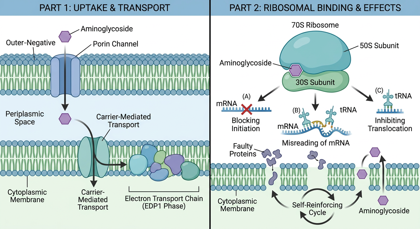
A clear educational diagram showing aminoglycoside antibiotic mechanism of action in two parts: Part 1 shows the drug crossing the gram-negative bacterial outer membrane through porin channels, entering the periplasmic space, then crossing the cytoplasmic membrane via carrier-mediated energy-dependent transport linked to the electron transport chain (EDP1 phase). Part 2 shows the aminoglycoside binding to the 30S ribosomal subunit causing three effects: (A) blocking initiation of protein synthesis, (B) causing misreading of mRNA leading to abnormal/faulty proteins, (C) inhibiting tRNA translocation. Arrows show faulty proteins inserting back into the membrane causing increased permeability and more drug entry (self-reinforcing cycle). Label key structures: outer membrane, porin channels, periplasmic space, cytoplasmic membrane, 30S subunit, 50S subunit, mRNA, tRNA, faulty proteins. Use clean medical illustration style with color coding.
🔬 Mechanism of Action — Visual Breakdown
Step 1: Penetration into the Bacterial Cell
| Stage | What Happens |
|---|---|
| Outer membrane | Drug diffuses through porin channels (passive, no energy needed) |
| Periplasmic space | Drug accumulates, waiting to cross the inner membrane |
| Cytoplasmic membrane | Crossed via carrier-mediated, energy-dependent transport — requires a polarized membrane and the electron transport chain (called EDP₁ entry) |
This is why aminoglycosides don't work on anaerobes — no active electron transport chain = no drug entry.
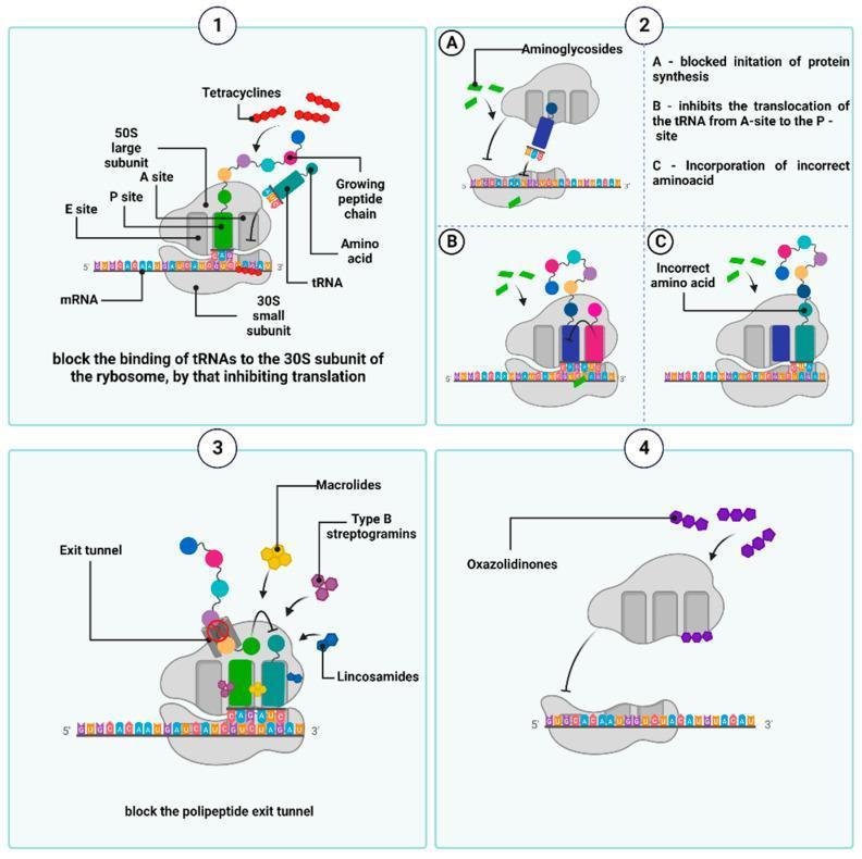
Step 2: Ribosomal Binding & Protein Synthesis Inhibition
| Effect | Result |
|---|---|
| (A) Block initiation | The 30S + 50S ribosome complex cannot form properly |
| (B) Inhibit tRNA translocation | tRNA cannot move from A-site → P-site; elongation stalls |
| (C) mRNA misreading | Incorrect amino acids are incorporated → faulty/non-functional proteins |
The Self-Amplifying Cycle (Key Concept!)
🧪 Common Properties at a Glance
| Property | Clinical Implication |
|---|---|
| Sulfate salts, highly water-soluble | Stable in solution for months |
| Not absorbed orally | Must be given parenterally (IM/IV) for systemic infections |
| Excreted unchanged by glomerular filtration | Dose adjustment required in renal failure |
| More active at alkaline pH | Less effective inside acidic abscesses |
| Active against aerobic gram-negatives only | No activity against anaerobes or penetrating CSF |
| Ototoxicity & nephrotoxicity | Major dose-limiting toxicities; monitor drug levels |
| Narrow safety margin | Requires therapeutic drug monitoring (TDM) |