
Make notes on topical steroid damaged facies for md dermatology theory exam
topical steroid damaged facies erythema telangiectasia perioral
"topical steroid damaged face"
topical steroid damaged facies TSDF clinical features management review
https://pmc.ncbi.nlm.nih.gov/articles/PMC4885178
steroid rosacea perioral dermatitis face telangiectasia
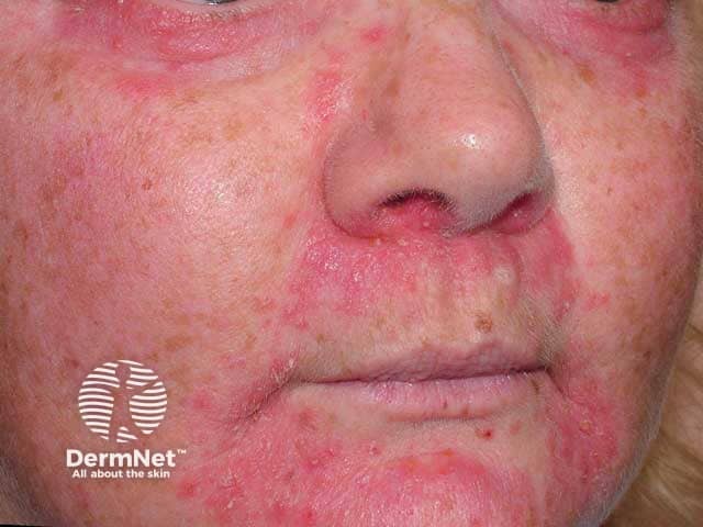
This is a frontal clinical photograph of an adult patient’s face illustrating steroid-induced facial rosacea with perioral dermatitis. Modality: Digital color photography, in vivo, captured under standard indoor lighting with routine color balance. Anatomical region: central face including cheeks, nasal area, perioral region and adjacent perinasal skin. The image shows diffuse erythema with a prominent papulopustular eruption, fine scaling, and subtle telangiectasia across the malar cheeks and nasal bridge, extending around the mouth. Bilateral distribution is evident, with accentuation along the nasolabial folds and perioral zone. Palpebral and eyelid skin not clearly involved. The lesions are small to moderate in size, with confluent erythematous patches interspersed by discrete papules and pustules; surface texture appears tender and mildly edematous. Clinically, the appearance is characteristic of corticosteroid-related facial dermatitis, specifically steroid-induced rosacea and perioral dermatitis, often resulting from recent or prolonged topical corticosteroid use. Diagnostic significance: history of steroid exposure with paradoxical facial inflammation; differential includes rosacea not steroid-related, acneiform dermatitis, seborrheic dermatitis, and contact dermatitis. Clinical correlation: cessation of potent steroids, gradual taper, and initiation of appropriate dermatologic therapy typically yield gradual improvement over weeks to months. Potential clinical use: education, documentation, disease monitoring, AI-training datasets for dermatology.
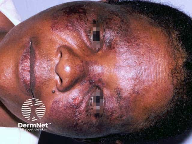
Clinical photograph of a middle-aged adult male presenting with facial steroid-induced rosacea and postinflammatory hyperpigmentation. Modality: Clinical photography; frontal view of the face under diffuse, non-diagnostic lighting. The centrofacial skin shows diffuse erythema with prominent telangiectasia, especially on the cheeks and nose. Superimposed papules and few pustules are evident around the nasolabial folds and forehead, with patchy hyperpigmented macules on the malar regions. The perioral area may be involved, and there is mild edema and skin fragility consistent with steroid-related epidermal thinning. Bilateral distribution is noted, with involvement of forehead, cheeks, and perioral skin. This pattern is characteristic of topical corticosteroid overuse leading to an inflammatory rosaceiform eruption rather than classic acne. The clinical diagnosis is steroid-induced rosacea with pigmentary changes; histopathology is not provided. Diagnostic significance includes recognizing steroid-related dermatosis to prevent progression and guide withdrawal and treatment. Differential diagnoses include rosacea (non-steroid–associated), acne vulgaris, perioral dermatitis, and dermatitis from irritants. Clinical correlation should focus on patient history of potent facial steroid use, duration, and taper strategy. This image is educational for dermatology training, medical education, and AI-assisted lesion recognition, highlighting pigmentary complications and telangiectasia in centrofacial dermatoses. Clinically, cessation of potent steroids is essential.
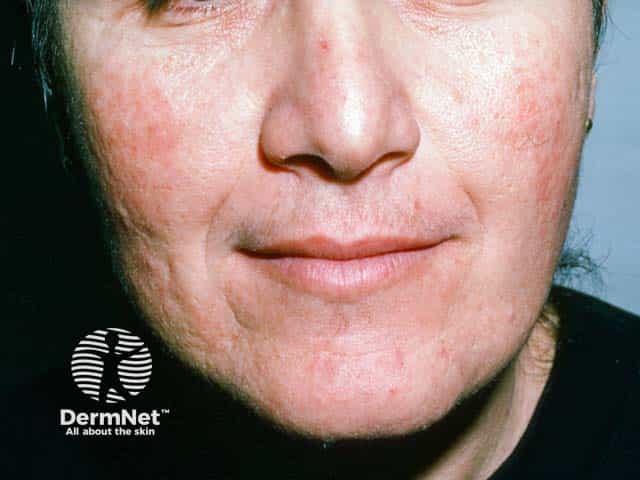
Clinical dermatology photograph of the face, frontal view, documenting steroid-induced rosacea involving the malar (cheek) and chin regions. Modality: clinical digital photography; technique: standardized facial lighting, color-balanced imaging; specimen: human skin; view: anterior facial aspect. Anatomical context: integumentary system; facial skin on the central face, cheeks (malar regions) and chin, bilateral distribution, anterior plane. Visual features: diffuse erythema with fine papules and minimal pustulation, subtle perifollicular accentuation, and mild visible telangiectasia in the affected zones; skin texture appears slightly roughened with reactive hyperemia; absence of comedones or nodular infiltration in this frame. Diagnostic implication: fits steroid-induced rosacea spectrum rather than classic acne vulgaris; triggers include potent topical corticosteroids; history of recent steroid use would corroborate; differential diagnoses include acne rosacea, steroid dermatitis, perioral dermatitis, contact dermatitis. Clinical significance: highlights adverse cutaneous effect of topical steroids, guides management (withdrawal or taper, barrier repair, anti-inflammatory strategies, calcineurin inhibitors, non-irritant skincare, photoprotection). Potential clinical use cases: educational reference for dermatology training, case-based learning on iatrogenic rosacea, monitoring response to withdrawal therapy, decision support for treatment planning, and aiding patient counseling about steroid safety and rosacea flares. This image supports standardized documentation, teaching symptom recognition, and guiding patient-tailored stop-start therapy.
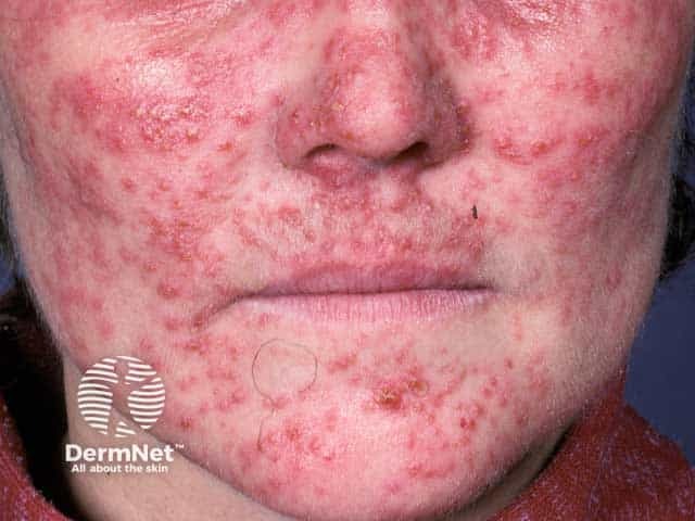
Clinical photography of an adult's face showing a diffuse erythematous papulopustular eruption consistent with steroid-induced rosacea. Modality: Clinical photography; anterior facial view. The cheeks, nasal bridge, perioral regions are symmetrically involved with diffuse erythema and scattered inflamed papules and pustules; mild edema and telangiectasia may be present but not clearly visible. Anatomical regions include the malar and nasolabial areas. The eruption reflects inflammatory infiltration of the epidermis and superficial dermis with vascular dilation. Pathophysiology likely relates to prolonged topical corticosteroid suppression of innate immunity and induction of vasodilation leading to rosacea-like eruption; withdrawal and gradual tapering of steroids are essential. Differential diagnoses include acne rosacea, acne vulgaris, perioral dermatitis, seborrheic dermatitis, and contact dermatitis. Diagnostic significance: steroid-induced facial rosacea is typically reversible with appropriate management after identifying and stopping the offending agent, though lingering erythema may persist. Clinical use cases include educational demonstration, differential diagnosis training, and guiding management decisions. The image supports recognition of steroid-induced rosacea patterns, informs patient counseling on prognosis, and assists in planning skin barrier restoration and anti-inflammatory therapy. High-quality color fidelity and standard lighting enhance lesion visualization for teaching and remote consultation. This description emphasizes morphological details such as papules, pustules, and diffuse erythema present.
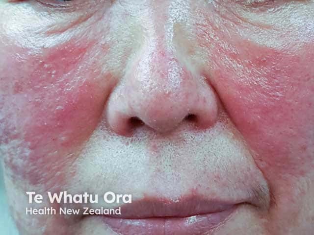
Modality and technique: high-resolution clinical photography of the face in frontal/anterior view, providing a close-up assessment of cutaneous inflammation. Anatomical focus: central facial skin encompassing the nasal dorsum, cheeks, nasolabial folds, and perioral region. Observed features: uniform facial erythema with prominent edema, particularly on the malar cheeks and nasal pyramid, accompanied by discrete papules and pustules within inflamed erythematous areas. The texture appears smooth to slightly glossy, with diffuse vascular blush and minor swelling around the nose and midface. The pattern is bilateral and symmetrical, consistent with steroid-induced rosacea (SR), a dermatosis precipitated by topical or systemic corticosteroid exposure. Differential considerations include acne rosacea, perioral dermatitis, dermatitis medicamentosa, and contact dermatitis; however, the history of steroid use and the central facial predominance favor SR. Pathophysiology involves disruption of pilosebaceous unit homeostasis and steroid-induced telangiectasia, leading to papulopustular flares. Clinical significance lies in the need to discontinue the offending steroid, initiate rosacea-appropriate therapy (e.g., metronidazole, doxycycline, brimonidine), and monitor for rebound flaring. Potential clinical uses include documenting treatment response, educating patients, and supporting differential diagnosis in photodermatology and cosmetic dermatology. This image exemplifies central facial inflammation with papulopustular lesions in SR. Interpretation aids patient counseling and targeted management planning for clinicians.
Topical Steroid Damaged Facies (TSDF) — MD Dermatology Exam Notes
1. Definition
- Older synonym: "Red Face Syndrome" (Rapaport, 1999) — erythema is considered the hallmark feature.
- Also described as an entity of cutaneous pharmacodependence — analogous to drug addiction, with psychological compulsion to keep applying steroid despite awareness of harm.
- Coined/popularized in India (Lahiri, 2008) due to an epidemic of OTC misuse; led to a pan-Indian IADVL multicentric study (2008-2009).
2. Epidemiology & Precipitating Factors
- Extremely common in India/South Asia due to over-the-counter (OTC) availability of potent/super-potent steroids without prescription.
- Fixed-dose combination creams widely misused: potent steroid (e.g., clobetasol propionate) + antifungal + antibacterial + tretinoin — marketed and self-used as "fairness creams" or for minor complaints (acne, melasma, insect bites, "pimples," tanning).
- Driven by quackery, cosmetic counter sales, peer/advertisement influence, and desire for fairness/glow.
- Facial skin is uniquely vulnerable: thinner stratum corneum, higher density of pilosebaceous units and blood vessels, greater percutaneous absorption than trunk/limb skin — hence lower threshold for steroid-induced damage.
3. Pathogenesis
- Chronic vasoconstriction from TC followed by rebound vasodilatation on stoppage -> persistent erythema and telangiectasia.
- Antimitotic/antiproliferative effect -> epidermal and dermal atrophy, decreased collagen/elastin synthesis -> thin, fragile, translucent skin with visible vessels.
- Local immunosuppression -> proliferation of Demodex folliculorum, subclinical dermatophyte infection (tinea incognito), and bacterial folliculitis.
- Comedogenic and follicular occlusive effect -> steroid acne/secondary comedones.
- Nitric oxide-mediated vascular dysregulation contributes to the rosacea-like papulopustular component (steroid rosacea).
4. Clinical Features
| Category | Features |
|---|---|
| Vascular | Diffuse erythema, telangiectasia, flushing, papulopustular rosacea-like eruption |
| Atrophic | Epidermal/dermal atrophy, thinning, striae (especially periorbital), easy bruising, shiny skin |
| Follicular | Steroid acne, secondary comedones, perioral dermatitis, folliculitis |
| Pigmentary | Mottled hyper- and hypopigmentation |
| Infective | Tinea incognito, demodicosis, bacterial superinfection |
| Sensory | Burning, stinging, tightness, photosensitivity |
| Adnexal | Hypertrichosis (steroid-induced facial hirsutism, especially cheeks/forehead) |
| Rebound/withdrawal | Severe flare of erythema/burning on stopping steroid — drives dependence; may spread beyond original application site |
| Systemic (rare, with prolonged widespread potent use) | HPA-axis suppression, cushingoid features; periocular use risk of glaucoma/cataract (warrants ophthalmology referral) |
5. Dermoscopy (high-yield for MD exam)
- Diffuse polygonal/linear branching (arborizing) vessels on a milky-red to pink-red background
- Follicular red dots and prominent follicular openings
- Perifollicular whitish halo
- Fine scaling; comedo-like structures in acneiform variant
- Dermoscopy is useful to differentiate TSDF from primary rosacea, seborrheic dermatitis, and connective tissue disease, and to objectively monitor response during steroid tapering.
6. Grading
7. Differential Diagnosis
- Primary rosacea (papulopustular/erythematotelangiectatic)
- Perioral dermatitis (non-steroid)
- Seborrheic dermatitis
- Photodermatitis / chronic actinic dermatitis
- Connective tissue disease (lupus malar rash)
- Contact dermatitis / status cosmeticus
- Tinea incognito (isolated)
8. Management
- Patient counseling and education — central to management given the psychological/behavioral dependence component; explain the rebound phenomenon in advance so patient does not restart steroid out of panic.
- Never abrupt stop of potent steroid — taper gradually:
- Step down potency (e.g., potent -> mild) and/or
- Reduce frequency (daily -> alternate day -> twice weekly -> stop) over weeks.
- Steroid-sparing bridge therapy: topical calcineurin inhibitors (tacrolimus 0.03-0.1%, pimecrolimus 1%) to control inflammation during taper without further atrophy.
- Barrier repair: bland emollients/ceramide-based moisturizers for xerosis and skin fragility.
- Photoprotection: broad-spectrum, preferably mineral/physical sunscreens (photosensitized skin).
- Treat secondary complications:
- Papulopustular/rosacea-like component: oral doxycycline/tetracycline, topical metronidazole or azelaic acid
- Demodicosis: topical permethrin/ivermectin
- Tinea incognito: appropriate oral/topical antifungal (avoid combination creams)
- Secondary bacterial infection: targeted antibiotics
- Cool compresses, avoidance of irritant cosmetics/cleansers during acute rebound phase.
- Psychiatric/psychological referral in cases with marked dependence behavior or comorbid anxiety/OCD traits.
- Systemic evaluation if extensive potent steroid use over prolonged period is suspected — morning cortisol/ACTH stimulation test for HPA-axis suppression; ophthalmology referral if periocular application.
- Public health/regulatory angle (favorite exam point): IADVL's advocacy led to India restricting OTC sale of potent topical corticosteroid combinations under Schedule H — highlights the socio-regulatory dimension of TSDF.
9. Prognosis
Exam-Focused High-Yield Points
- Erythema = hallmark sign; rebound phenomenon = defining feature of "dependence."
- Term coined by Lahiri and Coondoo (2008), India.
- Driven by OTC potent steroid + antifungal + antibacterial combination creams.
- Dermoscopy: polygonal/arborizing vessels + follicular red dots + perifollicular whitish halo.
- Management principle: taper, never abrupt stop; use TCI as bridge; counsel for dependence behavior.