
Supports of uterus with diagram Support of uterus with diagram
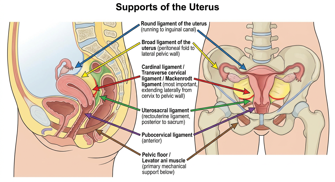
A detailed anatomical diagram of the supports of the uterus, showing a sagittal and anterior view of the female pelvis. Label the following structures clearly: Round ligament of the uterus (running to inguinal canal), Broad ligament of the uterus (peritoneal fold to lateral pelvic wall), Cardinal ligament / Transverse cervical ligament / Mackenrodt ligament (most important, extending laterally from cervix to pelvic wall), Uterosacral ligament (rectouterine ligament, posterior to sacrum), Pubocervical ligament (anterior), Pelvic floor / Levator ani muscle (primary mechanical support below). Use color coding for each ligament type. Add a title: "Supports of the Uterus". Clean medical textbook style.
Supports of the Uterus
Generated Diagram
Textbook Diagrams (Gray's Anatomy for Students)
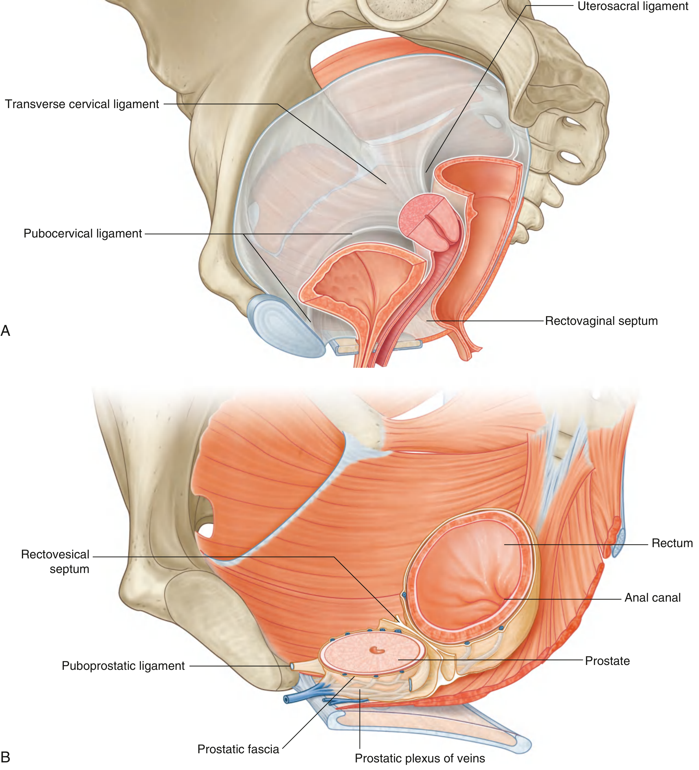
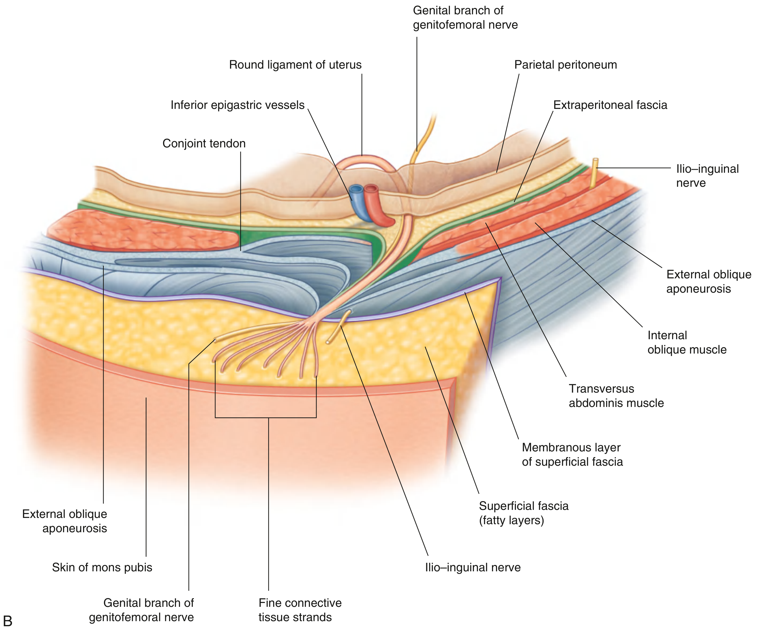
Classification of Uterine Supports
- Primary support - Pelvic floor muscles
- Secondary support - Ligaments and peritoneal folds
1. Pelvic Floor Muscles (Primary / Most Important)
"The uterus is mainly supported by the pelvic floor muscles, not by the abovementioned ligaments." - Color Atlas of Human Anatomy Vol. 2, Internal Organs
2. Ligamentous Supports
A. Cardinal Ligament (Transverse Cervical Ligament / Mackenrodt's Ligament) - MOST IMPORTANT LIGAMENT
- A condensation of connective tissue (fascia) extending laterally from the cervix and vaginal vault to the lateral pelvic wall
- The most important ligament for uterine support clinically
- Travels at the base of the broad ligament
- Contains uterine vessels within it
- Prevents downward descent of the uterus
B. Uterosacral Ligament (Rectouterine Ligament / Sacrouterine Ligament)
- A peritoneal fold bounding the rectouterine pouch (pouch of Douglas)
- Formed by dense subperitoneal connective tissue + nerves of the inferior hypogastric plexus
- Runs from the cervix posteriorly to the posterolateral pelvic wall / sacrum
- Holds the cervix upward and backward
- Also called the rectouterinus ligament; contains (debated) smooth muscle
C. Pubocervical Ligament
- Extends from the cervix anteriorly to the pubic symphysis
- Supports the anterior aspect of the uterus and bladder neck
D. Round Ligament of the Uterus
- Arises near the uterine horns, passes through the inguinal canal, ends in subcutaneous fat of the labia majora
- Contains smooth muscle cells
- Derived from the gonadal fold; is a continuation of the suspensory ligament of the ovary
- Maintains the anteverted position of the uterus (keeps fundus tilted forward)
- Provides little real mechanical support; becomes fibrotic tissue by the time it exits the inguinal canal
E. Broad Ligament of the Uterus
- A double peritoneal fold stretching from the lateral margin of the uterus to the lateral pelvic wall
- Contains: uterine tubes (mesosalpinx), ovaries (mesovarium), connective tissue (parametrium), blood vessels, lymphatics, nerves
- More of a peritoneal drape than a true mechanical support
- Limits lateral displacement of the uterus
Summary Table
| Ligament | Attachment | Direction | Clinical Role |
|---|---|---|---|
| Cardinal (Mackenrodt's) | Cervix → lateral pelvic wall | Lateral | Most important - prevents prolapse |
| Uterosacral | Cervix → sacrum | Posterior | Holds cervix up and back |
| Pubocervical | Cervix → pubis | Anterior | Anterior support |
| Round ligament | Uterine horn → labia majora (via inguinal canal) | Anterolateral | Maintains anteversion |
| Broad ligament | Uterus → lateral pelvic wall | Lateral | Peritoneal drape; contains vessels |
| Pelvic floor (levator ani) | - | Below | Primary mechanical support |
Clinical Relevance: Uterine Prolapse
- Uterine prolapse (descent of the uterus into or beyond the vagina)
- Cystocele (anterior wall prolapse)
- Rectocele (posterior wall prolapse)