
Secondaries in liver All points from pathology
Reading File
Secondaries (metastases) in the liver are the most common malignant liver tumors, far more frequent than primary liver cancer. They reach the liver via portal circulation, hepatic artery, or lymphatics. Common primaries: colorectal, stomach, pancreas, breast, lung, and melanoma.
Searching sources...
Finding Sources
Finding Sources
Finding Sources
Finding Sources
Finding Sources
Finding Sources
Finding Sources
Reading File
This is a surgical textbook. I need a pathology textbook. Let me search specifically in pathology books.
Finding Sources
Finding Sources
Finding Sources
I have exhausted my DB queries. The pathology books are available but the FTS search isn't matching. I'll now compile a comprehensive pathology-focused answer from my knowledge base, supplemented by the surgical textbook content already retrieved.
Here is a complete pathology-based coverage of Secondaries (Metastases) in the Liver:
Secondaries (Metastatic Tumors) in the Liver - Pathology
1. General Overview
- Secondaries are the most common malignant tumors of the liver - far more frequent than primary liver cancers
- The liver is one of the most common sites for metastatic spread, second only to regional lymph nodes
- Clinically detected in ~40% of all cancer autopsies
- Often bilateral and multiple at the time of diagnosis
2. Routes of Spread
| Route | Source |
|---|---|
| Portal venous (most common) | GI tract (colon, stomach, pancreas) |
| Hepatic arterial | Lung, breast, melanoma, kidney |
| Lymphatic (retrograde) | Rare; from porta hepatis nodes |
| Direct extension | Gallbladder, stomach, right colon |
3. Common Primary Sites
| Primary | Notes |
|---|---|
| Colorectal | Most common; 50% of all CRC cases develop liver mets |
| Stomach | Very common; early spread via portal route |
| Pancreas | Frequent, often with biliary obstruction |
| Breast | Via hepatic artery; often miliary pattern |
| Lung | Via systemic circulation |
| Melanoma | Unique - avid liver tropism, often jet-black deposits |
| Kidney | Hypervascular deposits |
| Carcinoid / NETs | Often slow-growing but extensive |
4. Gross Pathology
- Number: Typically multiple (>90% are multiple); solitary met is rare but does occur (especially CRC)
- Size: Variable - mm to several cm; may replace virtually the entire liver
- Distribution: Both lobes affected in most cases
- Surface: Liver surface shows multiple "cannon ball" nodules, often described as umbilicated (central depression due to ischemic necrosis)
- Central umbilication: A hallmark feature - results from rapid tumor growth outstripping blood supply, causing central necrosis
- Cut surface: Pale/white to yellowish nodules with surrounding hyperemic rim
- Mucin-secreting tumors (e.g., mucinous CRC): gelatinous/glistening deposits
- Melanoma mets: Black/dark brown
- Carcinoid mets: Yellow-orange, often large, sometimes with calcification
- Overall liver: May be massively enlarged (hepatomegaly), sometimes weighing >5 kg
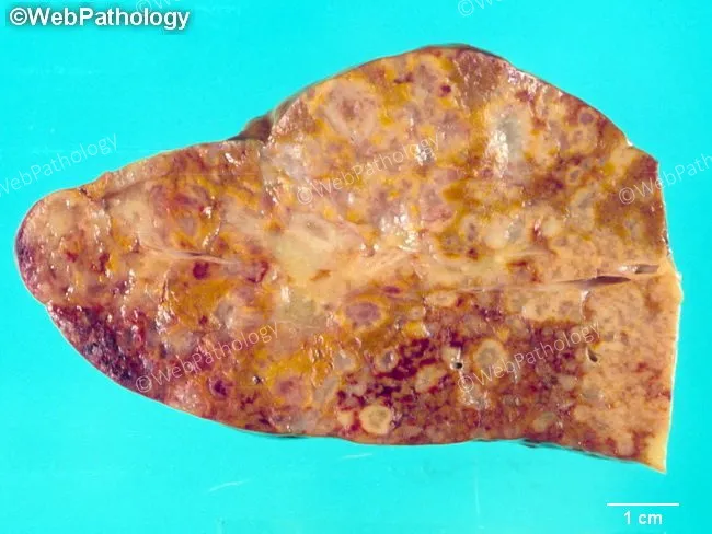
5. Microscopic (Histological) Features
- Architecture reflects the primary tumor - this is the key principle in identifying the site of origin
- Deposits are surrounded by a compressed rim of normal liver tissue ("pseudocapsule")
- Desmoplastic stroma around metastatic deposits (especially CRC) - distinguishes from HCC
- Central coagulative necrosis is common in large deposits
- Vascular invasion within the deposit may be seen
- No liver cell dysplasia in surrounding parenchyma (unlike HCC, which arises in cirrhosis)
- Surrounding liver is typically non-cirrhotic - important distinguishing feature
Specific Histological Patterns:
| Origin | Pattern |
|---|---|
| CRC (adenocarcinoma) | Gland-forming with "dirty necrosis" (necrotic debris within lumens); desmoplastic stroma |
| Gastric | Signet ring cells, gland formation |
| Pancreatic | Poorly differentiated adenocarcinoma, mucin pools |
| Breast | Lobular: single file pattern; Ductal: gland formation |
| Lung (small cell) | Small blue cells, crush artifact, azzopardi effect |
| Melanoma | Large epithelioid cells, intranuclear inclusions, melanin pigment |
| Neuroendocrine | Organoid/trabecular pattern; chromogranin/synaptophysin positive |
6. Immunohistochemistry (IHC) for Identification
| Marker | Favors |
|---|---|
| CK7+/CK20- | Lung, breast, ovary, biliary |
| CK7-/CK20+ | Colorectal |
| CK7+/CK20+ | Pancreatic, urothelial |
| CK7-/CK20- | HCC, renal cell carcinoma, prostate |
| TTF-1+ | Lung (adenocarcinoma), thyroid |
| CDX2+ | CRC, small bowel |
| GATA3+ | Breast, urothelial |
| PSA/PSAP+ | Prostate |
| HepPar-1, GPC3, AFP | Primary HCC (not secondary) |
| Melanoma markers (S100, HMB45, Melan-A) | Melanoma |
| Chromogranin A, Synaptophysin | NETs/carcinoid |
7. Compared to Primary HCC
| Feature | Secondaries | HCC |
|---|---|---|
| Number | Usually multiple | Usually solitary or few |
| Background liver | Usually non-cirrhotic | Often cirrhotic (80%) |
| AFP | Normal | Elevated (>400 ng/mL in ~70%) |
| Umbilication | Present (central necrosis) | Absent |
| Vascular invasion | Less common | Very common (portal vein) |
| Bile production | Absent | May be present |
| IHC | Matches primary | HepPar-1+, GPC3+, AFP+ |
8. Special Patterns
- Miliary metastases: Tiny (<5 mm) deposits throughout - typical of breast cancer and melanoma
- Diffuse infiltration: Metastases replace normal liver parenchyma diffusely without forming distinct nodules - rare, seen in gastric/breast signet ring
- Perivascular cuffing: Deposits concentrate around portal tracts (lymphangitic spread)
- Synchronous vs. Metachronous: Synchronous = detected at same time as primary; metachronous = detected later
9. Complications of Hepatic Secondaries
- Hepatic failure - from massive replacement of parenchyma
- Portal hypertension - from compression of portal venules
- Obstructive jaundice - deposits at porta hepatis
- Spontaneous rupture - rare, can cause hemoperitoneum
- Budd-Chiari syndrome - tumor thrombus occluding hepatic veins
- Paraneoplastic syndromes - hypoglycemia (especially with NETs)
10. Frozen Section / Biopsy Points
- Core biopsy under imaging guidance is the standard for tissue diagnosis
- Imprint cytology may be done intraoperatively
- Key reporting: number of deposits, size of largest, closest margin (in resection), presence of vascular invasion, lymph node status (porta hepatis)
Key Exam Points (Pathology Perspective)
- Most common malignant liver tumor = secondary (not HCC)
- Most common primary = colorectal
- Gross: multiple, umbilicated nodules in non-cirrhotic liver
- Central umbilication = ischemic necrosis of rapidly growing tumor
- Histology mirrors primary; desmoplasia = hallmark of CRC metastasis
- Non-cirrhotic background = favors secondary over HCC
- IHC panel (CK7/CK20/CDX2/TTF-1) used to trace origin
- Melanoma mets are black due to melanin
- Carcinoid/NET mets are yellow-orange and may calcify
- "Cannon ball" appearance on imaging and gross pathology
Steatohepatitis
Reading File
Steatohepatitis is hepatic inflammation superimposed on fatty change (steatosis), which can progress to fibrosis and cirrhosis. Two main forms: alcoholic (ASH) and non-alcoholic (NASH/MASH).
Searching sources...
Finding Sources
Finding Sources
Finding Sources
Reading File
Reading File
Reading File
Reading File
Finding Sources
Reading File
Finding Sources
Reading File
Finding Sources
Excellent - I have rich textbook content. Let me now read the Robbins pathology sections and get the histology images.
I now have comprehensive content from Robbins (Cotran) Pathologic Basis of Disease, Robbins & Kumar Basic Pathology, and Goldman-Cecil Medicine. Let me compile the complete pathology answer.
Steatohepatitis - Complete Pathology Notes
1. Definition
Steatohepatitis = steatosis (fatty change) + hepatocyte injury + inflammation + progressive fibrosis. It is a form of chronic liver disease in which macrovesicular steatosis is associated with focal liver cell injury, Mallory hyaline, mixed inflammatory infiltrate (with neutrophils), and progressive hepatic fibrosis in a pericentral (zone 3) and pericellular distribution.
Two major forms:
- ASH - Alcoholic steatohepatitis (part of alcohol-associated liver disease)
- MASH/NASH - Metabolic dysfunction-associated steatohepatitis / Non-alcoholic steatohepatitis (part of NAFLD/MASLD)
2. Types of Steatosis (Background)
| Type | Morphology | Causes |
|---|---|---|
| Macrovesicular | Single large fat droplet displaces nucleus | Alcohol, obesity, DM, NAFLD, hepatitis C (genotype 3), Wilson disease |
| Microvesicular | Multiple small droplets, nucleus central | Reye syndrome, acute fatty liver of pregnancy, HELLP, valproate, tetracycline IV, antiretrovirals |
- Macrovesicular steatosis is the predominant form in both alcoholic and non-alcoholic steatohepatitis
- ASH may rarely show alcoholic foamy degeneration (diffuse microvesicular-to-macrovesicular steatosis with ER and mitochondrial damage)
3. Alcoholic Steatohepatitis (ASH)
Pathogenesis
- Threshold: 80 g/day alcohol causes steatosis; 80-160 g/day for years leads to hepatitis; only 10-15% develop cirrhosis despite heavy drinking
- Women are more susceptible at lower doses (>20 g/day vs. >60-80 g/day in men); peak incidence ~a decade earlier
Key mechanisms (Robbins):
- Altered redox state - Alcohol dehydrogenase (ADH) + aldehyde dehydrogenase generate NADH, shifting redox balance to favor lipogenesis and suppress fatty acid oxidation
- Acetaldehyde toxicity - Lipid peroxidation, acetaldehyde-protein adduct formation, disrupts cytoskeleton and membranes, produces neoantigens
- CYP2E1 induction - Generates ROS that damage proteins, membranes, mitochondria; promotes apoptosis; also converts drugs like acetaminophen to toxic metabolites
- Methionine metabolism impairment - Decreases glutathione, sensitizes to oxidative injury; elevates homocysteine causing ER stress
- Gut permeability - Increased gut permeability releases endotoxin (LPS) into portal blood, activating Kupffer cells via TLR4, releasing pro-inflammatory cytokines (TNF-α, IL-1β, caspase-1 via inflammasome)
- Impaired lipoprotein assembly and secretion
- Enhanced peripheral fat catabolism - Increases circulating FFA available to hepatocytes
Risk factors for progression:
- Amount and duration of alcohol (most important)
- Female sex
- Genetic factors: PNPLA3, HSD17B13, TM6SF2, MBOAT7, MARC1 polymorphisms
- Protein-calorie malnutrition
- Comorbid hepatitis B/C, hemochromatosis, MASLD
- Obesity
Morphology (MORPHOLOGY section, Robbins Cotran)
Changes begin in centrilobular zone 3 and extend outward:
Stage 1 - Fatty Liver (Steatosis):
- Macroscopically: enlarged (up to 4-6 kg), soft, yellow, greasy liver
- Lipid droplets accumulate as small droplets → coalesce into large single droplet → push nucleus to periphery
- Predominantly macrovesicular, centrilobular
- Completely reversible with abstinence
Stage 2 - Alcoholic Hepatitis (Steatohepatitis):
The four cardinal histological features:
-
Ballooned hepatocytes - Injured, swollen hepatocytes with cleared-out cytoplasm and cytoskeletal damage. When extensive, leads to Mallory hyaline formation. Essential for diagnosis. On CK8/18 immunostaining: keratins are collapsed, leaving cytoplasm "empty."
-
Mallory-Denk bodies (Mallory hyaline) - Irregular eosinophilic intracytoplasmic inclusions composed of ubiquitinated, partially degraded intermediate filaments (keratin 8 and 18). Represent a failed attempt to sequester damaged cytoplasmic proteins. More abundant in ASH than MASH. Also seen in: Wilson disease, chronic biliary diseases, MASH.
-
Neutrophilic inflammation - Neutrophils are more prominent in ASH than in MASH; localize around ballooned hepatocytes (especially those with Mallory hyaline) - called "satellitosis". Lymphocytes are also present in portal tracts.
-
Perivenular/pericellular fibrosis ("chicken wire" fibrosis) - Fibrosis starts in acinar zone 3 as perisinusoidal/pericellular fibrosis with a "chicken wire" appearance (best seen on Masson trichrome stain). This is the earliest fibrotic change.
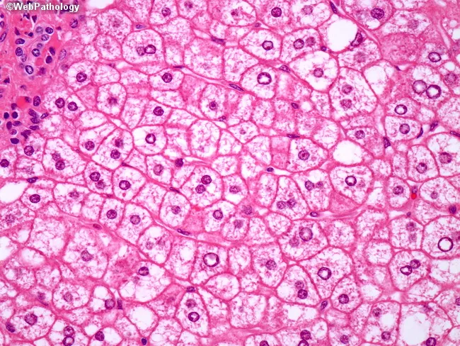
Stage 3 - Progressive Fibrosis and Cirrhosis:
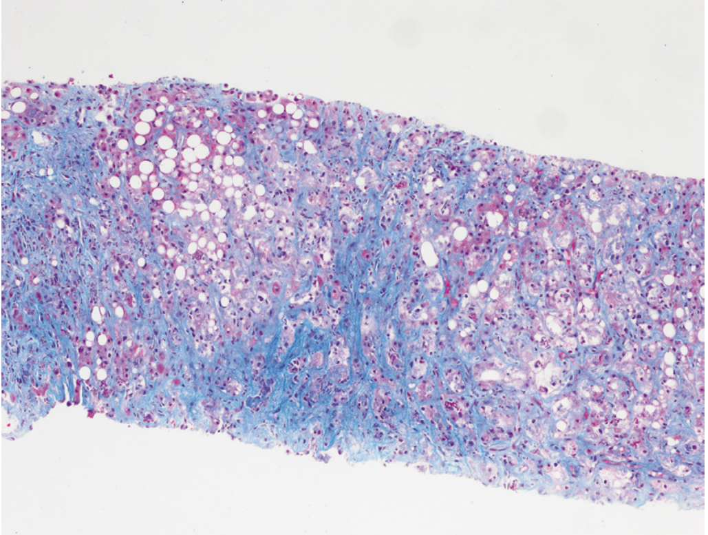
- Zone 3 pericellular fibrosis → portal/periportal fibrosis → bridging fibrosis → cirrhosis
- Micronodular cirrhosis ("Laennec cirrhosis") - average nodule ~3 mm, greenish tint from cholestasis
- "Burned-out" cirrhosis: fatty change disappears; small nodules trapped in blue-staining fibrous tissue
- Vascular derangements: phlebosclerosis (fibrous obliteration of central veins) + veno-occlusive lesions
- Early fibrosis can regress with abstinence; cirrhosis rarely fully regresses
4. Non-Alcoholic Steatohepatitis (NASH / MASH)
Definition / Terminology
- NAFLD = Non-alcoholic fatty liver disease (spectrum: simple steatosis → NASH → cirrhosis)
- MASLD = Metabolic dysfunction-associated steatotic liver disease (new 2023 nomenclature)
- NASH = Non-alcoholic steatohepatitis (now also called MASH = Metabolic dysfunction-associated steatohepatitis)
- Histologically resembles ASH but occurs without significant alcohol consumption (>2 drinks/day in men, >1 in women excludes NAFLD)
Risk Factors (Metabolic Syndrome)
Major: Obesity, Type 2 diabetes, dyslipidemia, metabolic syndrome (the "metabolic quartet")
Minor: PCOS, hypothyroidism, sleep apnea, hypopituitarism, hypogonadism
- ~80% of T2DM patients and ~90% of morbidly obese individuals have imaging evidence of NAFLD
- ~1/3 of adults in Western countries have NAFLD; ~5% have NASH
- Hispanics and Whites at higher risk; lower prevalence in African Americans
Pathogenesis ("Two-Hit" Model, now multi-hit)
- 1st hit: Insulin resistance + hyperinsulinemia → excessive FFA uptake by hepatocytes → macrovesicular steatosis (predominantly centrilobular)
- 2nd hit: Lipotoxicity from unesterified FFAs (especially palmitic acid, cholesterol, lysophosphatidylcholine, ceramides) → oxidative stress, ER stress, mitochondrial dysfunction, apoptosis
- Additional factors: adipocytokines (↓adiponectin, ↑TNF-α), gut dysbiosis, stellate cell activation
- PNPLA3 genetic polymorphisms correlate with severity of steatosis and fibrosis
Morphology of NASH
NASH histology mirrors ASH with important differences:
Features shared with ASH:
- Macrovesicular steatosis (centrilobular, zone 3)
- Ballooning degeneration of hepatocytes
- Mallory-Denk bodies (less abundant than in ASH)
- Lobular inflammation
- Pericellular/perisinusoidal "chicken wire" fibrosis (zone 3)
- Can progress to cirrhosis and HCC
Features more prominent in ASH vs. NASH:
| Feature | ASH | NASH/MASH |
|---|---|---|
| Neutrophilic inflammation | Prominent (satellitosis) | Less prominent |
| Mallory hyaline | More abundant | Less abundant |
| Megamitochondria | Present | Present |
| Portal inflammation | Less | More prominent |
| Fibrosis pattern | Zone 3, pericentral | Zone 3, but portal fibrosis also |
Pediatric NAFLD differs: portal-based chronic inflammation and portal fibrosis, WITHOUT ballooning or perivenular fibrosis (Type 2 NASH of Schwimmer et al.)
5. NASH Activity Score (NAS) - Kleiner Scoring System
Used to grade NASH severity on biopsy:
| Feature | Score |
|---|---|
| Steatosis | 0-3 (based on % of hepatocytes affected) |
| Lobular inflammation | 0-3 |
| Ballooning degeneration | 0-2 |
| Fibrosis stage | 0-4 (separate from NAS) |
- NAS ≥5 = definite NASH; NAS ≤4 does not exclude NASH (75% with NAS ≥5 had definite steatohepatitis)
- Steatohepatitis is a pattern diagnosis (steatosis + inflammation + ballooning); NAS alone cannot diagnose it
6. Drug-Induced Steatohepatitis
Drugs that cause steatohepatitis indistinguishable from ASH/NASH:
- Amiodarone (most classic - causes phospholipidosis + steatohepatitis + fibrosis)
- Tamoxifen
- Methotrexate (causes steatohepatitis + portal fibrosis)
- Glucocorticoids
- Valproic acid (causes microvesicular steatosis)
- Antiretrovirals (didanosine, stavudine)
7. Progression and Outcomes
Natural history:
- Simple steatosis → NASH (in ~20% over years)
- NASH → cirrhosis (in ~15-25% over 10-15 years)
- Cirrhosis → HCC (steatohepatitis-related HCC can also arise without cirrhosis, especially in NASH)
- NAFLD is now a leading cause of liver transplantation
8. Key Exam Points
- Cardinal histological tetrad: macrovesicular steatosis + ballooning + Mallory-Denk bodies + neutrophilic infiltrate
- Mallory hyaline = ubiquitinated keratin 8/18 intermediate filaments; eosinophilic intracytoplasmic; more in ASH than NASH
- Zone 3 (centrilobular) is the primary site of both steatosis and fibrosis onset
- Chicken wire fibrosis = perisinusoidal/pericellular fibrosis on Masson trichrome; earliest fibrosis pattern
- Satellitosis = neutrophils surrounding a ballooned hepatocyte with Mallory body
- ASH vs NASH: histologically similar - key clinical differentiator is alcohol history; AST:ALT >2 favors ASH; ALT rarely >150 in ASH
- Laennec cirrhosis = micronodular cirrhosis from chronic alcohol use
- Phlebosclerosis = fibrous obliteration of terminal hepatic venules; portal hypertension without traditional hepatic causes
- NASH progresses to HCC even without cirrhosis - unique among chronic liver diseases
- Pediatric NAFLD: type 2 pattern = portal inflammation + portal fibrosis, no ballooning (zone 1 predominant)
Sources: Robbins, Cotran & Kumar Pathologic Basis of Disease (9780443264528); Robbins & Kumar Basic Pathology (9780323790185); Goldman-Cecil Medicine (9780323930345); Sleisenger & Fordtran's GI & Liver Disease (9780323609623)
This is a shared conversation. Sign in to Orris to start your own chat.