
X ray C spine signs of Hirayama disease
Hirayama disease cervical spine X-ray radiograph signs findings
Hirayama disease cervical spine X-ray radiograph
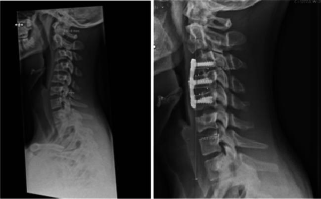
This side-by-side lateral cervical spine X-ray comparison illustrates the pre-operative and post-operative states of a patient with Hirayama disease and cervical kyphosis. The pre-operative image on the left demonstrates a significant kyphotic deformity centered between the C3 and C5 vertebral levels, characterized by an anterior curvature and defined intervertebral disc spaces. The post-operative image on the right shows the result of an anterior cervical discectomy and fusion (ACDF) procedure spanning C3 to C5. Visible instrumentation includes an anterior cervical plate secured with six screws into the C3, C4, and C5 vertebral bodies. The post-surgical alignment reveals a notable correction of the kyphosis toward a more neutral or lordotic curvature. Intervertebral spaces at the operated levels are no longer distinct, indicating the placement of fusion cages or bone grafts to promote solid osseous union. Measurements like the cervical sagittal vertical axis (cSVA) are indicated by vertical lines, showing the change in global alignment from 3 mm pre-operatively to 27 mm post-operatively at long-term follow-up.
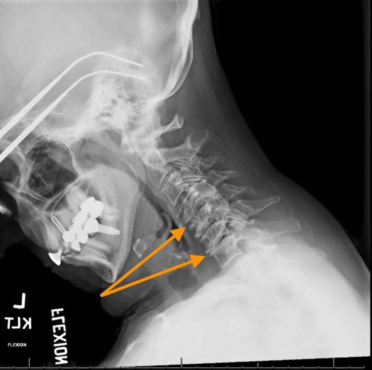
This lateral flexion X-ray of the cervical spine illustrates diagnostic signs of multilevel degenerative disc disease (cervical spondylosis). The radiograph captures the cervical vertebrae (C1-C7) in a forward flexed position to assess dynamic alignment and structural integrity. Key findings highlighted by orange arrows include significant intervertebral disc space narrowing at the C4-C5 and C6-C7 levels. While the overall spinal alignment is maintained without evidence of spondylolisthesis or dynamic instability in this view, the reduction in disc height suggests progressive discogenic degeneration. The image also displays the base of the skull, the mandible with dental hardware, and the posterior elements of the spine. This clinical imaging is essential for evaluating mechanical neck pain and neuroforaminal narrowing associated with chronic spinal wear.
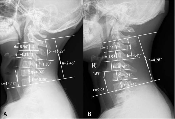
This diagnostic image displays two conventional sagittal cervical radiographs, labeled A and B, demonstrating different cervical spine curvatures in Hirayama disease. Radiograph A illustrates a 'reverse S-shape' pattern, characterized by upper cervical kyphosis (negative Cobb angles from C2-C4) and lower cervical lordosis (positive Cobb angles from C5-C7). The C2-4 Cobb angle (b) is -13.27°, while the C5-7 angle (c) is 14.43°. Radiograph B shows a relatively straight cervical spine typical of simple distal Hirayama disease, with a global C2-7 Cobb angle (a) of 4.78°. Both images include quantitative Cobb angle measurements for global (a), segmental groups (b, c), and individual levels (d: C2-3, e: C3-4, f: C4-5, g: C5-6, h: C6-7). These measurements highlight the postural differences between proximal and distal involvement, focusing on the loss of normal lordosis and the development of segmental kyphosis as key radiological markers for classification and diagnostic assessment in neurology and orthopedics.
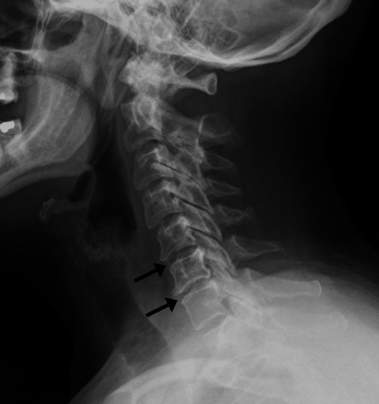
This lateral view diagnostic radiograph (X-ray) of the cervical spine demonstrates skeletal anatomy from the base of the skull to the upper thoracic region. The image reveals early-stage degenerative changes, specifically cervical spondylosis, localized to the mid-to-lower cervical levels. Key findings include a visible reduction in intervertebral disc height at the C5-C6 and C6-C7 levels. Black arrows highlight the presence of marginal anterior osteophyte formation (bony spurs) projecting from the anterior aspect of the C5 and C6 vertebral bodies. While the overall vertebral alignment and the spinal curvature appear maintained, the disc space narrowing and bone remodeling indicate chronic degenerative joint disease. The posterior elements, including the spinous processes and laminae, are visible without overt fracture or displacement. This image serves as a typical clinical example of age-related or post-traumatic spondylotic changes that can lead to symptoms like neck pain or radiculopathy through foraminal encroachment.
X-ray C-Spine Signs in Hirayama Disease
Key Point Up Front
X-ray Findings (Plain Radiograph, Lateral View)
1. Loss of Cervical Lordosis
- The most consistent and well-documented plain X-ray finding
- The normal cervical lordosis is flattened or straightened
- Present in a variable proportion of patients (stated in multiple guidelines)
- All major sources agree this is the primary X-ray finding: StatPearls on Monomelic Amyotrophy states "traditional x-rays and fluoroscopy usually only reveal loss of cervical lordosis"
2. Cervical Kyphosis
- More severe form of loss of lordosis, with actual forward angulation
- Particularly at the mid-to-lower cervical levels (C3-C7)
- Documented as a criterion in the Frontiers in Neurology diagnostic update
3. "Reverse S-Shape" Curvature Pattern
- A distinctive pattern seen in proximal Hirayama disease (less common variant)
- Upper cervical kyphosis (C2-C4) combined with lower cervical lordosis (C5-C7)
- This pattern on lateral X-ray helps differentiate proximal vs. distal disease involvement
- The image above (Panel A) shows this pattern with a C2-4 Cobb angle of -13.27° and C5-7 angle of +14.43°
4. Cervical Scoliosis
- Lateral curvature of the cervical spine may be seen, typically mild
- Usually corresponds to asymmetric muscle atrophy and tone
5. Abnormal Cobb Angle Measurements (on Lateral X-ray)
- Global C2-C7 Cobb angle is reduced or reversed
- Segmental Cobb angles at individual levels (C2-3, C3-4, etc.) may show focal angulation
- Quantitative measurement of cervical sagittal alignment helps classify disease severity and guide surgical planning (as shown in pre/post-op X-rays)
What X-ray Does NOT Show (but MRI Does)
| Finding | Requires |
|---|---|
| Localized cord atrophy at C5-C7 | MRI |
| Asymmetric cord flattening (pear-shaped cord on axial) | MRI |
| Loss of attachment (LOA) of posterior dural sac to lamina | MRI (axial T2, neutral position) |
| Forward displacement of posterior dural wall on flexion | Flexion MRI |
| Enlarged laminodural space (LDS) | Flexion MRI |
| Intramedullary T2 hyperintensity in anterior horn | MRI |
| Crescent-shaped posterior epidural enhancement | Post-gadolinium flexion MRI |
Diagnostic Criteria Role of X-ray
Illustrated Cases
Summary Table
| X-ray Sign | Frequency | Specificity |
|---|---|---|
| Loss of cervical lordosis (straightening) | Most common | Low (non-specific) |
| Cervical kyphosis (C3-C7) | Common | Moderate (supportive) |
| Reverse S-shape curvature | Proximal HD variant | More specific to HD type |
| Cervical scoliosis | Occasional | Non-specific |
| Abnormal segmental Cobb angles | Variable | Aids classification |
- Bradley and Daroff's Neurology in Clinical Practice - Benign Focal Amyotrophy section
- PMC MRI findings in Hirayama disease
- AJNR Flexion MRI in Hirayama disease