
The gut-brain axis and the relation between the fiber intake and deep sleep duration.
gut-brain axis dietary fiber sleep
gut-brain axis dietary fiber deep sleep NREM microbiome mechanisms 2024 2025
PMID: 36373848
PMID: 40613226
PMID: 39493112
gut-brain axis microbiome diagram
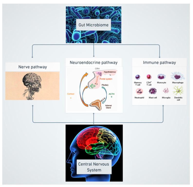
This pathophysiology diagram illustrates the bidirectional communication of the microbiome-gut-brain axis (MGBA) via three primary pathways: Nerve, Neuroendocrine, and Immune. At the top, a microscopic representation of the 'Gut Microbiome' originates signaling that flows downward through three parallel channels. The 'Nerve pathway' is represented by an anatomical illustration of the brain and brainstem. The 'Neuroendocrine pathway' depicts the Hypothalamic-Pituitary-Adrenal (HPA) axis, showing the hypothalamus releasing CRH, the portal system, the pituitary gland releasing ACTH, and the adrenal gland producing cortisol via MC2-R receptors; a cyclical arrow represents the hormonal feedback loop. The 'Immune pathway' displays various immune cells, including memory T cells, CD4+ T cells, monocytes, macrophages, neutrophils, mast cells, microglia, and dendritic cells. All three pathways converge at the 'Central Nervous System' (CNS) at the bottom, represented by a colorful lateral-view brain scan. The diagram demonstrates the integration of microbiological, hormonal, and immunological signals in human physiological regulation and disease mechanisms.
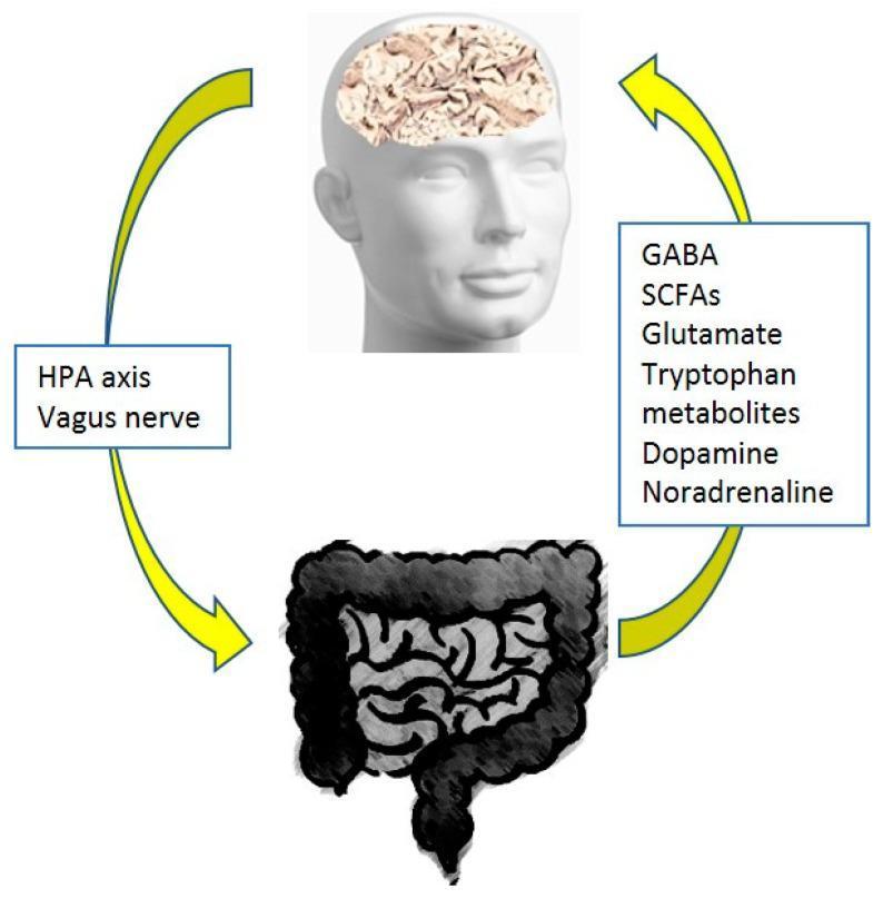
This pathophysiology diagram illustrates the bidirectional communication of the microbiome-gut-brain axis. The visual elements include a stylized human head with the brain visible, and a detailed illustration of the lower gastrointestinal tract (gut). Two large yellow curved arrows form a feedback loop between the two systems. The descending pathway (brain-to-gut) is mediated by the 'HPA axis' and the 'Vagus nerve,' representing the autonomic and endocrine influence on intestinal function. The ascending pathway (gut-to-brain) highlights biochemical signaling through microbial metabolites and neurotransmitters, explicitly listing GABA, Short-Chain Fatty Acids (SCFAs), Glutamate, Tryptophan metabolites, Dopamine, and Noradrenaline. This diagram serves as an educational summary of how the gut microbiome and its metabolic products influence central nervous system homeostasis and vice versa. It is relevant for medical study in neurology, gastroenterology, and psychiatry, specifically regarding the physiological and pathological links between intestinal health and neurochemical regulation.
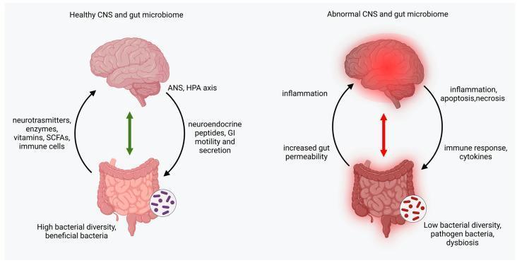
A pathophysiology diagram illustrating the bidirectional gut-brain axis in healthy versus abnormal states. The left side, 'Healthy CNS and gut microbiome,' depicts a standard anatomical brain and gut connected by a green bidirectional arrow. Communication involves neurotransmitters, enzymes, vitamins, short-chain fatty acids (SCFAs), and immune cells traveling from the gut to the brain, while neuroendocrine peptides, GI motility, and secretion signals move from the brain to the gut, mediated by the autonomic nervous system (ANS) and hypothalamic-pituitary-adrenal (HPA) axis. The gut displays high bacterial diversity with beneficial microbes. The right side, 'Abnormal CNS and gut microbiome,' shows the brain and gut with a red inflammatory glow, connected by a red bidirectional arrow signaling dysregulation. Key pathological features include inflammation, apoptosis, and necrosis in the brain, and increased gut permeability, immune responses, and cytokines in the gastrointestinal tract. The microbiome shows low diversity, pathogenic bacteria, and dysbiosis. This infographic highlights the role of the gut-brain axis in systemic inflammation and neurological health.
dietary fiber slow wave sleep SCFA microbiome
https://link.springer.com/article/10.1186/s41606-026-00187-6
The Gut-Brain Axis and Dietary Fiber's Influence on Deep Sleep
1. The Gut-Brain Axis: Overview
2. Deep Sleep (Slow-Wave Sleep / NREM N3) - A Brief Primer
3. How Dietary Fiber Acts Through the Gut-Brain Axis to Influence Deep Sleep
Step 1 - Fiber → Microbial Fermentation → SCFAs
Step 2 - SCFAs Signal the Gut-Brain Axis via Multiple Routes
| Mechanism | Detail |
|---|---|
| Gut barrier integrity | Butyrate is the preferred energy source of colonocytes; it tightens tight junctions, reducing "leaky gut." A permeable gut allows bacterial LPS to enter systemic circulation, triggering neuroinflammation that fragments SWS. |
| Serotonin production | SCFAs stimulate enterochromaffin cells to release serotonin (5-HT). About 90-95% of the body's serotonin is made in the gut. Serotonin is the precursor to melatonin via tryptophan hydroxylase → 5-HTP → 5-HT → melatonin in the pineal gland. More gut serotonin = more melatonin substrate. |
| Vagal activation | Butyrate and propionate bind free fatty acid receptors (FFAR2/FFAR3) on vagal afferent nerve terminals in the gut wall, sending signals to the nucleus tractus solitarius (NTS) in the brainstem, which projects to sleep-regulating areas including the hypothalamus and ventrolateral preoptic nucleus (VLPO). |
| Anti-inflammatory effect | SCFAs - especially butyrate - inhibit histone deacetylases (HDACs) and NF-κB signaling, reducing pro-inflammatory cytokines. Lower systemic inflammation correlates with longer, more consolidated slow-wave sleep. |
Step 3 - Tryptophan Pathway (Separate but Parallel)
Step 4 - Circadian Microbiota Rhythms Reinforce SWS Timing
4. The Evidence: What Studies Show
5. The Bidirectional Loop (Sleep Also Shapes the Microbiome)
- Elevates cortisol (HPA axis activation), which reduces Lactobacillus and Bifidobacterium populations
- Disrupts diurnal oscillations of the microbiota
- Reduces SCFA production the following day
- Increases intestinal permeability ("leaky gut") and systemic LPS, which then further fragments subsequent sleep
6. Practical Implications
- Fiber types that help most: Prebiotic fibers (inulin, FOS, galacto-oligosaccharides, arabinoxylan) preferentially feed SCFA-producing species. Beta-glucan (oats, barley) and resistant starch (cooled cooked potatoes, legumes) are particularly well-fermented.
- Timing matters: Evening consumption of fermentable fiber (e.g., legumes at dinner) aligns SCFA production with the natural evening melatonin rise, potentially amplifying sleep onset signals.
- Diet pattern context: Isolated fiber supplementation has smaller effects than whole dietary patterns (Mediterranean, MIND diet) that combine fiber with polyphenols, omega-3s, and limit ultra-processed foods that disrupt microbial diversity.
Summary
| Step | Mechanism |
|---|---|
| Fiber intake | Fermented by colonic microbiota |
| SCFA production | Butyrate, propionate, acetate generated |
| Gut barrier repair | Reduces LPS-driven neuroinflammation |
| Serotonin ↑ | Enterochromaffin cell stimulation → melatonin precursor |
| Vagus nerve signaling | SCFAs → FFAR2/3 → brainstem sleep centers |
| Tryptophan → melatonin | Microbiota shift pathway away from kynurenine |
| Anti-inflammation | HDAC inhibition, NF-κB suppression → less SWS fragmentation |
| Net effect | Longer, more consolidated slow-wave (deep) sleep |