
Explain pterygomaxilary fissure with relationship to nasal caviry and sphenopalatine fossa
Pterygomaxillary Fissure: Anatomy, Relationships, and Clinical Relevance
What Is the Pterygomaxillary Fissure?
- Anteriorly: the posterior (infratemporal) surface of the maxilla
- Posteriorly: the anterior surface of the pterygoid process of the sphenoid bone
The Pterygopalatine (Pterygomaxillary) Fossa
Bony Walls
| Wall | Bone |
|---|---|
| Anterior | Posterior surface of the maxilla |
| Posterior & roof | Pterygoid process + greater wing of sphenoid |
| Medial | Perpendicular plate of the palatine bone (abuts lateral nasal wall) |
| Lateral | Open - communicates via the pterygomaxillary fissure |
| Inferior (apex) | Narrows into the greater palatine canal |
| Superior | Opens into the inferior orbital fissure |
The Pterygomaxillary Fissure in Detail
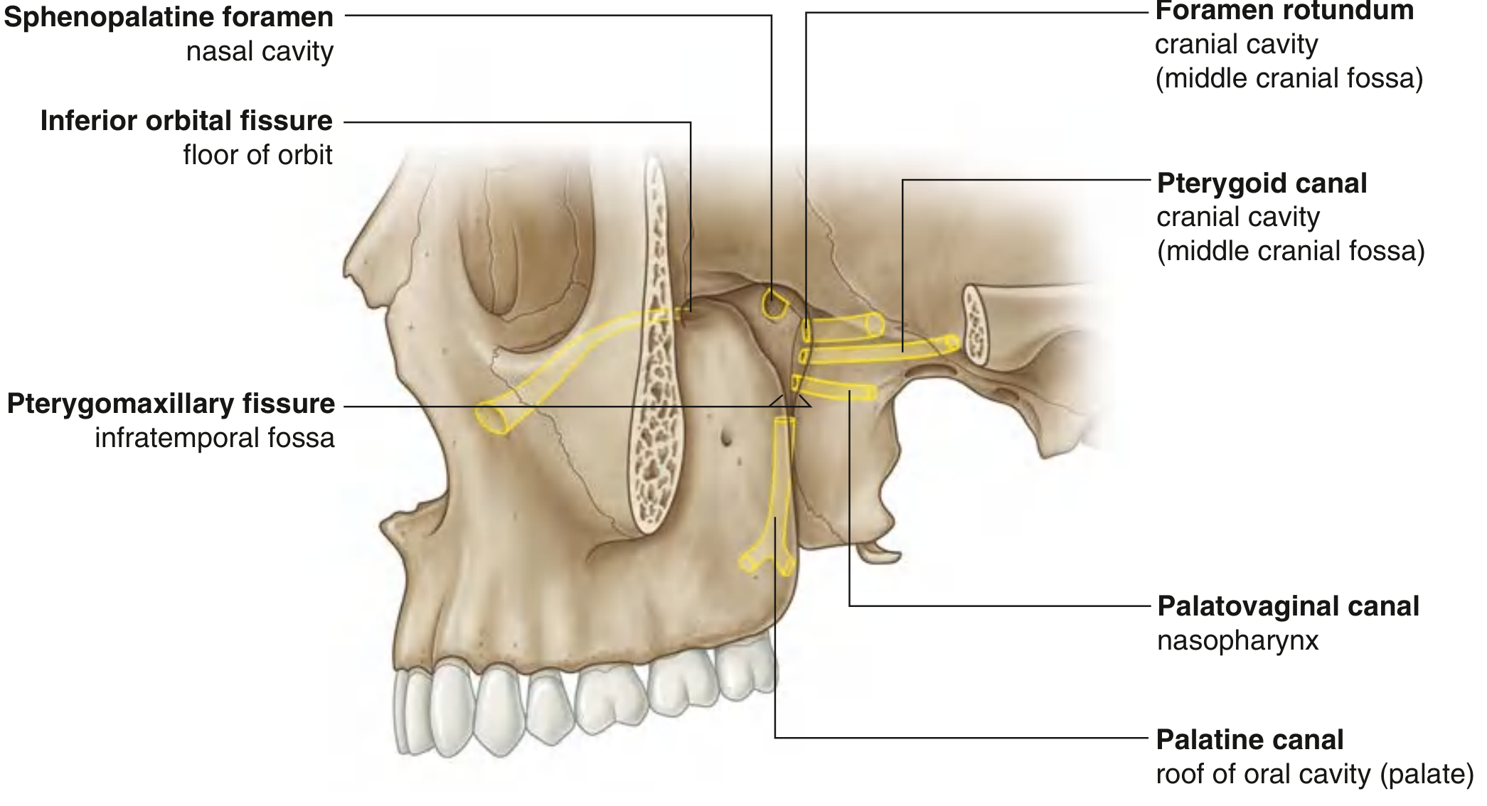
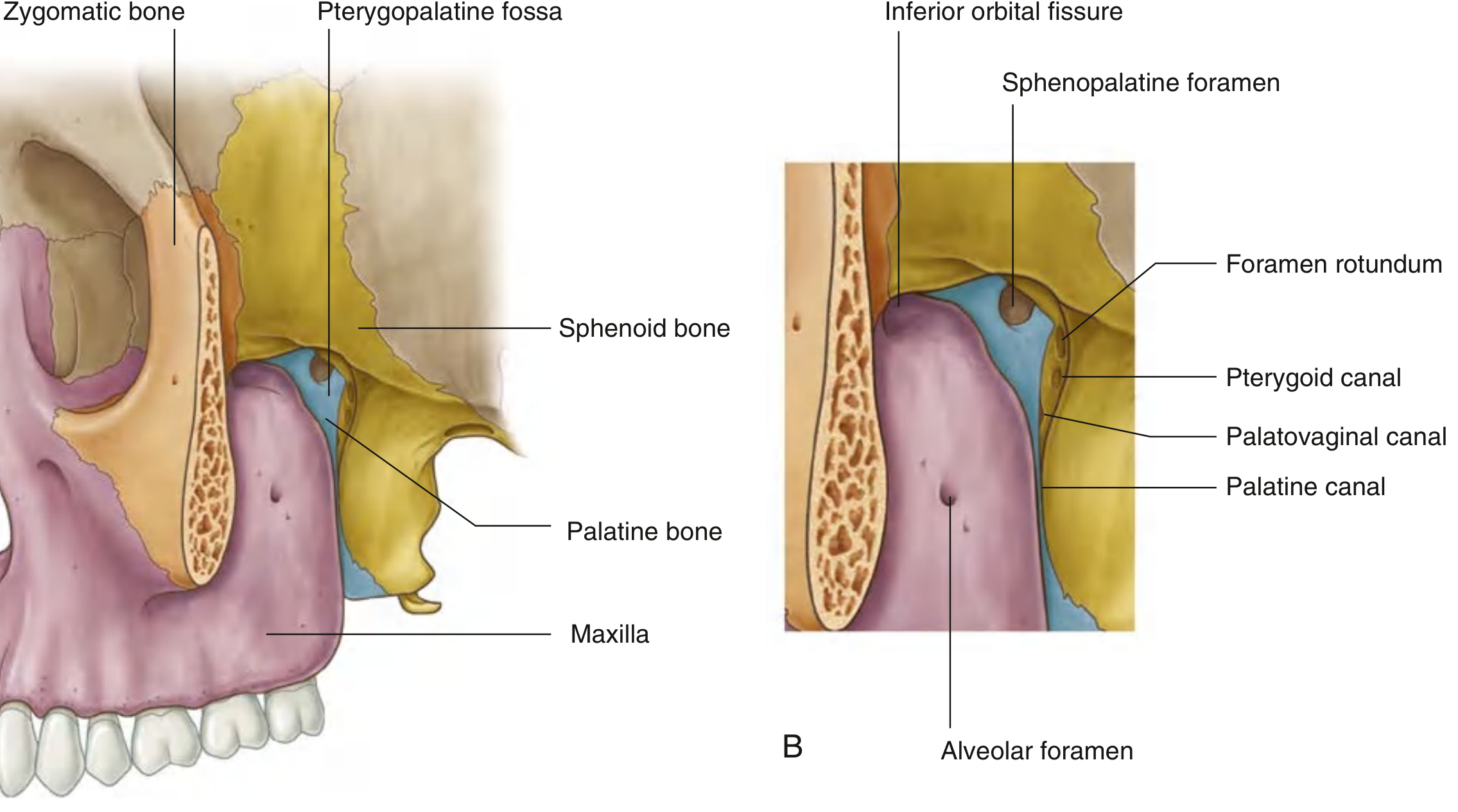
Key Structural Relationships
- Inferior orbital fissure (superiorly) → communicates with the floor of the orbit
- Pterygomaxillary fissure (inferiorly) → communicates with the infratemporal fossa
Structures Passing Through the Pterygomaxillary Fissure
- The maxillary artery (third/pterygomaxillary portion) ascends obliquely through the infratemporal fossa and enters the PPF via this fissure - it may travel either medial or lateral to the lower head of the lateral pterygoid muscle before entering
- The pterygoid venous plexus drains into veins that coalesce in the PPF and then pass laterally back through the fissure to join the pterygoid plexus in the infratemporal fossa
- The posterior superior alveolar nerve (from V2 in the PPF) exits via this fissure and descends on the posterolateral surface of the maxilla
All Seven Gateways of the Pterygopalatine Fossa
| Opening | Direction | Communicates with |
|---|---|---|
| Pterygomaxillary fissure | Lateral | Infratemporal fossa |
| Inferior orbital fissure | Superolateral | Floor of orbit |
| Sphenopalatine foramen | Medial | Lateral wall of nasal cavity |
| Foramen rotundum | Posterior | Middle cranial fossa (V2 enters) |
| Pterygoid (Vidian) canal | Posterior | Middle cranial fossa (Vidian nerve) |
| Palatovaginal canal | Posterior/inferior | Nasopharynx |
| Greater palatine canal | Inferior | Roof of oral cavity (hard palate) |
Relationship to the Nasal Cavity
- The pterygomaxillary fissure = lateral communication (to infratemporal fossa)
- The sphenopalatine foramen = medial communication (to nasal cavity)
The Sphenopalatine Foramen
- Sphenopalatine artery (terminal branch of the maxillary artery) - the most important arterial supply to the nasal cavity; divides into posterior lateral nasal and posterior septal branches
- Nasopalatine nerve (branch of V2)
- Superior nasal branches of the maxillary nerve (V2)
Infratemporal fossa → pterygomaxillary fissure → pterygopalatine fossa → sphenopalatine foramen → nasal cavity
Contents of the Pterygopalatine Fossa
- Maxillary nerve (V2) - enters via foramen rotundum; the most important sensory nerve for the upper teeth, palate, nasal cavity, and cheek
- Pterygopalatine (sphenopalatine) ganglion - a parasympathetic ganglion; preganglionic fibers from CN VII (via greater petrosal nerve → Vidian nerve) synapse here; postganglionic fibers hitch a ride on V2 branches to reach the lacrimal gland, nasal mucosa, and palate
- Terminal part of the maxillary artery (third portion) - enters via the pterygomaxillary fissure and branches into the sphenopalatine, infraorbital, descending palatine, and posterior superior alveolar arteries
Masticator Space Connection
Clinical Significance
-
Juvenile nasopharyngeal angiofibroma (JNA): These tumors classically arise near the sphenopalatine foramen and extend laterally through the pterygomaxillary fissure into the infratemporal fossa - widening of this fissure on imaging (Holman-Miller sign/antral sign) is a classic radiological finding.
-
Sphenopalatine ganglion (SPG) block: The lateral approach to the SPG targets the fossa via the pterygomaxillary fissure with a needle passed through the coronoid notch of the mandible - used for cluster headaches, trigeminal neuralgia, and nasal procedures.
-
Endoscopic skull base surgery: Medial access to the PPF is via the sphenopalatine foramen (medial wall), while lateral access requires going through the pterygomaxillary fissure - the approach chosen depends on tumor location and extension.
-
Spread of infection/tumor: The PPF acts as a crossroads - tumors of the nasal cavity, maxillary sinus, nasopharynx, or infratemporal fossa can all reach the middle cranial fossa by spreading through the PPF along nerve sheaths (perineural spread, especially along V2).
I have a doubt... The macilary artery first enters the nose via foramen rotundum and then goes into the sphenopalatine foramen right
Correcting the Misconception
| Maxillary Nerve (V2) | Maxillary Artery | |
|---|---|---|
| Origin | Trigeminal ganglion (middle cranial fossa) | External carotid artery (neck) |
| Route into PPF | Via foramen rotundum (posterior wall) | Via pterygomaxillary fissure (lateral wall) |
| Direction of travel | Cranial fossa → PPF (going forward/down) | Infratemporal fossa → PPF (going medially) |
The Correct Path of the Maxillary Artery
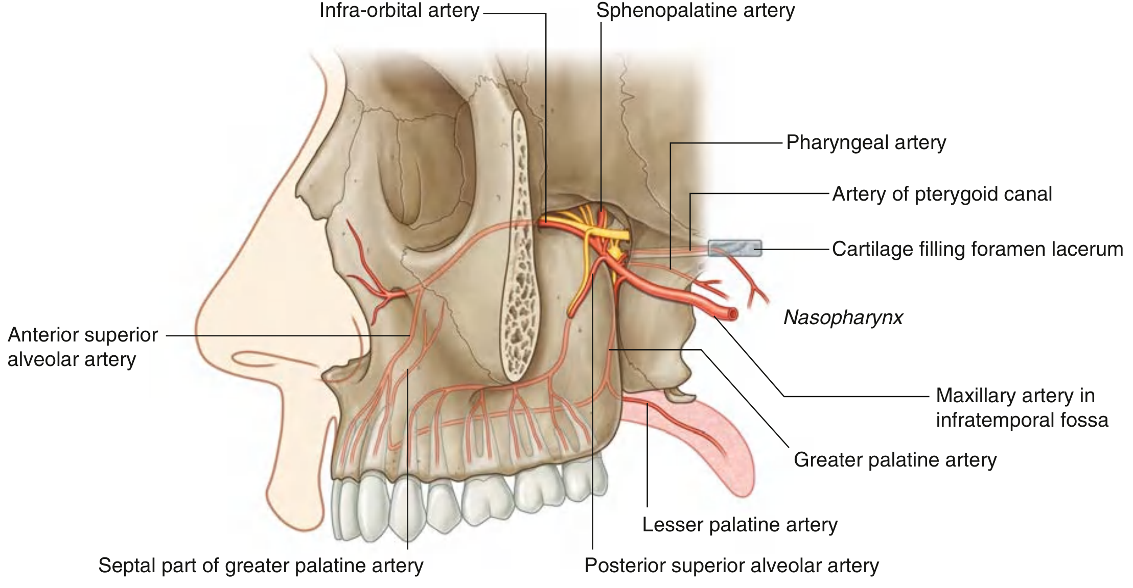
External carotid artery (neck, near neck of mandible) → passes forward through the infratemporal fossa (this is the 2nd part) → enters the pterygopalatine fossa via the pterygomaxillary fissure (this is the 3rd/pterygomaxillary part) → branches out inside the PPF → its terminal branch = sphenopalatine artery exits medially through the sphenopalatine foramen → enters the nasal cavity
Branches of the Maxillary Artery from the PPF
| Branch | Exits via | Supplies |
|---|---|---|
| Posterior superior alveolar a. | Pterygomaxillary fissure (back out) | Molar/premolar teeth, maxillary sinus |
| Infra-orbital a. | Inferior orbital fissure | Face, anterior teeth |
| Greater palatine a. | Palatine canal (inferiorly) | Hard palate, anterior nasal septum |
| Pharyngeal a. | Palatovaginal canal | Roof of nasal cavity, sphenoid sinus |
| Sphenopalatine a. (terminal) | Sphenopalatine foramen | Lateral nasal wall, nasal septum |
| Artery of pterygoid canal | Pterygoid canal (posteriorly) | Nasopharynx mucosa |
Can u generate an image explaining the pathwy from infratempiral fossa to nasal cavity
Medical anatomy diagram showing the pathway of the maxillary artery from the infratemporal fossa to the nasal cavity. Clean educational diagram with labeled arrows showing the step-by-step route: 1) External carotid artery branching into maxillary artery near neck of mandible, 2) Maxillary artery traveling through the infratemporal fossa (2nd part), 3) Entering the pterygopalatine fossa via the pterygomaxillary fissure (gap between posterior maxilla and pterygoid process of sphenoid), 4) Branching inside the pterygopalatine fossa into posterior superior alveolar artery, infraorbital artery, greater palatine artery, pharyngeal artery, and sphenopalatine artery, 5) Terminal sphenopalatine artery exiting medially through the sphenopalatine foramen into the nasal cavity where it divides into posterior lateral nasal branches and posterior septal branches. Also show the maxillary nerve V2 entering the pterygopalatine fossa via foramen rotundum separately, clearly labeled to distinguish it from the artery. Color coded: artery in red, nerve in yellow. Side cross-section view of skull showing bones: maxilla, sphenoid pterygoid process, palatine bone. Title: "Pathway: Infratemporal Fossa to Nasal Cavity via Pterygopalatine Fossa"
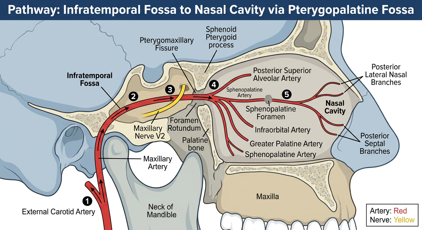
- Branches from the external carotid artery near the neck of the mandible
- Travels medially through the infratemporal fossa (2nd part)
- Enters the pterygopalatine fossa via the pterygomaxillary fissure (the lateral gap between the maxilla and pterygoid process)
- Branches out inside the PPF - posterior superior alveolar, infraorbital, greater palatine, pharyngeal, artery of pterygoid canal
- Its terminal branch (sphenopalatine artery) exits medially through the sphenopalatine foramen into the nasal cavity, dividing into posterior lateral nasal and posterior septal branches
- Comes from the middle cranial fossa
- Enters the PPF via foramen rotundum (posterior wall) - completely separate from the artery's route