
Urogenital development short notes
Reading File
Now let me search the medical library for urogenital development content in parallel.
Finding Sources
Finding Sources
Finding Sources
Finding Sources
Reading File
Reading File
Finding Sources
Finding Sources
Reading File
Reading File
Reading File
Finding Sources
Reading File
Finding Sources
Reading File
Reading File
Reading File
Reading File
I now have comprehensive content from multiple authoritative textbooks. Let me compile the short notes.
Loading Image
Loading Image
I now have all the content needed for comprehensive short notes. Here is the complete write-up:
Urogenital Development - Short Notes
1. Overview
The urogenital system arises from the intermediate mesoderm of the urogenital ridge along the posterior abdominal wall. It encompasses the urinary and genital systems, which are embryologically linked and often share congenital anomalies. Development proceeds in three overlapping phases: kidney formation, bladder/urethra formation, and gonadal/duct differentiation.
2. Development of the Kidney (Three Successive Stages)
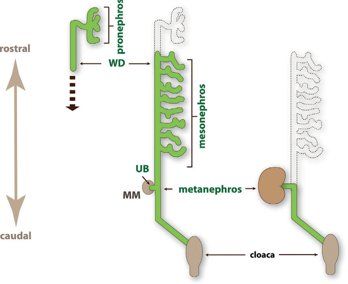
The kidney develops in a rostral-to-caudal sequence through three structures:
A. Pronephros
- Appears at day 22 of gestation (E8 in mice)
- Most rostral part of the urogenital ridge
- Consists of pronephric tubules and the pronephric duct (precursor of the Wolffian duct)
- Functional excretory organ in fish/amphibia larvae; vestigial in mammals
- Regresses by day 25
B. Mesonephros
- Appears at day 24 (E10 in mice); disappears by week 16
- Develops caudal to the pronephros in the midsection of the urogenital ridge
- Functional excretory organ in lower vertebrates; may filter during early embryonic life in mammals
- Before degeneration, endothelial, peritubular myoid, and steroidogenic cells migrate to the adrenogenadal primordium - abnormal migration causes gonadal dysgenesis
- In males: mesonephric (Wolffian) duct persists; in females: degenerates
C. Metanephros - The Definitive Kidney
- Begins at weeks 4-5 (day 28-32; E10.5 in mice)
- Results from reciprocal inductive interactions between:
- Ureteric bud (UB) - outgrowth from the distal Wolffian duct
- Metanephric mesenchyme (MM) - adjacent cluster of cells
Reciprocal Induction
- MM signals (GDNF, HGF) cause UB to branch into a T-tubule (E11.5) and then undergo iterative dichotomous branching → forms the entire collecting duct system
- UB sends reciprocal signals back to MM, inducing condensation
- Condensed MM forms pretubular aggregates → undergo mesenchymal-to-epithelial transition → form the renal vesicle → elongates into S-shaped body → becomes the nephron (from Bowman capsule to distal convoluted tubule)
Key Molecular Regulators
| Gene/Factor | Role |
|---|---|
| WT1 | Makes MM competent to respond to UB induction |
| PAX2 | Required for UB outgrowth |
| GDNF (from MM) | Stimulates UB branching via RET receptor |
| HGF | Supports metanephric growth |
| BMP7, Wnt/β-catenin | Proliferation of nephron progenitors |
Timeline of Nephron Formation
-
Glomeruli increase from weeks 10-18, then rapidly to week 36
-
Nephron formation is complete at birth: 200,000 to 2 million nephrons per kidney
-
No new nephrons form after birth - limited nephron number has lifelong health consequences
-
Glomerular filtration begins at ~9th fetal week
-
Brenner and Rector's The Kidney; The Developing Human - Clinically Oriented Embryology; Langman's Medical Embryology
3. Ascent of the Kidney
- Metanephros initially lies in the pelvis; ascends to the lumbar region by week 9
- Ascent occurs as the lumbar/sacral region grows caudally
- Arterial supply changes as the kidney ascends (original pelvic arteries degenerate; new vessels arise from the aorta)
- Failure to ascend → pelvic kidney
- Fusion of the lower poles before rotation → horseshoe kidney (caught on the inferior mesenteric artery, preventing ascent above L3)
- Crossed fused renal ectopia: both kidneys fused and on the same side of the midline
4. Development of the Urinary Bladder and Urethra
Cloaca and Urorectal Septum
- By week 5, the fetal cloaca is identifiable (common chamber receiving hindgut, allantois, and vestigial tailgut)
- The urorectal septum grows caudally (weeks 4-6) and divides the cloaca into:
- Ventral urogenital sinus
- Dorsal anorectal canal
- The urogenital sinus is then divided into three parts:
| Part | Derivative |
|---|---|
| Vesical (cranial) | Urinary bladder (epithelium from endoderm) |
| Pelvic | Female: entire urethra; Male: prostatic + membranous urethra |
| Phallic | Spongy urethra (male); vestibule + lower vagina (female) |
Bladder
- Epithelium: endoderm of the vesical part of the urogenital sinus
- Wall musculature: adjacent splanchnic mesenchyme
- Initially continuous with the allantois, which constricts → becomes the urachus (fibrous cord from bladder apex to umbilicus) → in adults = median umbilical ligament
- Distal mesonephric ducts are absorbed into the dorsal bladder wall → form connective tissue of the trigone
- Ureters open separately into the bladder; traction from ascending kidneys causes orifices to move superolaterally (oblique course)
In Males
-
Distal mesonephric duct ends → become ejaculatory ducts
-
Multiple endodermal outgrowths from prostatic urethra → prostate gland (glands from endoderm, stroma from mesenchyme)
-
Campbell-Walsh-Wein Urology; The Developing Human; Grainger & Allison's Diagnostic Radiology
5. Development of the Gonads
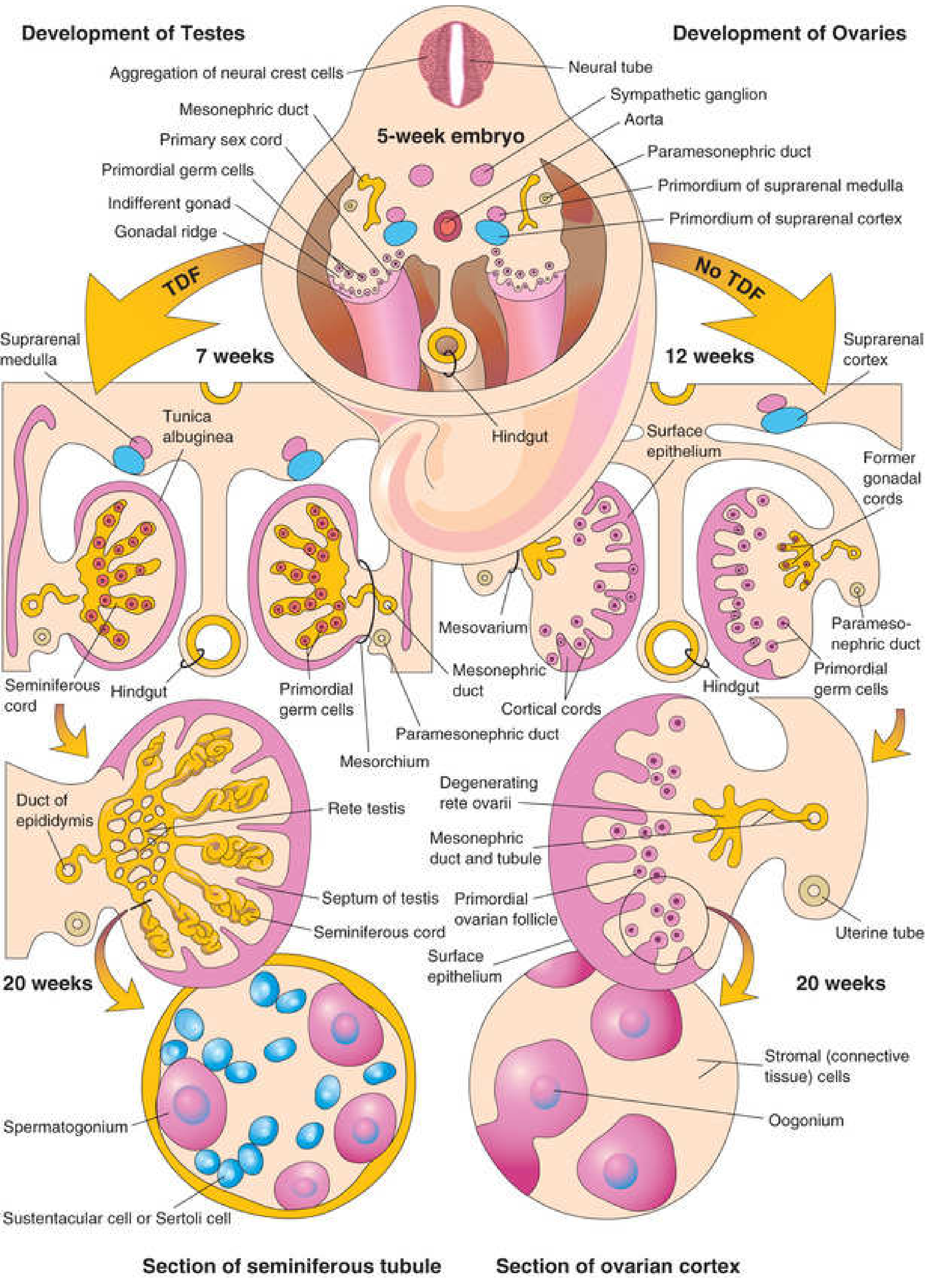
Origin of Gonads (Three Sources)
- Mesothelium lining the posterior abdominal wall (coelomic epithelium)
- Underlying mesenchyme
- Primordial germ cells (PGCs)
Primordial Germ Cells (PGCs)
- First recognizable at day 24 in the endoderm of the umbilical vesicle near the allantois
- Migrate along the dorsal mesentery of the hindgut to the gonadal ridges
- Enter gonadal cords by the 6th week
- Migration regulated by genes stella, fragilis, and BMP-4
Indifferent (Bipotential) Gonad
- Forms during week 5: mesothelium thickens on medial side of mesonephros → gonadal ridge with finger-like gonadal cords growing inward
- Consists of an outer cortex and inner medulla
- Transcription factors FOG2, WT1, NR5A1 required for bipotential gonad development
- Gonads remain morphologically identical until week 7 (indifferent stage)
Sex Determination
- Chromosomal sex determined at fertilization
- Morphological differentiation begins at week 7
- SRY gene (short arm of Y chromosome) encodes testis-determining factor (TDF)
| Chromosome | Gonadal Fate | Mechanism |
|---|---|---|
| XY | Medulla → testis; cortex regresses | SRY activates Sox9 → seminiferous cords form |
| XX | Cortex → ovary; medulla regresses | Absence of TDF; Wnt4, FoxL2, Rspo1 drive ovarian program |
Testis Development
- SRY activates Sox9 → gonadal cords become seminiferous cords
- Seminiferous cords: contain spermatogonia (from PGCs) + Sertoli cells (from mesenchyme)
- Cords enter medulla → rete testis
- Tunica albuginea forms, separating cords from surface epithelium
- Leydig cells (interstitial) produce testosterone from week 8
Ovary Development
-
No TDF → cortical cords extend from surface epithelium
-
PGCs within cortical cords → oogonia
-
Follicular (granulosa) cells derived from surface epithelium
-
Rete ovarii degenerates
-
The Developing Human - Clinically Oriented Embryology
6. Development of the Genital Ducts
Indifferent Stage
Both sexes have two duct systems:
- Mesonephric (Wolffian) ducts - drain the mesonephros
- Paramesonephric (Müllerian) ducts - form as evaginations in coelomic epithelium lateral to Wolffian duct (~week 6)
Male Differentiation (requires active hormonal signals)
| Hormone | Source | Effect |
|---|---|---|
| Testosterone | Leydig cells (from week 8) | Wolffian duct → epididymis, vas deferens, seminal vesicle, ejaculatory duct |
| Anti-Müllerian Hormone (AMH/MIS) | Sertoli cells | Müllerian duct regression |
| DHT (from testosterone via 5α-reductase) | Target tissues | External genitalia masculinization |
Wolffian duct derivatives in males:
- Proximal portion → head, body, tail of epididymis
- → Vas deferens
- Lateral outgrowth → seminal vesicle
- Distal segment → ejaculatory duct
- Multiple endodermal urethral outgrowths → prostate
Jost's classic 1953 experiment: castrating a male fetus before Wolffian maturation causes female development; castrating a female fetus has no effect on Müllerian development - proving male development requires active testicular factors, while female development is the "default" pathway.
Female Differentiation (default pathway - no active hormonal induction required)
- Absence of AMH → Müllerian ducts persist
- Absence of testosterone → Wolffian ducts degenerate
Müllerian duct derivatives in females:
-
Cranial portions remain separate → fallopian tubes (fimbriae from müllerian tunnels)
-
Midportions fuse → fundus and corpus of uterus
-
Distal fused portions → cervix + upper 1/3 of vagina (uterovaginal primordium)
-
Lower 2/3 of vagina + external female genitalia → from urogenital sinus
-
Medical Physiology (Boron & Boulpaep); Campbell-Walsh-Wein Urology; Color Atlas of Human Anatomy
7. Development of the Adrenal Glands
- Cortex: mesenchyme of the urogenital ridge (6th week); cells aggregate between dorsal mesentery root and developing gonad
- Medulla: neural crest cells migrating from adjacent sympathetic ganglion
- Fetal adrenal glands are 10-20x larger relative to body weight than adult glands
- Zona glomerulosa and fasciculata present at birth; zona reticularis not recognizable until year 3
- Key regulators: SF1, DAX1, Pbx1 for cortical development
8. Key Congenital Anomalies (Summary)
| Anomaly | Developmental Basis |
|---|---|
| Renal agenesis | Failure of UB outgrowth or MM induction |
| Horseshoe kidney | Lower poles fuse before rotation; arrested at IMA |
| Pelvic kidney | Failed renal ascent |
| Duplex ureter | Early UB bifurcation or two UB outgrowths |
| Patent urachus | Failure of allantois obliteration |
| Urachal cyst/sinus | Partial obliteration |
| Cryptorchidism | Failure of testicular descent |
| Gonadal dysgenesis | Abnormal mesonephric cell migration |
| Hypospadias | Failure of urethral folds to fuse (DHT deficiency/resistance) |
| Cloacal anomalies | Failure of urorectal septum division |
| Persistent Müllerian duct syndrome | AMH deficiency or AMH receptor mutation in males |
| Congenital adrenal hyperplasia | Enzyme defect in adrenal steroidogenesis |
9. Quick Summary Timeline
| Week | Event |
|---|---|
| 3-4 | Intermediate mesoderm forms urogenital ridge |
| 3.5 | Pronephros appears (day 22) |
| 4 | Mesonephros appears; pronephros regresses |
| 4-5 | UB outgrowth; metanephros begins |
| 5 | Gonadal ridges form; PGCs colonize; cloaca present |
| 6 | PGCs in gonadal cords; adrenal cortex begins; Müllerian ducts form |
| 7 | Urorectal septum complete; sex differentiation begins |
| 8-9 | Leydig cells active; testosterone production; AMH secreted |
| 9-10 | Kidney ascends to lumbar position |
| 12 | External genitalia differentiated |
| 16 | Mesonephros gone |
| 36 | Nephron formation complete |
| Birth | All nephrons present (200,000-2 million per kidney) |
Sources: The Developing Human - Clinically Oriented Embryology (Moore & Persaud); Brenner and Rector's The Kidney; Medical Physiology (Boron & Boulpaep); Campbell-Walsh-Wein Urology; Langman's Medical Embryology
Develpment of heart short notes
Finding Sources
Finding Sources
Finding Sources
Reading File
Finding Sources
Reading File
Finding Sources
Reading File
Finding Sources
Reading File
Reading File
Reading File
Reading File
Finding Sources
Finding Sources
Reading File
Reading File
Reading File
Loading Image
Loading Image
Reading File
I now have comprehensive content for the heart development short notes. Let me compile the full answer.
Development of the Heart - Short Notes
1. Overview and Timeline
The heart is the first functional organ to develop. The vascular system appears in the middle of the third week (day ~18) when diffusion alone can no longer satisfy the embryo's nutritional needs. The heart beats by day 22-23 and is largely complete by week 8.
2. Origin - Heart Fields
Progenitor heart cells lie in the epiblast adjacent to the cranial end of the primitive streak. They migrate through the streak into the splanchnic (visceral) layer of lateral plate mesoderm.
Two Heart Fields
| Field | Location | Contribution |
|---|---|---|
| Primary Heart Field (PHF) | Horseshoe-shaped cluster, cranial to neural folds | Parts of both atria + entire left ventricle |
| Secondary Heart Field (SHF) | Splanchnic mesoderm ventral to pharynx | Right ventricle + outflow tract (conus cordis + truncus arteriosus) + remainder of atria |
- Neural crest cells also contribute, migrating into the outflow tract region for septation and into the aortic arches
- Laterality (left-right patterning) is established simultaneously; the PITX2 transcription factor (master gene for left-sidedness) programs heart cells in both fields
Key Molecular Signals for Heart Induction
- BMPs (from endoderm and lateral plate mesoderm) + inhibition of WNT by CRESCENT → induce NKX2.5 in PHF
- NKX2.5 (homolog of Drosophila tinman) - master cardiac transcription factor
- TBX5 - expressed later; critical for septation
- HAND1/HAND2 - downstream of NKX2.5; expressed in future left and right ventricles respectively
- Looping regulated by: PITX2, NKX2.5, HAND1/HAND2, SONIC HEDGEHOG (SHH)
3. Formation of the Single Heart Tube
Days 17-22:
- PHF cells form blood islands in a horseshoe-shaped region (cardiogenic region) above the oropharyngeal membrane
- Islands unite → a horseshoe-shaped endothelial-lined tube surrounded by myoblasts
- With embryonic folding (cranial and lateral), the paired tubes fuse in the midline at the cephalic end (caudal ends remain initially separate)
- Result: a single primitive heart tube in the pericardial cavity
Layers of the Primitive Heart Tube
- Endocardium - inner endothelial lining
- Myocardium - thickens and secretes cardiac jelly (extracellular matrix rich in hyaluronic acid)
- Epicardium (visceral pericardium) - from the proepicardial organ (mesenchymal cells at caudal border of dorsal mesocardium); also gives rise to coronary artery endothelium and smooth muscle
The tube is attached to the dorsal pericardial wall by the dorsal mesocardium. The central part disappears → creates the transverse pericardial sinus.
Regions of the Heart Tube (Cranial → Caudal)
- Truncus arteriosus → roots/proximal portions of aorta and pulmonary artery
- Bulbus cordis (conus cordis) → outflow tracts of both ventricles
- Primitive ventricle → trabeculated portion of left ventricle (mainly)
- Primitive atrium → trabeculated portions of both atria
- Sinus venosus → smooth-walled right atrium (sinus venarum) + coronary sinus + oblique vein of left atrium
4. Cardiac Looping (Days 23-28)
On day 23, the heart tube begins to bend:
- Cephalic portion bends ventrally, caudally, and to the RIGHT
- Atrial (caudal) portion shifts dorsocranially to the LEFT
- Creates the cardiac loop (D-loop) - complete by day 28
Result: The atrium and sinus venosus lie dorsal to the truncus arteriosus and bulbus cordis. The atrioventricular canal connects the common atrium to the primitive ventricle.
Looping is the first morphological sign of left-right asymmetry. Abnormal looping → dextrocardia or situs inversus.
5. Development of the Sinus Venosus
- Mid-week 4: Sinus venosus drains blood from both right and left sinus horns via three veins each: vitelline, umbilical, and common cardinal veins
- Left-to-right shunts during weeks 4-5 → entrance of sinus shifts to the right
- Right umbilical vein + left vitelline vein obliterate
- Left common cardinal vein obliterates at week 10 → left sinus horn remnants become:
- Oblique vein of the left atrium
- Coronary sinus
- Right sinus horn incorporated into the posterior right atrial wall → smooth-walled sinus venarum
- Right and left venous valves form; right valve contributes to crista terminalis and the Eustachian (inferior vena caval) valve
6. Atrial Septation
Formation of Interatrial Septum (End of week 4 through week 6)
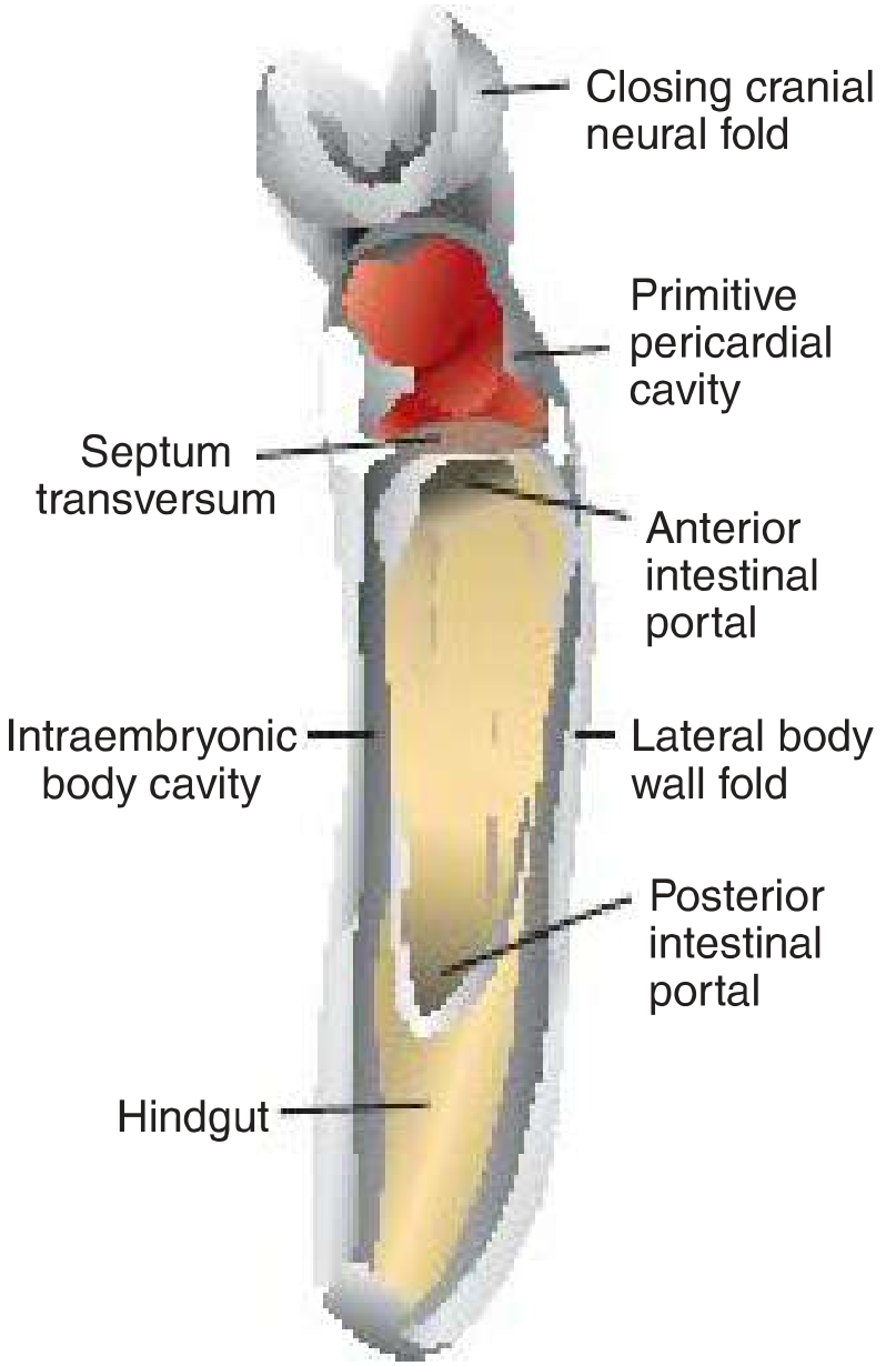
Step 1 - Septum Primum:
- A sickle-shaped crest grows from the roof of the common atrium downward toward the endocardial cushions
- The gap between its lower rim and the cushions = ostium primum (allows R→L flow)
- Endocardial cushion extensions grow up to close ostium primum
- Before closure: apoptosis in the upper septum primum creates perforations → coalesce into ostium secundum (maintains R→L flow)
Step 2 - Septum Secundum:
- A new crescent-shaped fold (septum secundum) descends from the roof of the right atrium, to the right of septum primum
- Never completes as a full partition
- Its free concave edge overlaps the ostium secundum → leaves the foramen ovale (oval foramen)
- Upper part of septum primum gradually disappears → remaining portion = valve of the foramen ovale
Fetal circulation: Blood passes from right atrium → through oblique cleft (foramen ovale) → left atrium (R→L)
At birth: Increased left atrial pressure → valve of foramen ovale pressed against septum secundum → foramen ovale closes (anatomically fuses in ~75% of people; probe patent in ~20% without hemodynamic shunting)
AV Canal Septation
- End of week 4: 4 endocardial cushions appear in the AV canal (superior, inferior, and 2 lateral)
- Superior and inferior cushions fuse → divide AV canal into right and left AV orifices (by end of week 5)
- AV canal enlarges to the right → blood now has access to both primitive ventricles
AV Valve Formation
- After cushion fusion, surrounding tissue is hollowed out from the ventricular side
- Forms the valve leaflets, attached to ventricular wall by muscular cords
- Muscular cords degenerate → chordae tendineae (dense connective tissue)
- Connected to papillary muscles
- Left AV canal → bicuspid (mitral) valve (2 leaflets)
- Right AV canal → tricuspid valve (3 leaflets)
Left Atrium
- Primitive left atrium expands
- A pulmonary vein appears from the dorsal mesocardium (not as an outgrowth of the atrium itself)
- Stem of pulmonary vein incorporated into left atrium → becomes the smooth-walled portion of the left atrium
- Original embryonic left atrium becomes only the trabeculated left atrial appendage
7. Ventricular Septation
End of week 4:
- Two primitive ventricles expand by myocardial growth + diverticulation/trabeculation
- Medial walls appose and merge → muscular interventricular septum (grows upward from apex)
- Gap between free rim and fused endocardial cushions = interventricular foramen (allows communication)
Closure of interventricular foramen (by week 7):
- Outgrowth of tissue from inferior endocardial cushion grows along top of muscular septum
- Fuses with parts of the conus (conotruncal) septum
- Forms the membranous part of the interventricular septum
8. Outflow Tract (Conotruncal) Septation
- Spiral conotruncal ridges (formed by neural crest cells + SHF-derived mesenchyme) appear in the conus cordis and truncus arteriosus
- These ridges spiral 180° and fuse to form the aorticopulmonary septum
- Spiral nature ensures: pulmonary artery wraps around the aorta
- Aorta → exits from left ventricle; Pulmonary trunk → exits from right ventricle
Semilunar Valves
- Small tubercles appear on the truncus swellings when partitioning is nearly complete
- Each of the two channels (aortic and pulmonary) gets 3 tubercles
- Tubercles hollow out on their upper surface → form 3 semilunar valve cusps each
- Neural crest cells contribute to formation of these valves
9. Congenital Heart Defects Summary
| Defect | Incidence | Embryologic Basis |
|---|---|---|
| VSD (membranous) | 12/10,000 - most common CHD | Failure of membranous septum formation (endocardial cushion tissue + conus septum fusion failure) |
| VSD (muscular) | 80% of VSDs, many spontaneously close | Incomplete merging of ventricular walls |
| ASD (secundum) | Common | Excessive resorption of septum primum or inadequate septum secundum |
| ASD (primum) | Endocardial cushion defect | Failure of endocardial cushions to close ostium primum |
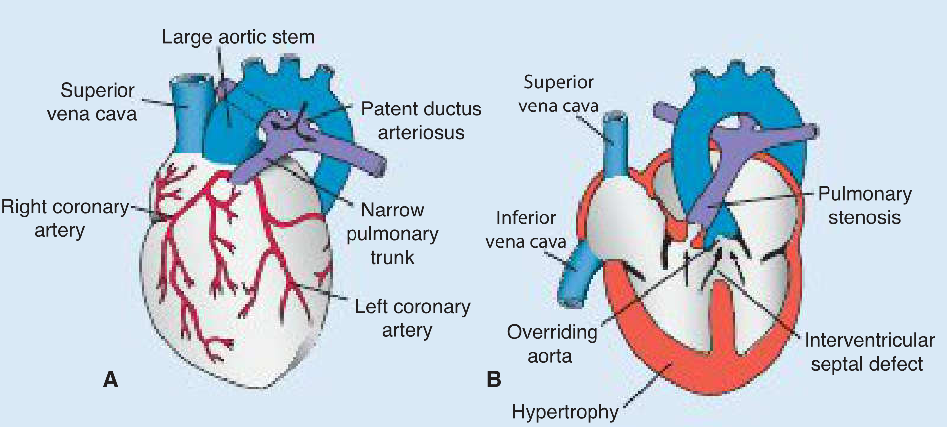
| Tetralogy of Fallot | 9.6/10,000 | Anterior displacement of conotruncal septum → (1) pulmonary stenosis, (2) VSD, (3) overriding aorta, (4) RV hypertrophy |
| Transposition of Great Arteries | 4.8/10,000 | Conotruncal septum fails to spiral (runs straight) → aorta from RV, PA from LV |
| Persistent Truncus Arteriosus | 0.8/10,000 | Conotruncal ridges fail to form; no outflow tract division; always with VSD |
| Patent Ductus Arteriosus (PDA) | Common | Failure of ductus arteriosus to close after birth |
| Dextrocardia | Rare | Abnormal cardiac looping (L-loop instead of D-loop) |
| DiGeorge syndrome | 22q11 deletion | Abnormal neural crest migration → truncus arteriosus, ToF + thymus/parathyroid aplasia |
| Holt-Oram syndrome | 1/100,000 | TBX5 mutation (autosomal dominant) → ASDs + radial limb defects |
| TAPVR | Rare | Aberrant positioning of dorsal mesocardium → pulmonary vein drains to right atrium/SVC |
10. Key Timeline
| Time | Event |
|---|---|
| Day 17-18 | Primary heart field established; cardiogenic region forms |
| Day 18-20 | Paired heart tubes fuse |
| Day 22-23 | Single heart tube pumps blood; looping begins |
| Day 28 | Cardiac loop complete |
| End of week 4 | Sinus venosus shifts right; endocardial cushions appear; septum primum begins |
| Week 5 | Ostium primum closes; ostium secundum forms; AV canal divides |
| Week 5-6 | Septum secundum + foramen ovale form; ventricular septation begins |
| Week 6-7 | Conotruncal septation; semilunar valves form |
| Week 7 | IVF closes (membranous septum complete) |
| Week 8 | Heart structurally complete |
| Birth | Foramen ovale closes; ductus arteriosus constricts |
Source: Langman's Medical Embryology; The Developing Human - Clinically Oriented Embryology (Moore & Persaud)
Development of heart short notes
I provided detailed short notes on Development of the Heart just above in our conversation. Here is the complete content again for easy reference:
Development of the Heart - Short Notes
1. Overview and Timeline
The heart is the first functional organ to develop. The vascular system appears in the middle of the third week (~day 18) when diffusion alone can no longer satisfy the embryo's nutritional needs. The heart beats by day 22-23 and is largely structurally complete by week 8.
2. Origin - Heart Fields
Progenitor heart cells lie in the epiblast adjacent to the cranial end of the primitive streak. They migrate through the streak into the splanchnic (visceral) layer of lateral plate mesoderm.
Two Heart Fields
| Field | Location | Contribution |
|---|---|---|
| Primary Heart Field (PHF) | Horseshoe-shaped cluster, cranial to neural folds | Parts of both atria + entire left ventricle |
| Secondary Heart Field (SHF) | Splanchnic mesoderm ventral to pharynx | Right ventricle + outflow tract (conus cordis + truncus arteriosus) + remainder of atria |
- Neural crest cells migrate into the outflow tract for conotruncal septation and into aortic arches
- Laterality is established simultaneously; PITX2 (master gene for left-sidedness) programs heart cells in both fields
Key Molecular Signals
| Gene/Signal | Role |
|---|---|
| BMPs + CRESCENT (WNT inhibitor) | Induce NKX2.5 in PHF |
| NKX2.5 (homolog of Drosophila tinman) | Master cardiac transcription factor; mutations → ASD, ToF |
| TBX5 | Expressed later; critical for septation; mutations → Holt-Oram syndrome |
| HAND1 / HAND2 | Downstream of NKX2.5; specify left and right ventricles respectively |
| SHH, NOTCH/JAG1 | SHF lengthening; neural crest regulation for outflow tract |
3. Formation of the Single Heart Tube (Days 17-22)
- PHF cells form blood islands in a horseshoe-shaped cardiogenic region cranial to the oropharyngeal membrane
- Islands unite → a horseshoe-shaped endothelial-lined tube surrounded by myoblasts
- With cranial and lateral embryonic folding, the paired tubes fuse in the midline
- Result: single primitive heart tube suspended in the pericardial cavity by the dorsal mesocardium
- Central dorsal mesocardium disappears → creates the transverse pericardial sinus
Layers of the Primitive Heart Tube
| Layer | Origin | Notes |
|---|---|---|
| Endocardium | Endothelial lining | Inner layer |
| Myocardium | Myoblasts | Secretes cardiac jelly (hyaluronic acid-rich ECM) |
| Epicardium | Proepicardial organ (mesenchyme at caudal dorsal mesocardium border) | Also gives rise to coronary artery endothelium and smooth muscle |
Regions of the Heart Tube (Cranial → Caudal)
- Truncus arteriosus → roots/proximal aorta and pulmonary artery
- Conus cordis (Bulbus cordis) → outflow tracts of both ventricles
- Primitive ventricle → trabeculated left ventricle (mainly)
- Primitive atrium → trabeculated portions of both atria
- Sinus venosus → smooth-walled right atrium, coronary sinus, oblique vein of left atrium
4. Cardiac Looping (Days 23-28)
On day 23, the heart tube begins to bend:
- Cephalic portion bends ventrally, caudally, and to the RIGHT
- Atrial (caudal) portion shifts dorsocranially to the LEFT
- Creates the D-loop - complete by day 28
Result: Atrium + sinus venosus lie dorsal to the truncus and bulbus cordis. The atrioventricular canal connects common atrium to the primitive ventricle.
Abnormal looping (L-loop) → dextrocardia / situs inversus. This is the first visible sign of left-right asymmetry.
5. Sinus Venosus Development
- Mid-week 4: Sinus receives blood from both sinus horns via 3 veins each: vitelline, umbilical, and common cardinal veins
- Left-to-right shunts (weeks 4-5) → entrance of sinus shifts rightward
- Left common cardinal vein obliterates at week 10 → left horn remnants become:
- Oblique vein of the left atrium
- Coronary sinus
- Right sinus horn → incorporated into the posterior right atrial wall = smooth-walled sinus venarum
- Right and left venous valves form; right valve contributes to crista terminalis and Eustachian valve (inferior vena caval valve)
6. Atrial Septation (Weeks 4-6)
Step 1 - Septum Primum
- Sickle-shaped crest grows downward from roof of the common atrium toward endocardial cushions
- Gap between lower rim and cushions = ostium primum (R→L flow)
- Endocardial cushion extensions grow up to close ostium primum
- Before closure: apoptosis in upper septum primum → perforations coalesce into ostium secundum (maintains R→L flow)
Step 2 - Septum Secundum
- Crescent-shaped fold descends from roof of right atrium, to the right of septum primum
- Never fully partitions atrium; free edge overlaps ostium secundum → leaves the foramen ovale
- Remaining septum primum = valve of the foramen ovale
Fetal state: R→L flow through foramen ovale (oblique cleft)
At birth: Left atrial pressure rises (lung circulation established) → valve pressed against septum secundum → foramen ovale closes
- Probe patent in ~20% of people (no hemodynamic shunting)
7. AV Canal Septation and Valve Formation
- End of week 4: 4 endocardial cushions appear (superior, inferior, and 2 lateral) in the AV canal
- Superior + inferior cushions fuse → divide AV canal into right and left AV orifices (by end of week 5)
- AV canal enlarges to right → blood accesses both primitive ventricles
AV Valve Formation
- Surrounding tissue hollowed out from the ventricular side
- Muscular cords degenerate → chordae tendineae (dense connective tissue)
- Connected to papillary muscles
- Left → Bicuspid (mitral) valve (2 leaflets)
- Right → Tricuspid valve (3 leaflets)
8. Ventricular Septation (Weeks 4-7)
| Step | Structure | Mechanism |
|---|---|---|
| 1 | Muscular IVS | Medial walls of expanding ventricles appose and merge from apex upward |
| 2 | Interventricular foramen | Gap above muscular septum - allows L-R communication initially |
| 3 | Membranous IVS | Endocardial cushion tissue + conus septum fuse to close the IVF by week 7 |
- Membranous VSD = most common isolated CHD (12/10,000) - failure of IVF closure
9. Outflow Tract (Conotruncal) Septation (Weeks 5-7)
- Neural crest cells + SHF cells form spiral conotruncal ridges in the conus cordis and truncus arteriosus
- Ridges spiral 180° and fuse → aorticopulmonary (conotruncal) septum
- Spiral nature ensures pulmonary trunk wraps around the aorta
- Aorta → exits left ventricle; Pulmonary trunk → exits right ventricle
Semilunar Valves
- Tubercles appear on truncal swellings → each channel (aortic + pulmonary) gets 3 tubercles
- Tubercles hollow out → 3 semilunar valve cusps per vessel
- Neural crest cells contribute to valve formation
10. Left Atrium and Pulmonary Veins
- A pulmonary vein arises from the dorsal mesocardium (not as an outgrowth of the left atrium)
- Stem of pulmonary vein is incorporated into the left atrium → smooth-walled portion of LA
- Original embryonic left atrium = only the trabeculated left atrial appendage
11. Key Congenital Heart Defects
| Defect | Incidence | Embryologic Basis |
|---|---|---|
| VSD (membranous) | 12/10,000 - most common CHD | Failure of membranous IVS (endocardial cushion + conus septum fusion) |
| VSD (muscular) | 80% of all VSDs; many close spontaneously | Incomplete ventricular wall merging |
| ASD (secundum) | Common | Excessive resorption of septum primum or inadequate septum secundum |
| ASD (primum) | Less common | Endocardial cushions fail to close ostium primum |
| Tetralogy of Fallot | 9.6/10,000 | Anterior displacement of conotruncal septum → (1) pulmonary stenosis, (2) VSD, (3) overriding aorta, (4) RV hypertrophy |
| Transposition of Great Arteries | 4.8/10,000 | Conotruncal septum fails to spiral (straight course) → aorta from RV, PA from LV |
| Persistent Truncus Arteriosus | 0.8/10,000 | Conotruncal ridges fail to form; always accompanied by VSD |
| Patent Ductus Arteriosus | Common | Ductus fails to close after birth |
| Dextrocardia | Rare | Abnormal L-loop instead of D-loop |
| DiGeorge syndrome | 22q11 deletion | Abnormal neural crest migration → truncus arteriosus, ToF + thymus/parathyroid aplasia |
| Holt-Oram syndrome | 1/100,000 | TBX5 mutation (autosomal dominant) → ASD + radial/thumb defects |
| TAPVR | Rare | Aberrant dorsal mesocardium positioning → pulmonary veins drain to RA or SVC |
12. Timeline Summary
| Time | Event |
|---|---|
| Day 17-18 | PHF established; cardiogenic region forms |
| Day 18-20 | Paired tubes fuse into single heart tube |
| Day 22-23 | Heart beats; looping begins |
| Day 28 | D-loop complete |
| End of week 4 | Sinus venosus shifts right; endocardial cushions appear; septum primum descends |
| Week 5 | Ostium primum closes; ostium secundum forms; AV canal divides |
| Weeks 5-6 | Septum secundum + foramen ovale established |
| Weeks 6-7 | Conotruncal septation; semilunar valves form |
| Week 7 | Membranous IVS complete; IVF closed |
| Week 8 | Heart structurally complete |
| Birth | Foramen ovale closes; ductus arteriosus constricts (closes by 1-3 months) |
Source: Langman's Medical Embryology; The Developing Human - Clinically Oriented Embryology (Moore & Persaud)
This is a shared conversation. Sign in to Orris to start your own chat.