
Interpret the report
python3 -c " import pdfplumber with pdfplumber.open('/tmp/workspace/attachments/89e229ed-c75f-4983-82e5-9c749ab6cbbd/labreportnew.aspx.pdf') as pdf: for i, page in enumerate(pdf.pages): print(f'=== PAGE {i+1} ===') text = page.extract_text() print(text) print() tables = page.extract_tables() for j, table in enumerate(tables): print(f'--- Table {j+1} ---') for row in table: print(row) "
Lab Report Interpretation - Mr. Vivek Srivastav (52M, 25 Jul 2026)
Patient Summary
- Name: Mr. Vivek Srivastav | Age: 52 years, Male
- Lab: Chandan Diagnostic Centre / Chandan Speciality Lab, Gonda (UP)
- Referring Doctor: Self-referred
Test Results at a Glance
| Test | Result | Reference Range | Status |
|---|---|---|---|
| Glucose Fasting (Plasma) | 73.80 mg/dL | < 100 | ✅ Normal |
| CRP (Quantitative) | 2.10 mg/L | < 6.0 | ✅ Normal |
| Eosinophils | 10.00% | 1 - 6% | ⚠️ HIGH |
| Total IgE | 1,421 IU/mL | < 158 | 🔴 VERY HIGH |
Detailed Interpretation
1. Glucose Fasting - 73.80 mg/dL ✅ NORMAL
- This is comfortably within the normal range (below 100 mg/dL).
- No diabetes, no pre-diabetes. The result effectively rules out hyperglycemia at the time of testing.
- It also rules out reactive hypoglycemia on its own (>70 mg/dL is acceptable).
- No follow-up needed for this parameter at this time.
2. CRP (C-Reactive Protein) - 2.10 mg/L ✅ NORMAL
- CRP is a non-specific marker of inflammation/infection. The result is negative (threshold: 6.0 mg/L).
- This means there is no significant acute systemic inflammation or active infection at the time of testing.
- This is useful context alongside the eosinophilia below - it suggests the elevation is not driven by an acute bacterial infection or inflammatory flare.
3. Eosinophils - 10.00% ⚠️ ELEVATED (Eosinophilia)
-
Normal range: 1-6%. A result of 10% is nearly double the upper limit - this is a significant finding.
-
This is reported as a differential percentage; the absolute count (AEC) would depend on the total WBC count, which was not included in this report. An absolute eosinophil count >500 cells/µL defines eosinophilia, and >1500 is considered significant.
-
Common causes of eosinophilia include:
- Allergic/Atopic disease - asthma, allergic rhinitis, urticaria, atopic dermatitis (most common in developed-world settings)
- Parasitic infections - especially helminths (roundworm, hookworm, filaria, toxocara) - a leading cause in endemic regions like India
- Drug reactions
- Less commonly: autoimmune disease, malignancy (lymphoma), eosinophilic GI disorders
-
In this patient's context, the eosinophilia is almost certainly connected to the markedly elevated IgE (see below). The two together point strongly toward allergy or parasitic infection.
4. Total IgE - 1,421 IU/mL 🔴 CRITICALLY ELEVATED (9x Upper Limit)
- Normal: < 158 IU/mL. This result is approximately 9 times the upper limit of normal - a very significant elevation.
- IgE is the primary immunoglobulin mediating allergic responses (Type I hypersensitivity).
| Category | Examples |
|---|---|
| Atopic/Allergic disease | Allergic asthma, allergic rhinitis, atopic dermatitis, urticaria |
| Parasitic infections | Helminths (Ascaris, hookworm, filaria, Toxocara) - very common in India |
| Fungal allergy | Allergic bronchopulmonary aspergillosis (ABPA) |
| Immunodeficiency | Hyper-IgE syndrome (Job's syndrome) |
| Other | IgE myeloma, Sezary syndrome, Hodgkin's lymphoma - rare |
- Helminthic (parasitic worm) infection - very prevalent in UP/rural India; helminths are the single most common cause of extreme IgE elevations globally
- Severe atopic/allergic disease (e.g., allergic asthma, atopic dermatitis, ABPA)
- Both can coexist
Summary & What Should Be Done Next
- Stool examination (for ova and parasites) and/or serology for specific helminths (Toxocara, filaria, Strongyloides, etc.) to rule out parasitic infection
- Complete Blood Count (CBC) with differential to get the absolute eosinophil count and check for other abnormalities
- Specific allergen IgE testing (RAST/ImmunoCAP) - to identify the exact allergen trigger if atopy is suspected
- Chest X-ray - to look for signs of ABPA or eosinophilic lung disease (pulmonary infiltrates)
- Clinical history - any symptoms of itching, rashes, breathing difficulty, wheezing, abdominal symptoms, or prior eczema/asthma
- Spirometry if respiratory symptoms are present (to assess for asthma)
Important note: This report does not replace a clinical consultation. The markedly elevated IgE and eosinophilia warrant evaluation by a physician - ideally an allergist or general medicine specialist. Early diagnosis of the underlying cause (parasitic vs. allergic) will guide targeted treatment.
Rashes itching
urticaria hives allergic skin rash
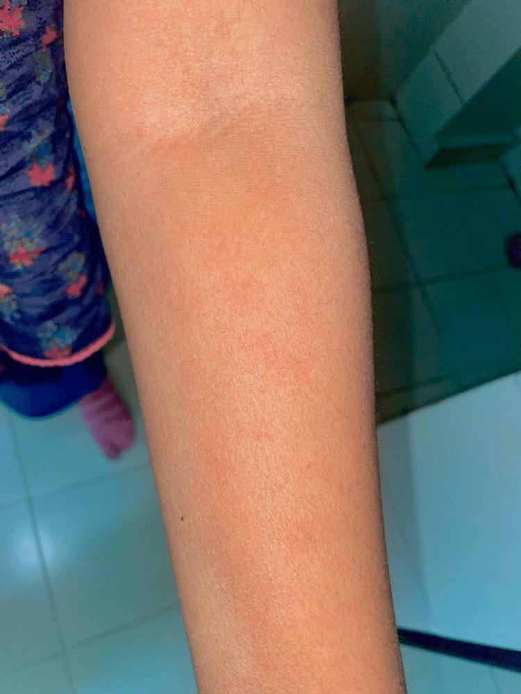
This clinical photograph displays a close-up view of a human upper extremity, specifically the forearm and elbow region, exhibiting a diffuse dermatological reaction. The skin presents with a generalized erythematous (reddish) hue and is covered by an extensive, confluent rash. Visible features include numerous small, raised, erythematous papules and classic wheals characteristic of urticaria (hives). The distribution is widespread across the limb, with a notable concentration of skin inflammation and swelling around the antecubital fossa (inner elbow). This presentation is clinically significant as it illustrates a hypersensitivity reaction or angioedema-like presentation, which in this medical context is associated with an adverse drug reaction following olanzapine administration. The image serves as an educational tool for identifying medication-induced cutaneous manifestations and acute allergic skin responses in clinical practice.
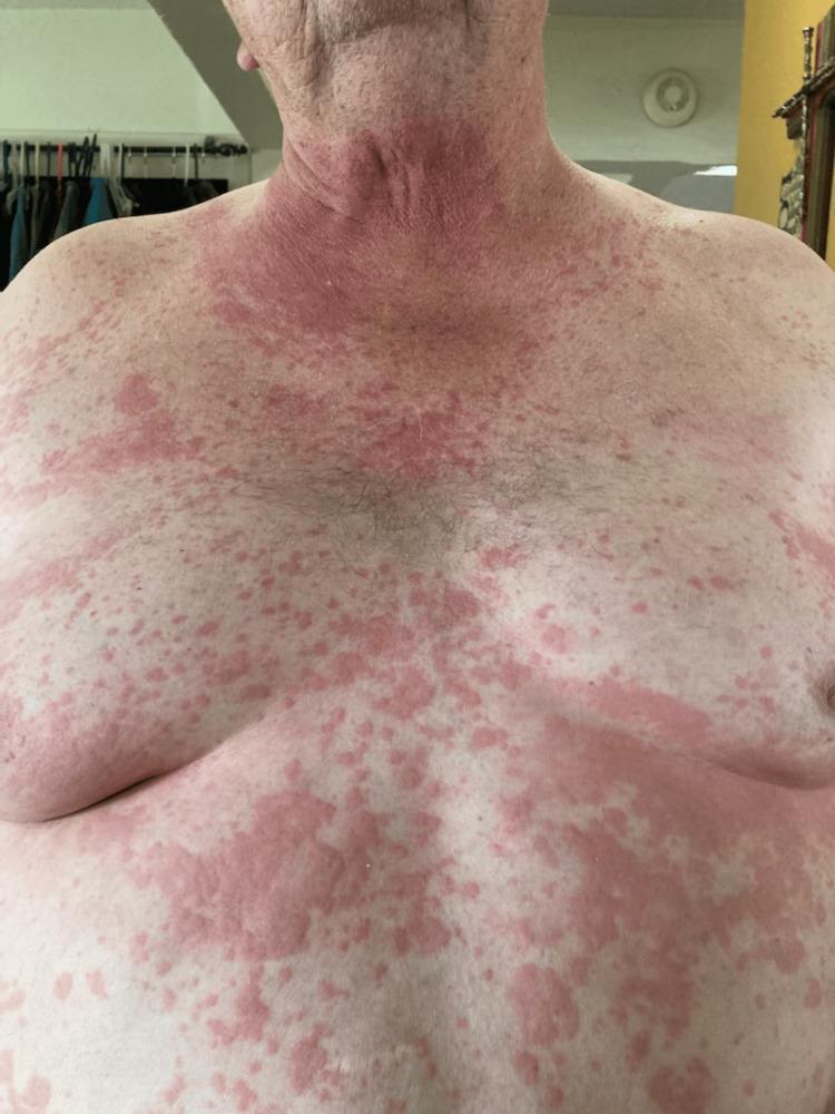
This clinical photograph displays a front-facing view of a male patient's neck, chest, and abdomen, showcasing a widespread inflammatory dermatologic reaction. The image demonstrates a mixed eruption characterized by urticaria (hives) and a maculopapular rash. The neck region exhibits intense, confluent erythema, while the chest and abdomen feature a more heterogeneous distribution of lesions. Visible skin findings include numerous erythematous macules, papules, and large, edematous, raised wheals that are beginning to coalesce into irregular, confluent patches. The lesions vary in morphology from small, discrete pink spots to larger, intensely red, elevated plaques with indistinct borders. This clinical presentation is typical of a systemic hypersensitivity reaction, specifically occurring in this context as a manifestation of alpha-gal syndrome (mammalian meat allergy) following beef consumption. The visual evidence emphasizes the extensive distribution and polymorphic nature of the rash, illustrating key diagnostic physical signs of IgE-mediated allergic responses in medical education.
Rashes & Itching - Connecting the Dots
The Full Picture
| Lab Finding | Value | Significance |
|---|---|---|
| IgE | 1,421 IU/mL (9x normal) | Massive allergic/parasitic response |
| Eosinophils | 10% (high) | Immune response to allergen or parasite |
| CRP | 2.10 (normal) | No acute bacterial infection |
| Glucose | 73.80 (normal) | Ruled out diabetes |
| Symptoms | Rashes + Itching | Skin manifestation of the above |
Two Most Likely Causes
1. 🪱 Parasitic (Helminth) Infection - HIGH SUSPICION in this context
- Strongyloides stercoralis - causes itching and urticaria after skin penetration, including during chronic infection; also causes itching around the anus
- Ascaris lumbricoides - urticaria and pruritus during larval migration (very common in UP)
- Hookworm (Ancylostoma/Necator) - itching and hives after skin entry and during larval migration
- Toxocara (from dogs/cats) - urticaria and pruritus during larval migration
- Schistosoma - transient urticaria with itching
2. 🌿 Allergic/Atopic Disease - ALSO POSSIBLE
- Chronic Spontaneous Urticaria (CSU) - recurring hives (wheals) lasting >6 weeks, driven by IgE
- Atopic Dermatitis (Eczema) - intensely itchy skin, strongly associated with high IgE
- Allergic contact dermatitis - reaction to a substance touching the skin
- Food allergy - certain foods (seafood, nuts, dairy) trigger IgE-mediated skin reactions
What the Rash Likely Looks Like
What Mr. Vivek Should Do Now
Immediate Relief (Symptomatic)
- Antihistamines (e.g., cetirizine, levocetirizine, fexofenadine) - taken under doctor's advice - are the first-line treatment for itch and hives from allergic reactions
- Avoid known triggers (certain foods, dust, pet contact) until evaluated
- Do not self-medicate with steroids without a doctor's guidance
Investigations to Confirm the Cause (with a doctor)
- Stool for ova & parasites (×3 samples on different days) - to identify helminth eggs
- Serology - blood tests for Strongyloides, Toxocara, filaria if stool is negative
- Absolute Eosinophil Count (AEC) - needs full CBC if not already done
- Specific IgE panel / skin prick test - to identify exact allergen if atopic cause is suspected
- Skin biopsy if rash morphology is atypical or diagnosis remains uncertain
Which Doctor to See
- Dermatologist - for the rash and itch (primary concern)
- General Physician / Internal Medicine - to coordinate parasite workup
- In Gonda/UP - a dermatologist at the district hospital or a private skin specialist would be appropriate
Bottom line: Rashes and itching in Mr. Vivek, combined with IgE of 1,421 and 10% eosinophils, strongly point to either a helminthic (worm) infection or allergic/atopic skin disease - or both. A stool test and a visit to a dermatologist/physician are the urgent next steps. The good news is both conditions are very treatable once the cause is confirmed.