
Make a schimatic diagram of polyporus and double ended pseudocomedones
polyporus fungus schematic diagram structure morphology
Schematic diagram of Polyporus (bracket fungus / Basidiomycete): labeled scientific illustration showing the external and internal structures including: pileus (cap) with concentric zones, pores on the underside (hymenophore), stipe (stalk), context (flesh), hyphal system with generative hyphae, skeletal hyphae, binding hyphae (trimitic system), basidiospores, basidia with sterigmata, cystidia, clamp connections, mycelium at base. Clean black and white educational diagram with clear labels and arrows, biology textbook style.
Schematic cross-section diagram of double-ended pseudocomedones (nevus comedonicus on palms): labeled histology diagram showing epidermis with papillomatous interfollicular surface, large dilated follicular invagination/infundibulum with orthokeratotic keratin plug (dark material), the infundibulum opens at both the surface and the base (double-ended), atrophic squamous epithelium lining the cyst wall, absence of hair shaft inside the dilated follicle, absence of sebaceous glands, surrounding dermis. Inset showing epithelial hyperplasia variant. Clean scientific educational diagram with labels and arrows, dermatology textbook style.
1. Polyporus (Bracket Fungus - Basidiomycete)
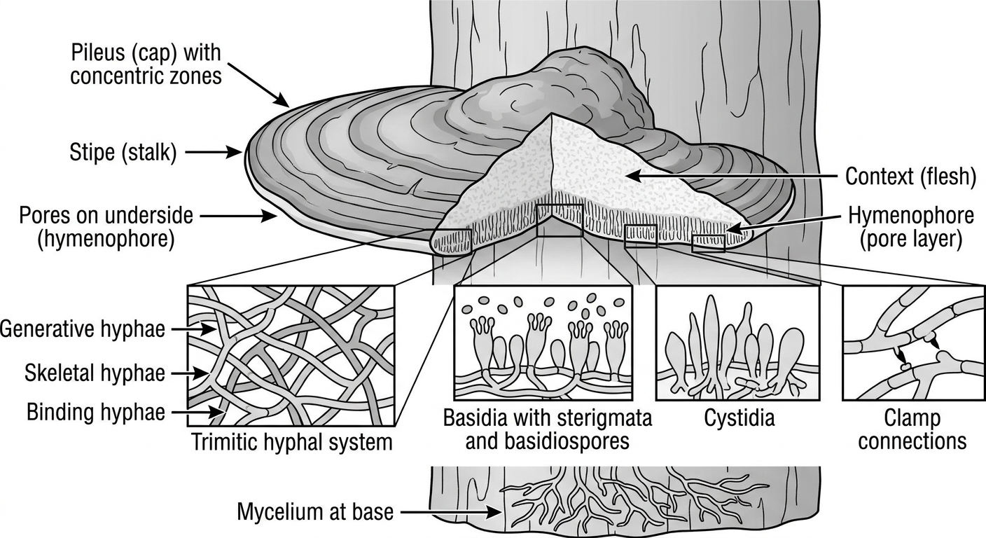
- Pileus (cap) - the bracket-shaped fruiting body with concentric growth zones
- Hymenophore - the pore-bearing undersurface where spores are released
- Stipe - the stalk attaching the fungus to substrate
- Context - the fleshy interior tissue
- Trimitic hyphal system - generative hyphae (with clamp connections), skeletal hyphae, and binding hyphae
- Basidia - club-shaped reproductive cells bearing sterigmata
- Basidiospores - the sexual spores
- Cystidia - sterile cells on the hymenium
2. Double-Ended Pseudocomedones (Nevus Comedonicus - Palmar Type)
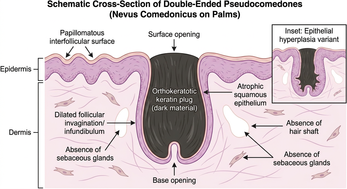
- Dilated follicular invagination - an undeveloped hair follicle opening at both the surface and base (the "double-ended" character)
- Orthokeratotic keratin plug - the dark, compacted horny material that mimics a comedone
- Atrophic squamous epithelium - thin lining of the infundibulum
- Papillomatous interfollicular epidermis - the surface epidermis between plugs
- Absent hair shaft - pseudocomedones contain no hair (distinguishing them from true comedones)
- Absent sebaceous glands - a diagnostic hallmark
Make a schimatic diagram of polyporus and double ended pseudocomedones
Detailed labeled schematic diagram of Polyporus fungus (bracket fungus, Basidiomycete). Show: pileus (cap) with concentric surface zones, pore surface (hymenophore) on underside with labeled pores and tubes, lateral stipe (stalk), context (flesh/trama), cross-section inset showing: basidia with 4 sterigmata and basidiospores, trimitic hyphal system with generative hyphae (with clamp connections), skeletal hyphae, binding hyphae, cystidia, mycelium threads at base anchoring to wood substrate. Black and white scientific educational textbook diagram with clean arrows and bold labels throughout.
Detailed labeled schematic histology cross-section diagram of double-ended pseudocomedones in nevus comedonicus. Show two adjacent structures side by side: LEFT - normal hair follicle with hair shaft, sebaceous gland, inner and outer root sheath, infundibulum opening at surface. RIGHT - double-ended pseudocomedone: dilated infundibular invagination opening at skin surface (top) AND at base (bottom, double-ended), packed with orthokeratotic laminated keratin plug (no hair shaft inside), thin atrophic squamous epithelial lining, no sebaceous gland, papillomatous epidermis between comedones, dermis and subcutis below. Labels: epidermis, cornified plug, dilated infundibulum, atrophic lining epithelium, absent hair shaft, absent sebaceous gland, dermis. Inset box showing epithelial hyperplasia variant. Clean black and white dermatopathology educational diagram with arrows and bold labels.
400, message: Got more than 4194304 bytes when reading: b'data: {"candidates": [{"content": {"role": "model","parts": [{"inlineData": {"mimeType": "image/png"...'.
We encountered an error while processing your request. Please try again or reach out to us for help.