
Pathphysiology of thyrotoxicosis related thyroid lesion pathogenesis morphology for md pathology exam 10 marks
Reading File
Finding Sources
Finding Sources
Reading File
Reading File
Finding Sources
Finding Sources
Reading File
Searching PubMed
"Graves disease" pathogenesis
Thyrotoxicosis - Pathophysiology, Pathogenesis and Morphology
(Model Answer - 10 Marks, MD Pathology)
1. Definition
Thyrotoxicosis is the hypermetabolic clinical syndrome produced by elevated circulating levels of free thyroid hormones (T3/T4), irrespective of source. When it results from hyperfunction of the thyroid gland itself, it is called hyperthyroidism (true thyrotoxicosis) - Robbins & Kumar Basic Pathology, p. 1866-1868.
Note the distinction asked frequently in exams: thyrotoxicosis can occur without hyperthyroidism (e.g., subacute thyroiditis releasing preformed hormone, or exogenous thyroxine excess), but hyperthyroidism always causes thyrotoxicosis.
2. Etiology (Causes)
| Category | Examples |
|---|---|
| Primary hyperthyroidism (thyroid overactivity) | Graves disease (~85% of cases), toxic multinodular goiter, toxic adenoma |
| Thyroiditis (transient release, not true hyperfunction) | Subacute (de Quervain) thyroiditis, painless (silent/postpartum) thyroiditis, Hashimoto "hashitoxicosis" |
| Secondary | TSH-secreting pituitary adenoma |
| Exogenous/ectopic | Factitious thyroxine ingestion, struma ovarii, metastatic follicular carcinoma |
3. Pathogenesis
A. General mechanism of thyrotoxic state
Excess circulating T3/T4 increases basal metabolic rate and potentiates catecholamine/sympathetic action on peripheral tissues, producing the hypermetabolic and hyperadrenergic clinical picture (tachycardia, heat intolerance, weight loss, tremor, diarrhea).
B. Pathogenesis of Graves disease (the prototype and commonest cause - focus of most exam answers)
Graves disease is an organ-specific autoimmune disorder, occurring at peak incidence 20-40 years, women affected up to 7 times more than men (Robbins Basic Pathology, p. 2009-2013).
Key immunologic events:
- Loss of self-tolerance to thyroid antigens, particularly the TSH receptor (TSHR), in a genetically susceptible individual (associated HLA alleles and CTLA4 polymorphisms; increased concordance in monozygotic twins).
- B cells produce thyroid-stimulating immunoglobulin (TSI) - an IgG autoantibody against the TSHR that binds and activates the receptor independent of TSH, driving continuous cAMP-mediated stimulation of follicular cells → increased synthesis and release of T3/T4 and gland hypertrophy/hyperplasia.
- Other TSHR antibodies may block TSH binding (TSH-binding inhibitory immunoglobulins) - coexistence of stimulating and blocking antibodies in the same patient can explain intermittent swings toward hypothyroidism.
- Ophthalmopathy: TSH receptor is also expressed on orbital fibroblasts and adipocytes. Activated CD4+ T cells secrete cytokines that stimulate fibroblasts to produce excess glycosaminoglycans/extracellular matrix, which accumulate in the retro-orbital space along with lymphocytic infiltration, producing proptosis and extraocular muscle edema/fibrosis.
- Dermopathy (pretibial myxedema): similar glycosaminoglycan deposition and lymphocytic infiltration in the dermis.
Recent literature adds nuance to classical teaching: meta-analyses continue to support strong HLA-DRB1 allele associations in Asian populations (PMID: 38698581) and an association between vitamin D deficiency and Graves disease risk (PMID: 38849834), supporting the multifactorial genetic-environmental model taught in Robbins, without contradicting it.
C. Pathogenesis of other causes (brief, for completeness)
- Toxic multinodular goiter: autonomous, TSH-independent nodules (often with activating somatic mutations in the TSHR or Gsα gene) that secrete hormone independent of pituitary control.
- Toxic adenoma: monoclonal proliferation of follicular cells, frequently harboring activating point mutations in the TSHR gene, causing constitutive receptor activation.
- Subacute/painless thyroiditis: follicular disruption (viral or autoimmune) causes leakage of stored colloid/hormone - a transient thyrotoxic phase without true glandular hyperfunction.
4. Morphology
Gross
- Thyroid is symmetrically, diffusely enlarged (in Graves disease) due to hypertrophy and hyperplasia of follicular cells.
- Gland is smooth, soft, and the capsule is intact; cut surface is fleshy and resembles muscle ("meaty").
- (Contrast: toxic multinodular goiter shows an irregular, multinodular gland; toxic adenoma shows a single encapsulated nodule.)
Microscopy (Graves disease - MORPHOLOGY section, Robbins p. 2018-2022)
- Follicular epithelial cells are tall, columnar, and crowded, often forming small papillary infoldings projecting into the follicular lumen - these papillae lack fibrovascular cores, distinguishing them from true papillary carcinoma.
- Colloid is scant and pale, with scalloped ("moth-eaten") margins due to active resorption at the apical cell border.
- Lymphoid infiltrate in the interstitium, predominantly T cells with fewer B cells/plasma cells; scattered germinal centers may be present.
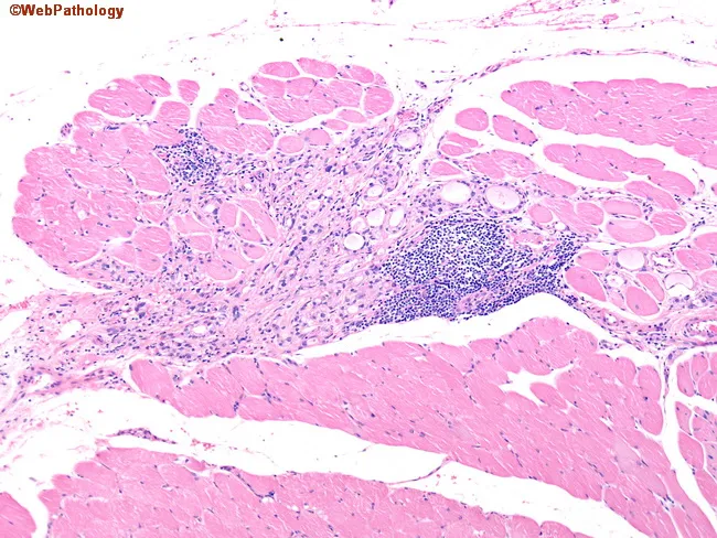
- Extrathyroidal changes: generalized lymphoid hyperplasia; orbital tissue shows edema (mucopolysaccharide/glycosaminoglycan deposition), lymphocytic infiltration (mostly T cells), and later fibrosis of extraocular muscles; dermis in pretibial myxedema shows glycosaminoglycan deposition with lymphocytic infiltrate.
5. Clinical correlation (brief, high-yield for exam)
- Constitutional: weight loss despite increased appetite, heat intolerance, warm moist skin.
- CVS: tachycardia, palpitations, atrial fibrillation, high-output cardiac failure in the elderly.
- GI: hypermotility, diarrhea.
- Neuromuscular: tremor, anxiety, proximal myopathy.
- Ocular: lid lag, wide staring gaze (sympathetic overactivity); true infiltrative exophthalmos is specific to Graves disease.
- Thyroid storm: abrupt, severe exacerbation of thyrotoxicosis, a medical emergency with risk of fatal cardiac arrhythmia, precipitated by infection, surgery, or abrupt withdrawal of anti-thyroid drugs.
- Apathetic hyperthyroidism: blunted classic features in the elderly, presenting as unexplained weight loss or worsening cardiac disease.
- Diagnosis: low serum TSH (most sensitive screening test) with elevated free T4 (or T3 in "T3 toxicosis"); radioiodine uptake scan differentiates Graves (diffuse uptake) from toxic adenoma (focal uptake) from thyroiditis (low uptake).
Suggested diagram for exam: A flow diagram showing genetic susceptibility (HLA, CTLA4) + environmental trigger -> loss of tolerance -> B cell production of TSI -> TSHR activation on follicular cells -> increased T3/T4 -> systemic hypermetabolic effects; with a side branch showing TSHR expression on orbital fibroblasts -> cytokine-driven GAG deposition -> ophthalmopathy. Draw this in the actual exam for extra marks.
Source: Robbins & Kumar Basic Pathology (10th ed.), Chapter 18 - "Hyperthyroidism" and "Graves Disease," p. 1866-1868, 2009-2022.
This is a shared conversation. Sign in to Orris to start your own chat.