
Carpal tunnel syndrome and arthroscopic carpal tunnel release structured answer
"carpal tunnel syndrome" AND "endoscopic release"
arthroscopic carpal tunnel release endoscopic vs open outcomes
endoscopic carpal tunnel release transverse carpal ligament median nerve
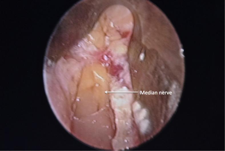
This endoscopic clinical photograph provides an intraoperative view of a carpal tunnel release procedure. The image depicts the surgical field after the successful division of the flexor retinaculum (transverse carpal ligament). Centrally located is the median nerve, which appears as a light yellow, elongated structure with a smooth surface texture. A prominent, brighter yellowish-white fat pad is visible overlying the distal portion of the nerve, serving as a critical anatomical landmark for confirming complete ligamentous release. The surrounding surgical field shows reddish-pink soft tissue and muscular layers, indicating local vascularity. The image demonstrates the specialized supraretinacular endoscopic approach, allowing for direct visualization and decompression of the median nerve within the carpal tunnel while minimizing trauma to superficial structures. This visual is intended for orthopedic and neurosurgical education regarding the anatomy of the wrist and endoscopic surgical techniques for carpal tunnel syndrome.
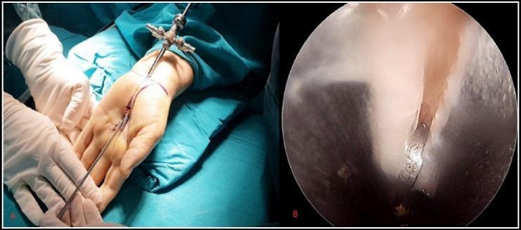
This composite clinical and diagnostic image illustrates Endoscopic Carpal Tunnel Release (ECTR) for the treatment of carpal tunnel syndrome. Image A is a clinical photograph showing the surgical setup: a patient's hand is positioned supine on a sterile blue drape with surgical markings indicating Kaplan's cardinal line. A small proximal wrist incision is visible, through which an endoscopic cannula and obturator assembly are inserted into the carpal canal. A surgeon's gloved hands are shown stabilizing the instrument. Image B provides the corresponding intraoperative endoscopic view from a 4-mm rigid scope. It depicts the pale, fibrous internal surface of the transverse carpal ligament (TCL) with a clear longitudinal division or cut, demonstrating the release of the ligament to decompress the median nerve. This content is intended for orthopedic and neurosurgical education, highlighting the minimally invasive endoscopic approach versus open carpal tunnel release.
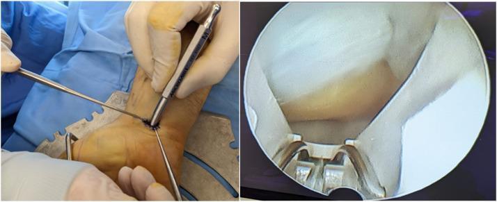
A composite image illustrating a minimally invasive endoscopic carpal tunnel release (ECTR) procedure. The left panel shows the external surgical setup: a patient's hand is positioned on a metallic hand table with blue sterile draping. A small transverse incision is visible at the proximal wrist crease, through which an endoscope and a specialized blade assembly are inserted. A surgeon's gloved hand is seen manipulating the instrumentation. The right panel displays the concurrent internal endoscopic view (endoview) within the carpal tunnel. The image highlights the dense, white, fibrous transverse carpal ligament (flexor retinaculum) and the positioning of the metallic blade and scope sheath underneath it. This visual demonstrates the surgical technique used to transect the ligament and decompress the median nerve, typically indicated for severe carpal tunnel syndrome. The procedure is categorized under orthopedic or hand surgery, focusing on compressive neuropathy management.
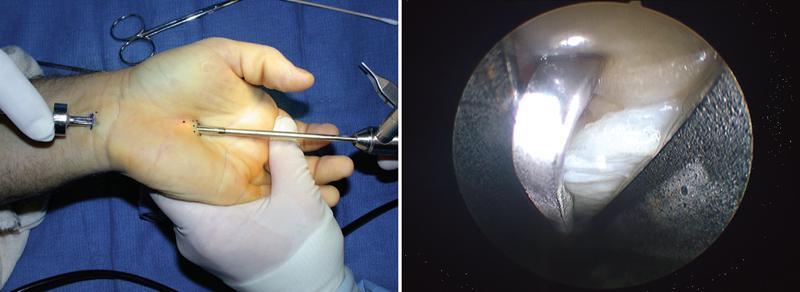
A composite image demonstrating biportal endoscopic carpal tunnel release (ECTR). The left side is a clinical photograph showing the surgical setup on a supinated human hand. Two portals are visible: a proximal port at the distal wrist crease and a distal port in the palm. An endoscope is inserted into the proximal portal, while a metallic surgical dissector is introduced through the distal portal, illustrating the 'two-portal' or Brown technique. The right side displays the corresponding internal endoscopic view of the carpal tunnel. The image shows the bright, white, fibrous bands of the transverse carpal ligament (TCL) and a metallic retrograde blade or dissector engaged with the tissue for transection. This visual captures the minimally invasive approach used to decompress the median nerve while minimizing surgical trauma to the palmar fascia and skin. The educational focus is on the instrumentation, portal placement, and internal visualization required for safe ligament release.
Carpal Tunnel Syndrome and Endoscopic ("Arthroscopic") Carpal Tunnel Release
1. Overview
2. Etiology / Risk Factors
- Fluid balance changes: pregnancy, menopause, hypothyroidism, renal failure, long-term hemodialysis
- Inflammatory conditions: rheumatoid arthritis, gout, tenosynovitis, infection
- Autoimmune/systemic disease: amyloidosis, lupus, scleroderma, multiple sclerosis, Paget disease
- External forces: vibration exposure, repetitive forceful wrist flexion/extension, sustained wrist flexion, direct pressure, trauma
- Rarely in children: congenital bone abnormalities, hypothyroidism, lysosomal storage disease, strong family history
3. Diagnosis
- Primarily clinical: paresthesia in the median nerve distribution (thumb, index, middle, radial half of ring finger), classically nocturnal, relieved by shaking the hand
- Provocative tests: Phalen test, Tinel sign, carpal compression test
- CTS-6 clinical diagnostic score weighs numbness in the median distribution, nocturnal symptoms, thenar atrophy/weakness, Tinel sign, Phalen sign, and loss of two-point discrimination
- Electrodiagnostic studies (nerve conduction studies/EMG) confirm and grade severity
- Imaging (ultrasound/MRI) can assess nerve cross-sectional area or exclude space-occupying lesions
- Late/severe disease: thenar muscle atrophy, absent provocative signs (nerve too damaged to trigger them)
4. Treatment Overview
5. Endoscopic Carpal Tunnel Release (ECTR)
- Incision at the proximal wrist flexion crease between palmaris longus/FCR and FCU
- Blunt dissection to protect subcutaneous nerves, expose forearm fascia
- Elevate a U-shaped fascial flap to create an opening at the proximal carpal tunnel
- Advance the blade assembly aligned with the ring finger, hugging the hamate hook, staying between median and ulnar nerves
- Confirm the distal TCL edge by video, ballottement, and transillumination
- Elevate the blade to divide the ligament under direct endoscopic view
- Know the anatomy
- Never overcommit to the procedure
- Confirm equipment works properly
- If scope insertion is obstructed, abort
- Confirm the blade assembly is in the carpal tunnel, not Guyon's canal
- If a clear view cannot be obtained, abort
- Do not explore the carpal canal with the scope
- If the view is abnormal, abort
- Stay in line with the ring finger
- "When in doubt, get out"
- Need for neurolysis, tenosynovectomy, TCL Z-plasty, or Guyon's canal decompression
- Suspected space-occupying lesion or severe tendon/muscle/vessel abnormality
- Localized infection, severe hand edema, or compromised vascular status
- Revision surgery for recurrent/unresolved CTS
- Anatomic median nerve variation (thenar wasting without sensory changes)
- Prior tendon surgery/flexor injury causing carpal tunnel scarring
- Limited wrist extension preventing instrument introduction
6. Endoscopic vs. Open Release - Evidence Summary
- Comparable long-term outcomes between open (OCTR) and endoscopic (ECTR) release in symptom relief and grip strength recovery
- Faster short-term recovery with ECTR: less palmar pain, earlier return to work (one RCT found ~8 days sooner), quicker return to activities of daily living
- Complication profile differs: OCTR carries more wound-related complications (scar tenderness, pillar pain); ECTR carries a slightly higher risk of transient/reversible nerve injury (neuropraxia) related to cannula insertion or canal pressure, though major irreversible nerve injury is rare with either technique
- ECTR is technically demanding and equipment-dependent; many surgeons reserve open technique for revision cases