
from Guyton and Hall textbook of medical physiology, 4th South Asia edition write a 10 mark long answer on Discuss mechanical events of cardiac cycle.
Reading File
Finding Sources
Finding Sources
Searching PubMed
"cardiac cycle" AND physiology
Searching the Web
Guyton Hall medical physiology cardiac cycle mechanical events heart sounds ventricular pressure volume
Finding Sources
Finding Sources
Finding Sources
Reading File
Reading File
Reading File
Reading File
Reading File
Mechanical Events of the Cardiac Cycle
The cardiac cycle is the sequence of events occurring in the heart from the beginning of one heartbeat to the beginning of the next. It includes alternate periods of atrial and ventricular systole (contraction) and diastole (relaxation). At a heart rate of 72/min, one cycle lasts about 0.83 second. The left and right sides follow the same sequence, but pressures are much higher on the left side. - Guyton and Hall Textbook of Medical Physiology, p. 126
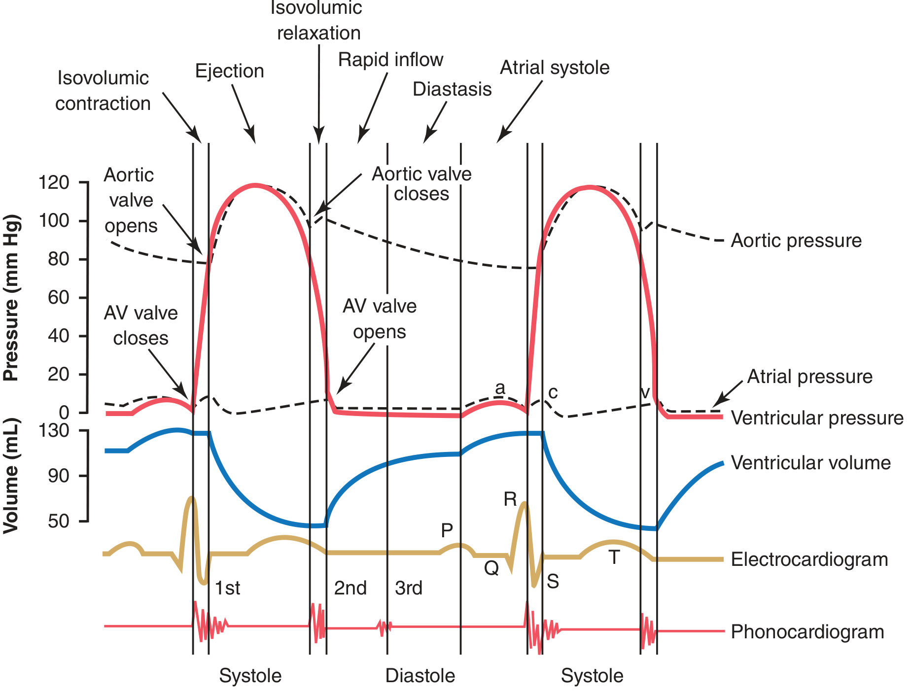
Phases of the cardiac cycle
| Phase | Valves | Main mechanical event |
|---|---|---|
| Atrial systole | AV valves open; semilunar valves closed | Final ventricular filling |
| Isovolumic ventricular contraction | All valves closed | Ventricular pressure rises without change in volume |
| Ventricular ejection | Semilunar valves open | Blood is expelled into aorta and pulmonary artery |
| Isovolumic relaxation | All valves closed | Ventricular pressure falls without change in volume |
| Ventricular filling | AV valves open; semilunar valves closed | Rapid filling, slow filling, then atrial contribution |
1. Atrial systole
Atrial systole begins after atrial depolarization, represented by the P wave of ECG. The atria contract before the ventricles because of the AV nodal delay. This pushes additional blood into the ventricles.
- During early ventricular diastole, about 70% to 80% of ventricular filling occurs passively as blood flows through the atria into the ventricles.
- Atrial contraction supplies the final 20% to 30% of ventricular filling. Hence, atria act as primer pumps for the ventricles.
- Atrial contraction produces the a wave in the atrial pressure curve.
- At the end of atrial systole, ventricular volume reaches its maximum, called the end-diastolic volume (EDV), normally about 110 to 120 mL.
Atrial systole becomes particularly important when ventricular filling is impaired, such as in a stiff or hypertrophied ventricle. - Guyton and Hall Textbook of Medical Physiology, pp. 127-128
2. Isovolumic ventricular contraction
Ventricular systole starts just after the QRS complex.
- Ventricular contraction causes a sharp increase in intraventricular pressure.
- When ventricular pressure exceeds atrial pressure, the mitral and tricuspid valves close, producing the first heart sound (S1).
- Initially, the aortic and pulmonary valves remain closed because ventricular pressure has not yet exceeded arterial pressure.
- Therefore, all four valves are closed. Ventricular muscle develops tension, but ventricular volume does not change. This is called isovolumic or isometric contraction.
- It lasts approximately 0.02 to 0.03 second.
- Bulging of the closed AV valves into the atria causes the c wave in the atrial pressure curve.
3. Ventricular ejection phase
When left ventricular pressure rises slightly above aortic pressure, approximately 80 mm Hg, the aortic valve opens. On the right side, pulmonary valve opens when right ventricular pressure exceeds pulmonary artery pressure, approximately 8 mm Hg.
Blood is ejected from the ventricles in two parts:
a. Rapid ejection phase
- Occurs during the first one-third of the ejection period.
- Ventricular pressure rises further and blood flow into the aorta and pulmonary artery is maximum.
- About 70% of the stroke volume is ejected in this phase.
b. Reduced ejection phase
- Ventricular contraction becomes less forceful.
- The rate of ejection decreases, although blood continues to leave the ventricles.
- The remaining approximately 30% of the stroke volume is ejected.
Normally, about 60% of EDV is ejected during systole. This is the ejection fraction, normally about 60%.
At the end of systole:
- Stroke volume is about 70 mL.
- End-systolic volume (ESV) is about 40 to 50 mL.
- Ventricular repolarization, indicated by the T wave, begins shortly before the end of ventricular contraction. - Guyton and Hall Textbook of Medical Physiology, p. 128
4. Isovolumic ventricular relaxation
At the end of ejection, ventricular muscle relaxes and intraventricular pressure falls rapidly.
- When ventricular pressure becomes less than pressure in the aorta or pulmonary artery, a brief backward flow of blood closes the semilunar valves.
- Closure of the aortic and pulmonary valves produces the second heart sound (S2).
- For about 0.03 to 0.06 second, all valves are closed.
- Ventricular volume remains constant at the end-systolic volume, while ventricular pressure falls rapidly. Hence, this phase is termed isovolumic relaxation.
- During ventricular systole, venous return continues to fill the atria while AV valves are closed. This produces the v wave in the atrial pressure curve.
5. Ventricular filling phase
When ventricular pressure falls below atrial pressure, the AV valves open and ventricular filling begins. It has three components:
a. Rapid ventricular filling
- This occurs in the first third of diastole.
- Blood that accumulated in the atria during ventricular systole rapidly enters the ventricles.
- Ventricular volume rises quickly.
- A third heart sound, S3, may be heard during rapid filling. It can be normal in children and young adults but may indicate ventricular failure in older adults.
b. Reduced filling or diastasis
- During the middle third of diastole, ventricular filling becomes slow.
- Blood returning from the veins passes through the atria directly into the ventricles.
- This period becomes shorter as heart rate rises.
c. Atrial systole
- The final one-third of diastole includes atrial contraction, which adds the last 20% to 30% of ventricular filling.
- The next cardiac cycle then begins.
Relation to heart rate
The duration of the cardiac cycle is inversely related to heart rate. When heart rate increases, both systole and diastole shorten, but diastole shortens proportionately more. Thus, at very high heart rates, the time available for ventricular filling decreases and cardiac output may be affected. - Guyton and Hall Textbook of Medical Physiology, p. 126
Conclusion
Mechanical events of the cardiac cycle result from orderly changes in atrial and ventricular contraction, pressure, volume, and valve position. Atrial contraction completes ventricular filling; ventricular systole produces isovolumic contraction followed by ejection; ventricular relaxation produces isovolumic relaxation followed by rapid and slow filling. The opening and closure of AV and semilunar valves ensure one-way flow of blood through the heart.
This is a shared conversation. Sign in to Orris to start your own chat.