
Using millers anaesthesiology book and barasch anaesthesiology book discuss in detail about local anaesthetics and most important concepts of local/regional anaesthesia
Reading File
Finding Sources
Finding Sources
Now let me search for local anaesthetic headings in both books simultaneously:
Finding Sources
Finding Sources
Excellent! Now I have the key file paths. Let me read the most important sections from both books in parallel:
Reading File
Finding Sources
Excellent content from Miller's! Let me continue reading Miller's block 10 for more content and simultaneously read Barash's local anaesthetic sections:
Reading File
Finding Sources
Reading File
Reading File
Reading File
Reading File
Reading File
Finding Sources
Reading File
Reading File
Reading File
Reading File
Reading File
Reading File
I now have comprehensive material from both textbooks. Let me do one more targeted read to get the differential blockade and adjuvants sections, then synthesize:
Reading File
Reading File
Here is a comprehensive, integrated discussion of local anaesthetics and the most important concepts of local/regional anaesthesia, synthesised directly from Miller's Anesthesia, 10th Edition and Barash, Cullen & Stoelting's Clinical Anesthesia, 9th Edition.
Local Anaesthetics and Regional Anaesthesia: A Comprehensive Discussion
1. CHEMISTRY AND CLASSIFICATION
The Local Anaesthetic Molecule
The typical local anaesthetic molecule contains a tertiary amine attached to a substituted aromatic ring by an intermediate chain that is either an ester (–C–O–) or an amide (–NHC–) linkage. The aromatic ring confers lipophilicity to one end of the molecule, while the tertiary amine end is relatively hydrophilic and partially protonated at physiological pH.
"The aromatic ring system gives a lipophilic (membrane-liking) character to its portion of the molecule, whereas the tertiary amine end is relatively hydrophilic, particularly since it is partially protonated and thus bears some positive charge in the physiologic pH range." — Miller's Anesthesia, 10e, p. 3545
Aminoesters (procaine, chloroprocaine, tetracaine, cocaine) are hydrolysed in plasma by pseudocholinesterase (plasma cholinesterase). A metabolite, para-aminobenzoic acid (PABA), is responsible for their slightly greater potential for true allergic reactions.
Aminoamides (lidocaine, bupivacaine, ropivacaine, mepivacaine, prilocaine, levobupivacaine) are extremely stable and undergo hepatic metabolism via carboxylesterases and cytochrome P450 enzymes. The classic mnemonic: amide local anaesthetics contain two "i"s in their name.
"Most clinically relevant local anesthetics consist of a lipid-soluble, aromatic benzene ring connected to an amide group via either an amide or an ester moiety… Aminoesters are hydrolyzed by plasma cholinesterases and aminoamides are degraded by hepatic carboxylesterases." — Barash Clinical Anesthesia, 9e, p. 1688
2. STRUCTURE–ACTIVITY RELATIONSHIPS
Lipophilicity
The most important physicochemical property. High lipid solubility (measured by the octanol:buffer partition coefficient) correlates positively with potency and duration of action. Bupivacaine and ropivacaine are highly lipophilic, accounting for their long duration. The first level of action is at the cellular membrane (facilitating entry), and the second is at the hydrophobic binding pocket within sodium channels (van der Waals interactions).
pKa and Onset
Local anaesthetics are weak bases. In aqueous solution they exist in equilibrium between the protonated cationic form (BH⁺) and the lipid-soluble neutral base form (B). The ratio is governed by the Henderson-Hasselbalch relationship and the drug's pKa. A lower pKa means a greater fraction of neutral form at physiological pH 7.4, which penetrates the nerve membrane faster → faster onset.
- Lidocaine pKa ≈ 7.9 → faster onset
- Bupivacaine pKa ≈ 8.1 → slower onset
- Chloroprocaine pKa ≈ 8.7 → slowest onset but rapid hydrolysis compensates
"The fraction of local anesthetic in the protonated, cationic form of an aqueous solution at physiologic pH (7.4) as a function of the pKa of the drug. Lidocaine, the drug with the lowest pKa, has the smallest fraction of its molecules protonated, the largest in the neutral form..." — Miller's Anesthesia, 10e, p. 3546
Once inside the cell, the neutral form re-protonates to the cationic form — it is the cationic form that principally blocks the sodium channel from inside.
Protein Binding
High protein binding (particularly to α1-acid glycoprotein) prolongs duration of action. Bupivacaine and ropivacaine have ~95% protein binding vs. lidocaine ~65%.
Stereoisomerism
Bupivacaine is a racemic mixture. The S(–)-enantiomers (levobupivacaine, ropivacaine) are less cardiotoxic than the R(+)-enantiomer. Ropivacaine is the pure S(–)-enantiomer and shows slightly less potency but a significantly improved cardiac safety profile.
3. NERVE FIBRE ANATOMY AND PHARMACOLOGY
Nerve Sheath Barriers
To reach its site of action, a local anaesthetic must traverse the paraneurium → epineurium → perineurium → endoneurium → axonal membrane. The perineurium is the main diffusion barrier. In desheathed nerves in vitro, 100-fold lower concentrations (0.7–0.9 mM lidocaine) are needed than clinically (75 mM in 2% lidocaine). Approximately only 1–2% of injected local anaesthetic ultimately penetrates the nerve.
"Even with close proximity of deposition, only about 1% to 2% of injected local anesthetics ultimately penetrate into the nerve." — Barash Clinical Anesthesia, 9e, p. 1685
Nerve Fibre Classification
| Fibre Type | Diameter | Myelination | Function | Sensitivity to LA |
|---|---|---|---|---|
| Aα | 12–20 µm | Heavy | Motor, proprioception | Least sensitive |
| Aβ | 5–12 µm | Heavy | Touch, pressure | Intermediate |
| Aγ | 3–6 µm | Moderate | Muscle spindle tone | — |
| Aδ | 1–4 µm | Light | Pain (fast), cold, touch | Sensitive |
| B | <3 µm | Light | Preganglionic autonomic | Very sensitive |
| C | 0.3–1.3 µm | None | Pain (slow), warmth, autonomic | Sensitive (but require broader exposure) |
"Na+ channels that serve generation and propagation of impulses are highly concentrated at the nodes of Ranvier of myelinated fibres but are distributed all along the axon of nonmyelinated fibres." — Miller's Anesthesia, 10e, p. 3550
Myelinated vs Unmyelinated Conduction
Myelinated fibres conduct via saltatory conduction (current jumps between nodes of Ranvier), which is faster but also means local anaesthetic must block at least 2–3 successive nodes to extinguish the impulse. In unmyelinated C fibres, a sufficient length of axon must be blocked — explaining why adequate volume is as important as concentration.
4. MECHANISM OF ACTION
Sodium Channel Blockade — The Core Mechanism
Local anaesthetics produce nerve blockade by binding to voltage-gated Na⁺ channels, preventing their activation (channel opening) and blocking Na⁺ influx. This abolishes the depolarising limb of the action potential.
The sodium channel has three states:
- Resting (closed) — at normal resting potential (–85 mV)
- Activated (open) — during depolarisation
- Inactivated — rapid automatic closure after activation
Local anaesthetics bind with much higher affinity to the open and inactivated states than to the resting state. This explains use-dependent (phasic) blockade: repeatedly firing fibres accumulate progressively more block because more channels are cycled into open/inactivated states.
"Binding of local anesthetic to sites on voltage-gated Na+ channels prevents opening of the channels by inhibiting the conformational changes that underlie channel activation… During onset of and recovery from local anesthesia, impulse blockade is incomplete and partially blocked fibres are further inhibited by repetitive stimulation, which produces an additional, use-dependent binding to Na+ channels." — Miller's Anesthesia, 10e, p. 3576
Decremental Conduction
As membrane depolarisation from an action potential passively decays along the nerve, local anaesthetics reduce the ability of adjacent nodes/membrane to regenerate the impulse. Transmission stops once depolarisation falls below threshold. If drug exposure is over too short a segment, impulses can "skip" over blocked areas and resume — the key concept of needing a minimum blocking length of nerve.
"If the exposure distance is inadequate, action potentials can 'skip' over blocked segments and resume nerve conduction. In contrast, exposure over a long segment of nerve to even a relatively low drug concentration can still result in gradual extinction of impulse by decremental decay." — Barash Clinical Anesthesia, 9e, p. 1686
Chronological Summary of Impulse Blockade (Miller's)
- Local anaesthetic is deposited near the nerve; penetration of nerve sheath by free drug
- Drug permeates axonal membranes — speed depends on pKa and lipophilicity
- Binding to voltage-gated Na⁺ channels prevents channel activation
- During onset/recovery, use-dependent (phasic) blockade from repetitive stimulation
- One binding site may account for both tonic and phasic actions
- Clinically effective block lasting hours is governed by slow diffusion of drug into/out of the whole nerve — not by the faster (seconds) binding/dissociation from ion channels
5. DIFFERENTIAL NERVE BLOCK
The sequence of clinical blockade typically proceeds: autonomic (B fibres) → pain (C and Aδ) → temperature → touch → proprioception → motor (Aα). Recovery occurs in the reverse order.
This is not simply due to fibre size, as was once thought. Barash emphasises that sodium channel isoform expression matters: Nav1.7 and Nav1.8 are highly expressed on dorsal root ganglia sensory neurons and show distinct sensitivities to lidocaine. The transient receptor potential channels TRPV1 and TRPA1, found predominantly on sensory neurons, can be activated by local anaesthetics, possibly facilitating selective drug entry into pain-mediating fibres.
"Sodium channel isoforms NaV 1.7 and NaV 1.8 are highly expressed on the dorsal root ganglia, and have been shown to have distinct sensitivities to lidocaine... Because these transient receptor potential channels are found predominantly on sensory neurons mediating specific stimulus modalities, their activation may selectively facilitate entry of local anesthetics, thereby resulting in the observed progression of sensory, autonomic, and motor blockade." — Barash Clinical Anesthesia, 9e, p. 1687
6. PHARMACOKINETICS
Absorption
Rate of systemic absorption (and peak blood levels) depends primarily on site vascularity:
Intercostal > caudal > epidural > brachial plexus > intravenous regional > lower extremity
Distribution, Metabolism, Elimination
| Drug | Vdss (L/kg) | CL (L/kg/hr) | T½ (hrs) |
|---|---|---|---|
| Bupivacaine | 1.02 | 0.41 | 3.5 |
| Ropivacaine | 0.84 | 0.63 | 1.9 |
| Lidocaine | 1.3 | 0.85 | 1.6 |
| Prilocaine | 2.73 | 2.03 | 1.6 |
| Chloroprocaine | 0.50 | 2.96 | 0.11 |
| Procaine | 0.93 | 5.62 | 0.14 |
(— Barash Clinical Anesthesia, 9e, Table 22-7, p. 1701)
The very short half-life of chloroprocaine (0.11 hr) reflects rapid plasma hydrolysis. Severe liver disease significantly slows clearance of all amide local anaesthetics, risking accumulation.
Effect of Site on Duration
Equivalent doses may produce 3–4 hours of anaesthesia in the epidural space but 12–14 hours in the arm and 24–36 hours along the sciatic nerve — reflecting differences in blood flow and sequestration in tissue.
7. ADJUVANTS AND ADDITIVES
Epinephrine
The most widely used adjuvant. Acts by causing vasoconstriction, reducing vascular absorption, allowing more drug to reach the nerve. Effects:
- Clinically used at 1:200,000 (5 µg/mL)
- Markedly prolongs shorter-acting agents (lidocaine, mepivacaine): increases duration by up to 50%
- Minimal effect on long-acting agents (bupivacaine, ropivacaine)
- Also used as a marker for inadvertent intravascular injection (HR ↑ ≥20 bpm after 15 µg IV, though false positives/negatives occur in beta-blocked or pregnant patients)
- Contraindicated near terminal vessels (digits, penis, ear, nose) and in IV regional anaesthesia
"Vasoconstrictors, usually epinephrine, are frequently included in local anesthetic solutions to decrease the rate of vascular absorption, thereby allowing more anesthetic molecules to reach the nerve membrane and thus improve the depth and duration of anesthesia." — Miller's Anesthesia, 10e, p. 3583
Alkalinisation
Commercial local anaesthetic preparations have pH 3.9–6.5 (more acidic when pre-packaged with epinephrine). Alkalinisation (typically adding sodium bicarbonate) increases the proportion of the lipid-soluble neutral form, which can theoretically accelerate onset. In practice, clinical benefit is modest — maximum pH before precipitation occurs is 6.05–8, yielding only ~10% neutral form. Clinically, alkalinisation hastens onset by less than 3 minutes in most studies.
"Clinical studies on the association between alkalinization of local anesthetics and hastening of block onset have shown an improvement of less than [3 minutes]." — Barash Clinical Anesthesia, 9e, p. 1691
Other Adjuvants (Miller's, p. 3583–3586)
| Adjuvant | Mechanism | Use |
|---|---|---|
| Dexamethasone | Anti-inflammatory, suppresses nociceptor activity | Prolongs PNB duration significantly |
| Dexmedetomidine (α₂ agonist) | Hyperpolarises nerve via GIRK channels | Prolongs sensory/motor block |
| Clonidine | α₂ agonist; intrinsic analgesic effect | Spinal/epidural/PNB adjuvant |
| Opioids (intrathecal/epidural) | Spinal μ-receptor agonism | Neuraxial analgesia |
| Neostigmine (intrathecal) | Inhibits acetylcholinesterase → ↑ Ach | Analgesic, not first line |
8. INDIVIDUAL LOCAL ANAESTHETIC AGENTS
Short-Acting
- Procaine (ester): low potency, slow onset; mainly historical for spinal use
- Chloroprocaine 2–3%: fastest onset (rapid hydrolysis), shortest duration; ideal for surgical epidural; risk of transient neurological symptoms (TNS) and adhesive arachnoiditis with preservative-containing formulations
Intermediate-Acting
- Lidocaine 0.5–2%: the most versatile agent; gold-standard for topical, infiltration, spinal, epidural, nerve blocks; risk of TNS with spinal use (especially lithotomy position, dose-dependent)
- Mepivacaine 0.5–2%: similar to lidocaine, slightly longer duration; safe in neonates (unlike bupivacaine), less vasodilatory
Long-Acting
- Bupivacaine 0.125–0.75%: workhorse of obstetric and postoperative epidural analgesia; profound differential sensory/motor block at low concentrations; 0.5% hyperbaric widely used for spinal; high cardiac toxicity (particularly R(+)-enantiomer)
- Levobupivacaine: pure S(–)-enantiomer of bupivacaine; similar clinical profile, lower cardiac toxicity
- Ropivacaine 0.2–1%: pure S(–)-enantiomer; similar to bupivacaine but less potent and significantly less cardiotoxic; intrinsic vasoconstrictive properties (unlike bupivacaine which vasodilates)
- Tetracaine 0.5% hyperbaric: long-acting spinal agent; rarely used epidurally due to slow onset
Unique Agents
- Cocaine: only agent with intrinsic vasoconstrictive properties; used solely for topical nasal/airway anaesthesia
- Prilocaine: lowest CNS toxicity; however, its metabolite o-toluidine causes methaemoglobinaemia — contraindicated in neonates and compromised patients
9. DOSAGE AND VOLUME PRINCIPLES
Concentration vs Volume
Increasing concentration at the same volume improves depth of block and onset; increasing volume at the same total dose improves spread. For example:
- 30 mL of 1% lidocaine epidurally spreads >4 dermatomes higher than 10 mL of 3% lidocaine
- Increasing epidural bupivacaine from 0.125% to 0.5% (same 10 mL) → shorter latency, longer sensory analgesia
Ultrasound Era and Minimum Volumes
Precise needle placement with ultrasound guidance has revealed that minimum local anaesthetic volumes (MLAV) for peripheral nerve block are approximately 0.1–0.15 mL/mm² of nerve diameter. In randomised trials, ultrasound-guided femoral nerve blockade required 54–57% of the volume needed with nerve stimulation.
However, reducing dose may shorten block duration — relevant for postoperative pain coverage beyond the acute surgical period.
10. LOCAL ANAESTHETIC SYSTEMIC TOXICITY (LAST)
Mechanism
Systemic absorption (most commonly from inadvertent intravascular injection) leads to toxic blood levels. Amide local anaesthetics block Na⁺ channels in both the CNS and cardiovascular system.
CNS (more sensitive — lower blood levels required):
- Early: circumoral tingling, tinnitus, dizziness, metallic taste, restlessness, slurred speech
- Progressive: visual/auditory disturbance, muscle twitching
- Severe: generalised seizures → CNS depression → respiratory arrest
Cardiovascular (at higher blood levels):
- Conduction disturbances, bradycardia, PR prolongation, QRS widening
- Re-entrant arrhythmias, ventricular tachycardia/fibrillation
- Cardiac depression and cardiovascular collapse
Bupivacaine cardiotoxicity is especially dangerous because it dissociates from Na⁺ channels very slowly ("fast-in, slow-out"), causing prolonged and potentially irreversible cardiac depression. The ratio of cardiotoxic dose to CNS-toxic dose (CC/CNS ratio) is much lower for bupivacaine than lidocaine.
"LAST is a potential complication of local anesthetic administration by any route and most commonly manifests clinically as neurologic or cardiovascular sequelae… Despite these precautions, life-threatening convulsions and, rarely, cardiovascular collapse may occur." — Barash Clinical Anesthesia, 9e, p. 3503–3504
Incidence
Overall incidence of major CNS or cardiovascular LAST: 0.004–0.18%. Trends show decreasing incidence with ultrasound guidance and awareness. Upper extremity blocks carry higher odds than lower extremity; penile blocks and tissue infiltrations account for a disproportionate number of events.
Risk Factors
- Extremes of age (infants <6 months: 6× higher risk)
- Low α1-acid glycoprotein (pregnant women, neonates, liver failure)
- Cardiac disease
- Metabolic acidosis (reduces protein binding and slows elimination)
- High-vascularity injection sites
Prevention
- Aspiration before each injection (does not reliably exclude intravascular placement)
- Fractional/incremental injection (3–5 mL aliquots)
- Epinephrine test dose (detects intravascular injection — HR rise ≥20 bpm)
- Ultrasound guidance to visualise needle tip and drug spread
- Respect maximum safe doses (adjusted for patient weight, site, and comorbidity)
Treatment — ASRA Practice Advisory
- Get help immediately, stop injection
- Airway management — 100% O₂, prevent hypoxia and acidosis (worsen toxicity)
- Seizure suppression — IV midazolam (1–5 mg) or propofol (small doses, avoid in haemodynamic compromise)
- Lipid emulsion therapy (20% Intralipid) — 1.5 mL/kg bolus over 1 minute, then 0.25 mL/kg/min infusion for ≥10 minutes after haemodynamic stability
- ACLS if cardiac arrest — prolonged resuscitation (local anaesthetic may clear over time)
- Epinephrine: limit to <1 µg/kg boluses (higher doses may trigger malignant arrhythmias)
- Avoid: vasopressin, calcium channel blockers, beta-blockers
- Amiodarone for ventricular dysrhythmias (avoid other Na⁺/Ca²⁺ channel–blocking antiarrhythmics)
- Cardiopulmonary bypass if refractory cardiovascular collapse
The mechanism of lipid emulsion rescue: "lipid shuttle" effect (scavenging tissue-bound drug) and direct cardiotonic effect via alternative mitochondrial energy substrate.
"Current society practice advisories unequivocally recommend initiation of lipid resuscitation therapy at the first sign of dysrhythmia from suspected LAST." — Barash Clinical Anesthesia, 9e, p. 1720
11. NEURAXIAL ANAESTHESIA — KEY CONCEPTS
Spinal Anaesthesia
- Drug injected into the subarachnoid space (CSF)
- Key determinants of spread: baricity, patient position, dose (mg), volume, speed of injection, age, height, spinal anatomy
- Hyperbaric bupivacaine 0.75% (in 8.25% dextrose) is the most widely used spinal agent worldwide
- Tetracaine (hyperbaric) offers long surgical anaesthesia
- Lidocaine spinal: rapid onset/offset but TNS risk (buttock and posterior leg pain) especially in lithotomy; dosing strategies now favour chloroprocaine for short procedures
Epidural Anaesthesia
- Denser drug concentration needed vs. spinal (no CSF dilution; large volume, high vascularity of epidural space)
- Onset: 5–15 min for chloroprocaine, lidocaine, mepivacaine; slower for bupivacaine
- Epinephrine significantly prolongs lidocaine/mepivacaine epidural duration; minimal effect on bupivacaine
- Dilute bupivacaine (0.0625–0.1%) ± opioid for labour epidural analgesia — exploits differential block
- Bolus injections spread more cephalocaudally than continuous infusions
Hypotension with Spinal Anaesthesia
Sympathetic blockade (T1–L2) causes vasodilation and bradycardia (if cardiac accelerators at T1–4 are blocked). Management:
- Prophylactic phenylephrine infusion reduces incidence and physician interventions vs. reactive bolus treatment
- Norepinephrine (potency ratio ≈11:1 to phenylephrine) — emerging agent; dose ~6 µg IV bolus; maintains cardiac output better due to β₁ agonism
Postdural Puncture Headache (PDPH)
- Caused by CSF leakage through dural puncture → intracranial hypotension
- Incidence: >70% with 16G cutting needle → <1% with 25G pencil-point needle
- Pencil-point needles (Whitacre, Sprotte) substantially reduce PDPH vs. cutting (Quincke) needles
- Treatment: conservative (rest, hydration, caffeine, analgesics) → epidural blood patch (15–20 mL autologous blood) for severe/refractory cases
- Pregnant women at higher risk due to age, sex, and post-delivery factors
Total Spinal Anaesthesia
- Excessive cephalad spread of LA in subarachnoid/epidural space
- Rapid ascending sensory-motor blockade, dyspnoea, inability to phonate, cardiovascular collapse, loss of consciousness
- Immediate management: vasopressors, fluid, left uterine displacement (obstetric), rapid airway control
12. PERIPHERAL NERVE BLOCKS (PNB) — KEY CONCEPTS
Nerve Localization Techniques
- Paraesthesia technique: historical; higher risk of nerve injury
- Nerve stimulation: motor twitch response at 0.2–0.5 mA confirms proximity; still useful as adjunct
- Ultrasound guidance: real-time visualisation of nerve, needle, and LA spread; reduces minimum effective volume; improves safety; now considered standard of care
Concentration Selection
Lower concentrations are appropriate for peripheral nerves vs. central neuraxial blocks:
- Lidocaine 1–1.5%, bupivacaine 0.125–0.5%
- Neural toxicity is concentration-dependent; reducing concentration when using large volumes is important to limit total dose
"Lower concentrations of local anesthetic (e.g., 1% to 1.5% lidocaine, 0.125% to 0.5% bupivacaine) compared to those used for epidural anesthesia are appropriate for peripheral nerves. The neural toxicity of these anesthetics appears to be concentration dependent." — Barash Clinical Anesthesia, 9e, p. 2901
Intraneural Injection and Nerve Injury
- Intrafascicular injection carries the highest risk of neural injury (endoneurial ischemia, Wallerian degeneration, demyelination)
- High injection pressure is the key correlate of nerve injury: injection pressure monitoring and resistance monitoring are recommended safety adjuncts
- Extrafascicular placement carries the mildest injury risk
- Most peripheral nerve injuries resolve spontaneously but may take months
Systemic Absorption Rates by Block Site
Equivalent doses produce peak blood levels in this descending order:
Intercostal > Caudal > Epidural > Brachial plexus > IV regional > Lower extremity
Intravenous Regional Anaesthesia (Bier Block)
- Lidocaine 0.5% (3 mg/kg, max 250 mg) into isolated limb vein
- Requires reliable double-cuff tourniquet to prevent systemic toxicity
- Epinephrine absolutely contraindicated — would cause distal ischaemia without blood flow
- Block dissipates immediately upon tourniquet release (cuff must remain inflated ≥20–25 min)
13. NEURAL TOXICITY OF LOCAL ANAESTHETICS
All clinically important local anaesthetics can produce dose-dependent abnormalities in nerve fibres at large concentrations. At clinically relevant concentrations, they appear generally safe. Mechanisms of direct neural injury include:
- Demyelination and Wallerian degeneration
- Dysregulation of axonal transport
- Disruption of the blood-nerve barrier
- Decreased blood flow to the vasa nervorum
- Loss of cell membrane integrity
Cauda equina syndrome has been reported with repeated spinal dosing of concentrated lidocaine (particularly with microcatheters), attributed to pooling of drug around sacral roots. This led to withdrawal of hyperbaric 5% lidocaine from the spinal formulary in many countries.
14. REGIONAL vs GENERAL ANAESTHESIA — CONSIDERATIONS
Regional anaesthesia avoids the systemic effects of general agents and provides excellent postoperative analgesia. Evidence supports regional techniques in reducing:
- Opioid consumption and related side-effects
- Pulmonary complications (especially neuraxial blocks for thoracoabdominal surgery)
- Postoperative nausea and vomiting
- Stress response and immunosuppression
However, regional techniques carry specific risks (LAST, nerve injury, haematoma, infection) and are not universally appropriate.
KEY TAKEAWAYS
| Concept | Core Point |
|---|---|
| Mechanism | Na⁺ channel blockade (open/inactivated state preference; use-dependent) |
| pKa | Lower pKa → faster onset (more neutral form at pH 7.4) |
| Lipophilicity | Higher → more potent and longer acting |
| Classification | Aminoester (plasma hydrolysis) vs Aminoamide (hepatic metabolism) |
| Differential block | Autonomic → pain/temp → touch → motor; multifactorial, not just fibre size |
| Epinephrine | Prolongs short-acting agents markedly; minimal effect on bupivacaine/ropivacaine |
| LAST | CNS before CVS; bupivacaine most cardiotoxic; treat with 20% Intralipid |
| Spinal hypotension | Sympathectomy; treat with prophylactic phenylephrine infusion |
| PDPH | Reduce with pencil-point needles; treat severe cases with epidural blood patch |
| Cardiotoxicity | Bupivacaine >> ropivacaine/levobupivacaine; avoid in high-risk situations |
Sources:
- Miller's Anesthesia, 2-Volume Set, 10th Edition (Elsevier) — Chapter 25 (Local Anesthetics); Chapter 41 (Neuraxial Anesthesia); Chapter 58 (Obstetric Anesthesia)
- Barash, Cullen & Stoelting's Clinical Anesthesia, 9th Edition (Wolters Kluwer) — Chapter 22 (Local Anesthetics); Chapter 12 (Peripheral Nerve Blocks); Chapter 14 (Neuraxial Anesthesia)This comprehensive discussion draws directly from both textbooks across 14 major topic areas. Here's a quick orientation to what was covered:
Summary of What's Covered
- Chemistry & Classification — ester vs. amide structure, metabolic pathways, allergy risk
- Structure-Activity Relationships — lipophilicity, pKa, protein binding, stereoisomerism (levobupivacaine, ropivacaine)
- Nerve Anatomy — barriers to diffusion (perineurium as the main barrier), nerve fibre classification table, myelinated vs unmyelinated conduction
- Mechanism of Action — Na⁺ channel state theory (resting/open/inactivated), use-dependent blockade, decremental conduction, Miller's 6-step chronology
- Differential Nerve Block — sequence of blockade, role of Nav1.7/Nav1.8 isoforms and TRPV1/TRPA1 channels (Barash)
- Pharmacokinetics — absorption by site, elimination half-lives table, hepatic vs plasma hydrolysis
- Adjuvants — epinephrine (mechanism, doses, effects per agent), alkalinisation, dexamethasone, dexmedetomidine, clonidine
- Individual Agents — short/intermediate/long-acting, TNS risk, cardiac safety hierarchy
- Dosage Principles — concentration vs volume, ultrasound-era MLAV, ED95 rationale
- LAST — mechanism, incidence, risk factors, prevention, full ASRA treatment protocol with lipid emulsion
- Neuraxial Anaesthesia — spinal factors, epidural principles, hypotension management (phenylephrine vs norepinephrine), PDPH, total spinal
- Peripheral Nerve Blocks — localisation techniques, concentration selection, intraneural injection, absorption rates by site, Bier block
- Neural Toxicity — direct neurotoxicity, cauda equina syndrome
- Regional vs General — evidence-based benefits
Using millers anaesthesiology book and barasch anaesthesiology book discuss in detail about local anaesthetics and most important concepts of local/regional anaesthesia
Reading File
Finding Sources
Finding Sources
Finding Sources
Finding Sources
Finding Sources
Now I'll read all the key sections in parallel:
Reading File
Reading File
Reading File
Reading File
Reading File
Reading File
Reading File
Reading File
Reading File
Reading File
Reading File
Reading File
Reading File
Reading File
Reading File
Reading File
I now have rich, comprehensive content from both textbooks. Let me synthesize this into the full detailed response.---
Local Anaesthetics and Regional Anaesthesia
A Detailed Discussion from Miller's Anesthesia (10e) and Barash's Clinical Anesthesia (9e)
SECTION 1 — CHEMISTRY AND CLASSIFICATION
1.1 The Local Anaesthetic Molecule (Miller's, Ch. 25)
The fundamental local anaesthetic molecule contains three parts:
- A hydrophobic aromatic ring (benzene derivative) — confers lipophilicity
- A hydrophilic tertiary amine — partially protonated at physiological pH, bears positive charge
- An intermediate chain — either an ester (–C–O–) or an amide (–NHC–) linkage, classifying all local anaesthetics into two families
"The typical local anesthetic molecule… contains a tertiary amine attached to a substituted aromatic ring by an intermediate chain that almost always contains either an ester or an amide linkage; local anesthetics may therefore be classified as aminoester or aminoamide compounds. The aromatic ring system gives a lipophilic (membrane-liking) character to its portion of the molecule, whereas the tertiary amine end is relatively hydrophilic." — Miller's Anesthesia, 10e, p. 3545
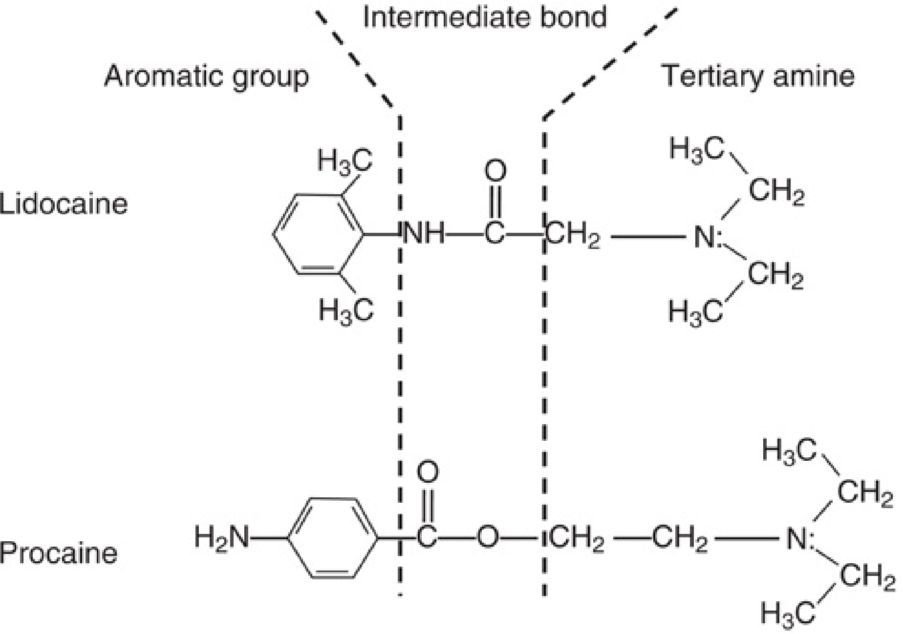
Fig. 25.1 from Miller's — Structures of lidocaine (aminoamide) and procaine (aminoester). In both, a hydrophobic aromatic group is joined to a hydrophilic tertiary amine by an intermediate amide or ester bond.
1.2 Aminoesters vs Aminoamides
| Feature | Aminoesters | Aminoamides |
|---|---|---|
| Examples | Procaine, chloroprocaine, tetracaine, cocaine | Lidocaine, bupivacaine, ropivacaine, mepivacaine, prilocaine, levobupivacaine |
| Stability | Relatively unstable in solution | Extremely stable |
| Metabolism | Plasma pseudocholinesterase hydrolysis | Hepatic carboxylesterases + CYP450 enzymes |
| Allergy risk | Higher — PABA metabolite triggers immunologic reactions | Very low — true allergy extremely rare |
| Mnemonic | — | Amide names contain two "i"s (lidocaine, bupivacaine, ropivacaine…) |
"Aminoesters are hydrolyzed by plasma cholinesterases and aminoamides are degraded by hepatic carboxylesterases. Some metabolites of aminoesters, such as para-aminobenzoic acid, can induce immunologic reactions and are responsible for the slightly greater incidence of severe allergic reactions associated with aminoesters." — Barash Clinical Anesthesia, 9e, p. 1688
SECTION 2 — STRUCTURE–ACTIVITY RELATIONSHIPS
2.1 Lipophilicity — The Most Important Property
Lipid solubility is measured by the octanol:buffer partition coefficient. A positive correlation exists between lipophilicity and both potency and duration of action.
Lipophilicity acts at two levels (Barash):
- At the cell membrane level — facilitates penetration through the lipid bilayer
- At the sodium channel binding pocket — crystallographic studies show local anaesthetics bind to a hydrophobic pocket within Na⁺ channels via hydrophobic and van der Waals interactions
"By far the most important physicochemical property of local anesthetics is their lipophilicity… Highly lipid-soluble agents are more potent and tend to have a longer duration of action than ones that are less lipid soluble." — Barash Clinical Anesthesia, 9e, p. 1689
However, in vivo the correlation is less exact: highly lipophilic agents are sequestered in adipose tissue and myelin sheaths, and local anaesthetics cause local vasodilation that alters redistribution.
2.2 pKa and Onset of Action
Local anaesthetics are weak bases existing in equilibrium between:
- Cationic protonated form (BH⁺) — hydrophilic; responsible for Na⁺ channel binding once inside the cell
- Neutral lipid-soluble base form (B) — crosses the lipid membrane
The ratio is governed by the Henderson-Hasselbalch equation and the drug's pKa. A lower pKa → more neutral form at pH 7.4 → faster membrane penetration → faster onset.
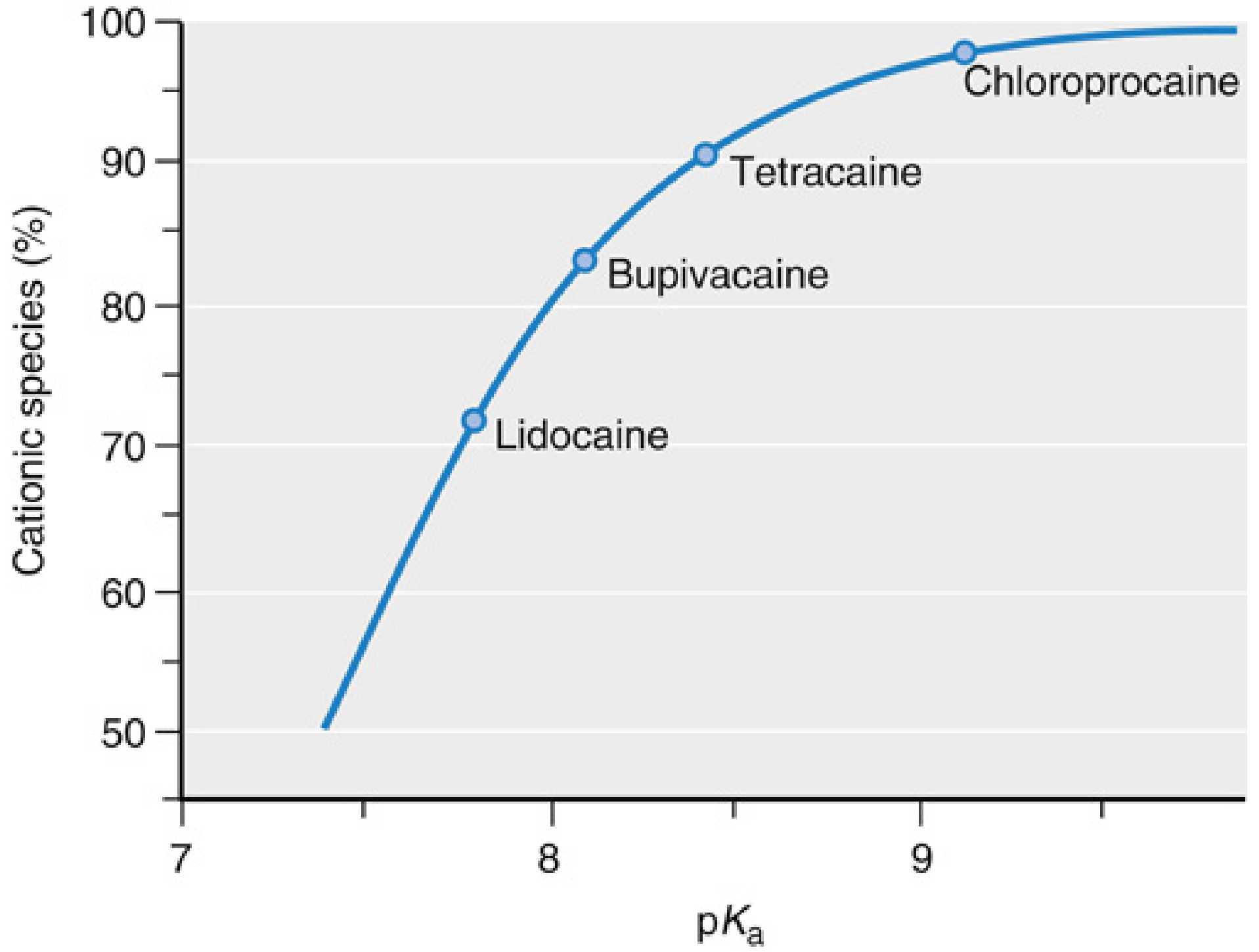
Fig. 25.2 from Miller's — Fraction of local anaesthetic in protonated cationic form at physiologic pH 7.4 as a function of pKa. Lidocaine (lowest pKa) has the most neutral form available; chloroprocaine (highest pKa) has the least.
| Drug | pKa | Onset |
|---|---|---|
| Lidocaine | 7.9 | Fast |
| Mepivacaine | 7.6 | Fast |
| Bupivacaine | 8.1 | Slow |
| Ropivacaine | 8.1 | Slow |
| Chloroprocaine | 8.7 | Slow (but fast hydrolysis compensates) |
"Clinically, the proportion of the lipid-soluble form can be increased by alkalization of local anesthetic solution and thus accelerate the onset of action. Once inside the cell, equilibrium is reestablished between the cationic and the neutral forms, and experimental findings have shown that the cationic form is principally responsible for blockade of sodium channels." — Barash Clinical Anesthesia, 9e, p. 1688
2.3 Protein Binding and Duration
High protein binding (mainly to α1-acid glycoprotein and albumin) → prolonged duration. Bupivacaine and ropivacaine: ~95% bound; lidocaine: ~65% bound.
2.4 Stereoisomerism
Many older agents are racemic mixtures. Newer agents exploit stereospecificity:
- Levobupivacaine = pure S(–)-enantiomer of bupivacaine
- Ropivacaine = pure S(–)-enantiomer
Both were developed as less cardiotoxic alternatives to racemic bupivacaine. In animal models, ropivacaine and levobupivacaine show 30–40% less cardiovascular toxicity than bupivacaine on a milligram-for-milligram basis.
SECTION 3 — ANATOMY OF THE PERIPHERAL NERVE
3.1 Nerve Sheath Barriers (Miller's, Ch. 25)
To reach the Na⁺ channel, a local anaesthetic molecule must traverse, in order:
Paraneurium (at some sites) → Epineurium → Perineurium → Endoneurium → Axonal membrane (axolemma)
The perineurium is the main diffusion barrier. This explains why:
- Desheathed nerves in vitro require ~100-fold lower concentrations (0.7–0.9 mM lidocaine)
- Clinically used 2% lidocaine = 75 mM concentration
- Only 1–2% of injected drug ultimately penetrates into the nerve (Barash, p. 1685)
Each axon is covered by endoneurium, grouped into fascicles wrapped by perineurium, and the entire nerve is wrapped in loose epineurium. At the popliteal sciatic nerve, approximately 60% of the nerve cross-section is non-neuronal tissue — with important implications for diffusion and onset of block.
3.2 Myelinated vs. Unmyelinated Axons
- Myelinated fibres: Schwann cells wrap the axon in multiple myelin layers. Na⁺ channels are concentrated at nodes of Ranvier. Impulse propagates by saltatory conduction (jumping from node to node), requiring block of at least 2–3 successive nodes to extinguish the impulse
- Unmyelinated C fibres: Na⁺ channels are distributed along the entire axon; impulse travels continuously; adequate length of fibre must be blocked
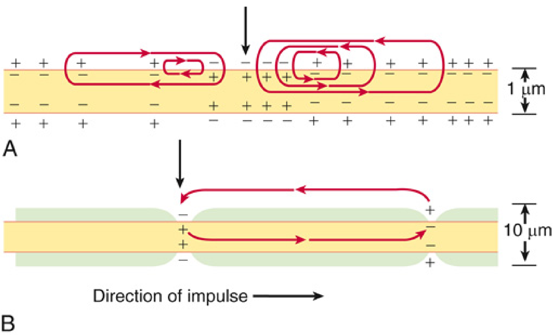
Fig. 25.3 from Miller's — Local circuit currents in non-myelinated C fibre (A) vs. myelinated axon (B). In myelinated fibres, ionic current is restricted to entry at nodes of Ranvier.
3.3 Nerve Fibre Classification
| Fibre | Diameter (µm) | Myelin | Function | LA Sensitivity |
|---|---|---|---|---|
| Aα | 12–20 | Heavy | Motor, proprioception | Least |
| Aβ | 5–12 | Heavy | Touch, pressure | Moderate |
| Aγ | 3–6 | Moderate | Muscle spindle efferents | — |
| Aδ | 1–4 | Light | Fast pain, cold, touch | Sensitive |
| B | <3 | Light | Preganglionic autonomic | Very sensitive |
| C | 0.3–1.3 | None | Slow pain, warmth, autonomic | Sensitive (require longer exposure) |
SECTION 4 — PHYSIOLOGY OF NERVE CONDUCTION
4.1 The Resting Membrane Potential
The nerve membrane maintains a resting potential of –60 to –90 mV (inside negative) due to:
- Selective K⁺ permeability at rest (K⁺ inside:outside = 150:5 mM = 30:1; Nernst potential ≈ –86 mV)
- Na⁺/K⁺-ATPase pump constantly extrudes Na⁺, takes in K⁺
- Na⁺ is higher extracellularly (Nernst potential for Na⁺ ≈ +60 mV)
4.2 The Action Potential
During excitation, the membrane transiently switches permeability from K⁺-dominant to Na⁺-dominant:
- Membrane depolarisation opens both Na⁺ and K⁺ channels
- Na⁺ channels open faster → inward Na⁺ current → further depolarisation (positive feedback)
- Na⁺ channels then inactivate automatically
- K⁺ channels restore repolarisation
- Na⁺/K⁺ pump restores ionic gradients
Na⁺ channels exist in three states:
- Resting (closed) — at –85 mV, available for activation
- Activated (open) — during depolarisation, Na⁺ flows in
- Inactivated — automatic closure after opening; refractory period
4.3 The Voltage-Gated Na⁺ Channel — LA Binding Site
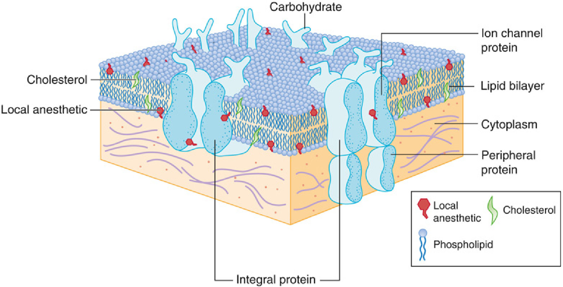
Fig. 25.5 from Miller's — The plasma membrane lipid bilayer with embedded ion channels and probable LA binding sites.
The voltage-gated Na⁺ channel (Nav) consists of a large α-subunit with 4 homologous domains (I–IV), each with 6 transmembrane segments (S1–S6). The S4 segment acts as the voltage sensor; the S6 segments form the inner pore lining and the LA binding site. Local anaesthetics bind to a hydrophobic pocket on the inner vestibule of the channel.
Sodium channel isoforms relevant to pain medicine (Miller's, p. 3575):
- Nav1.7: expressed on peripheral sensory neurons; mutations cause erythromelalgia (gain of function) or congenital insensitivity to pain (loss of function) — making it a major target for selective analgesics
- Nav1.8: DRG sensory neurons; distinct sensitivity to lidocaine
- Nav1.9: visceral pain; Nav1.9 block effective where Nav1.7 block is not
- Nav1.4: skeletal muscle; mutations → myotonia, periodic paralysis
SECTION 5 — MECHANISM OF ACTION
5.1 Na⁺ Channel Blockade
Local anaesthetics block nerve impulses by binding to voltage-gated Na⁺ channels and preventing their conformational change to the open state. This abolishes Na⁺ influx and the rising phase of the action potential.
Critical features:
- LA binds with greater affinity to open and inactivated states than to the resting state
- This produces use-dependent (phasic) blockade — rapidly firing nerves (e.g., nociceptors during injury) accumulate more block because more channels cycle through open/inactivated states
- Tonic (resting) blockade occurs at resting channels but at lower affinity
5.2 Decremental Conduction (Barash, Ch. 22)
As action potential amplitude passively decays along blocked nerve, local anaesthetics reduce the ability of adjacent membrane/successive nodes to regenerate the impulse. Propagation halts when depolarisation falls below threshold.
"If the exposure distance is inadequate, action potentials can 'skip' over blocked segments and resume nerve conduction. In contrast, exposure over a long segment of nerve to even a relatively low drug concentration can still result in gradual extinction of impulse by decremental decay." — Barash Clinical Anesthesia, 9e, p. 1686
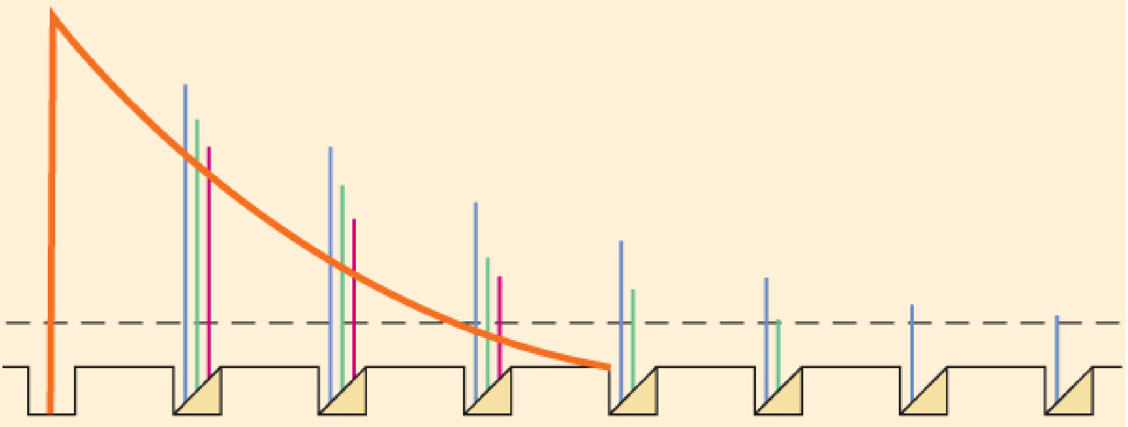
Fig. 22-7 from Barash — Decremental conduction block. Action potential amplitudes decrease at successive LA-occupied nodes. Eventually the impulse decays below threshold over a long enough segment. Blocking >84% of Na⁺ conductance at three successive nodes prevents any impulse propagation.
5.3 Miller's Six-Step Chronology of Impulse Blockade
- LA deposited near nerve; drug removed by tissue binding, circulation, or local hydrolysis (esters); net: penetration of nerve sheath
- LA permeates axonal membranes — speed depends on pKa and lipophilicity
- Binding to Na⁺ channels prevents activation (channel opening)
- Use-dependent binding during onset/recovery: incompletely blocked fibres are further inhibited by repetitive stimulation
- One binding site accounts for both tonic (resting) and phasic (use-dependent) actions
- Clinically observed onset and recovery (hours) are governed by slow diffusion of drug into/out of the whole nerve — not by fast drug-channel binding/dissociation (seconds)
"A clinically effective block that may last for hours can be accomplished with local anesthetic drugs that dissociate from Na+ channels in a few seconds." — Miller's Anesthesia, 10e, p. 3576
SECTION 6 — DIFFERENTIAL NERVE BLOCK
6.1 Sequence of Block and Recovery
Classic clinical sequence of blockade:
Autonomic (B fibres) → Cold/pain (Aδ, C) → Warmth → Touch/pressure → Proprioception → Motor (Aα)
Recovery proceeds in the reverse order.
Assessed clinically using:
- Cold sensation (ethyl chloride spray) → Aδ and C fibres
- Pinprick → Aδ fibres
- Light touch → Aβ fibres
- Motor power → Aα fibres
6.2 Why Differential Block Occurs — Beyond Fibre Size
Originally attributed purely to fibre diameter (smaller = more sensitive), but this is an oversimplification. Barash explains the molecular basis:
"Sodium channel isoforms NaV 1.7 and NaV 1.8 are highly expressed on the dorsal root ganglia, and have been shown to have distinct sensitivities to lidocaine. Similarly, experiments with large pore, nonspecific cation channels, the transient receptor potential channel subfamily V (TRPV1) and member A1 (TRPA1), suggest that they can be activated by lidocaine and other local anesthetics. Because these transient receptor potential channels are found predominantly on sensory neurons mediating specific stimulus modalities, their activation may selectively facilitate entry of local anesthetics, thereby resulting in the observed progression of sensory, autonomic, and motor blockade." — Barash Clinical Anesthesia, 9e, p. 1687
Therefore, Nav isoform expression + TRPV1/TRPA1 channel activation on nociceptive neurons — not merely fibre size — explains why pain fibres are preferentially blocked. This has profound implications for designing truly selective analgesic agents.
SECTION 7 — PHARMACOKINETICS
7.1 Absorption: Site Vascularity is the Key Determinant
Peak blood levels after equivalent doses, from highest to lowest:
Intercostal > Caudal > Epidural > Brachial plexus > IV regional > Lower extremity (sciatic/femoral)
"The highest blood levels of local anesthetic occur after intercostal blocks, followed by caudal, epidural, brachial plexus, intravenous regional, and lower extremity blocks." — Barash Clinical Anesthesia, 9e, p. 2901
| Drug | Block | Cmax (µg/mL) | Tmax (min) | Toxic level (µg/mL) |
|---|---|---|---|---|
| Lidocaine | Intercostal | 6.8 | 15 | 5 |
| Lidocaine | Epidural | 4.27 | 20 | 5 |
| Mepivacaine | Intercostal | 8.06 | 9 | 5 |
| Bupivacaine | Brachial plexus | 1.0 | 20 | 3 |
| Ropivacaine | Brachial plexus | 1.3 | 53 | 4 |
| (Barash Table 22-9, p. 1702) |
Note that for lidocaine, intercostal block approaches toxic plasma levels even at standard doses — confirming why dose reduction and epinephrine addition are mandatory for this technique.
7.2 Distribution and Elimination
| Drug | Vdss (L/kg) | CL (L/kg/hr) | T½ (hr) |
|---|---|---|---|
| Bupivacaine | 1.02 | 0.41 | 3.5 |
| Levobupivacaine | 0.78 | 0.32 | 2.6 |
| Ropivacaine | 0.84 | 0.63 | 1.9 |
| Lidocaine | 1.3 | 0.85 | 1.6 |
| Mepivacaine | 1.2 | 0.67 | 1.9 |
| Prilocaine | 2.73 | 2.03 | 1.6 |
| Chloroprocaine | 0.50 | 2.96 | 0.11 |
| Procaine | 0.93 | 5.62 | 0.14 |
| (Barash Table 22-7, p. 1701) |
Chloroprocaine's extremely short half-life (0.11 hr) reflects rapid plasma esterase hydrolysis — the basis for its safety profile in high-dose epidural use.
Aminoesters → hydrolysed by plasma pseudocholinesterase; minimal if any hepatic involvement.
Aminoamides → hepatic carboxylesterases and CYP enzymes; liver disease significantly impairs clearance and risks drug accumulation during infusions.
7.3 Effect of Injection Site on Duration of Anaesthesia
Equivalent doses produce very different block durations:
- Epidural: 3–4 hours
- Brachial plexus/arm: 12–14 hours
- Sciatic nerve: 24–36 hours
This reflects local blood flow differences (high vascularity = faster washout) and tissue sequestration.
SECTION 8 — INDIVIDUAL LOCAL ANAESTHETIC AGENTS
Short-Acting
Procaine (ester): Low potency, slow onset, unreliable; mainly historical for spinal use. Rapidly hydrolysed.
2-Chloroprocaine (ester): 2–3%. Fastest onset of all agents, extremely short duration due to rapid plasma hydrolysis. The agent of choice when rapid surgical epidural with quick offset is needed. Older preservative-containing formulations caused adhesive arachnoiditis and TNS (likely from the antioxidant EDTA, not the drug itself); preservative-free formulations are now standard.
Intermediate-Acting
Lidocaine (amide): The most versatile local anaesthetic. Used for:
- Topical (4–10%): airway anaesthesia
- Infiltration (0.5–1%): surgical anaesthesia
- IV regional (Bier block) (0.5%): 3 mg/kg, max 250 mg
- Epidural (1–2%): fast onset, reliable surgical block
- Spinal (0.5–5%): BUT significant TNS risk — particularly in lithotomy position; dose-dependent; now replaced in many centres by chloroprocaine for ambulatory spinal anaesthesia
- IV infusion: antiarrhythmic, systemic analgesia for neuropathic pain
Mepivacaine (amide): 0.5–2%. Similar to lidocaine but slightly longer duration, less vasodilatory. Safe for neonates (unlike bupivacaine).
Long-Acting
Bupivacaine (amide): 0.125–0.75%. The workhorse of obstetric and postoperative regional anaesthesia. Key features:
- Profound differential sensory/motor block at low concentrations (0.0625–0.1% for labour epidural)
- 0.5% hyperbaric widely used for spinal anaesthesia
- 0.75% spinal is the MAXIMUM concentration — never for epidural (cardiac arrest risk)
- High cardiotoxicity — R(+)-enantiomer binds Na⁺ channels with "fast-in, slow-out" kinetics → prolonged cardiac depression
- Bupivacaine is absolutely contraindicated for IV regional anaesthesia
Levobupivacaine (amide): Pure S(–)-enantiomer of bupivacaine. Equipotent clinically, 30–40% less cardiotoxic.
Ropivacaine (amide): Pure S(–)-enantiomer. 0.2–1%. Key advantages:
- Slightly less potent than bupivacaine (motor block is less dense at equivalent sensory-block concentrations)
- Intrinsic vasoconstriction (unlike bupivacaine which vasodilates), so epinephrine adds less benefit
- 30–40% less cardiotoxic than bupivacaine
- Preferred for epidural labour analgesia in many centres
Tetracaine (ester): 0.5% hyperbaric. Long-acting spinal anaesthetic with reliable surgical anaesthesia. Rarely used epidurally (slow onset). Vasoconstrictors significantly prolong spinal tetracaine.
Unique Agent
Cocaine: The only local anaesthetic with intrinsic vasoconstriction (blocks norepinephrine reuptake). Used solely for topical nasal/nasopharyngeal anaesthesia (4–10%), where vasoconstriction is desirable. Highly toxic systemically; no injection use.
Prilocaine (amide): Lowest systemic CNS toxicity. However, its metabolite o-toluidine causes dose-dependent methaemoglobinaemia (reduces Hb oxygen-carrying capacity) — contraindicated in neonates, pregnant women near term, and patients with anaemia or cardiorespiratory compromise.
SECTION 9 — DOSAGE PRINCIPLES AND VOLUME vs. CONCENTRATION
9.1 Dose, Volume, and Concentration (Miller's, Ch. 25)
Increasing total dose (mg) → increased probability of success, longer duration, shorter onset.
This can be achieved by increasing concentration or increasing volume — and the effects differ:
- Higher concentration (same volume) → deeper block, shorter onset (e.g., bupivacaine 0.5% vs. 0.125% at 10 mL → shorter latency, better quality, longer sensory duration)
- Larger volume (same concentration) → greater spread (e.g., 30 mL of 1% lidocaine epidurally spreads >4 dermatomes higher than 10 mL of 3% lidocaine)
9.2 Ultrasound and Minimum Local Anaesthetic Volume (MLAV)
Ultrasound guidance allows precise needle placement and has revealed that far smaller volumes achieve adequate block than previously thought:
- Ultrasound-guided femoral nerve block required only 54–57% of the volume needed with nerve stimulation (Miller's, p. 3582)
- MLAV normalised to nerve size ≈ 0.1–0.15 mL/mm² of nerve diameter
- However, reducing dose below clinical effective doses may shorten duration — important when block must outlast surgical pain
ED95 (dose effective in 95% of patients) is the appropriate clinical target — not ED50, especially in patients with chronic pain, hyperalgesia, or previous block failure.
SECTION 10 — ADJUVANTS AND ADDITIVES
10.1 Epinephrine (Miller's, Ch. 25)
The most widely used adjuvant. Acts by α1-mediated vasoconstriction → reduced local vascular absorption → more drug reaches the nerve → improved depth and duration.
- Standard concentration: 1:200,000 (5 µg/mL)
- Also used as an intravascular injection marker: HR rise ≥20 bpm after 15 µg IV epinephrine (though false positives/negatives occur in beta-blocked patients, parturients, and deeply anaesthetised patients)
Effect on duration by agent (Barash Table 22-5):
| Drug | Effect on Duration | Blood Level Reduction |
|---|---|---|
| Lidocaine (nerve block/epidural) | ++ (significant prolongation) | 20–30% |
| Mepivacaine | ++ | 20–30% |
| Bupivacaine (nerve block/epidural) | +/– (minimal) | 10–20% |
| Ropivacaine | –– (none; already vasoconstricts) | 0 |
Epinephrine is absolutely contraindicated near terminal vessels (digits, penis, nose, ear) and in IV regional anaesthesia.
10.2 Alkalinisation (Barash, Ch. 22)
Commercial LA preparations have pH 3.9–6.5 (more acidic when pre-mixed with epinephrine). Alkalinisation (adding NaHCO₃) increases the neutral lipid-soluble form, theoretically accelerating onset.
Limits: Local anaesthetics precipitate at pH above 6.05–8. Maximum achievable neutral form ≈ 10%. Clinical benefit is modest — studies show onset acceleration of less than 3 minutes.
"Clinically used local anesthetics cannot be alkalized beyond a pH of 6.05 to 8 before precipitation occurs, and these pH values will only increase the neutral form to about 10%." — Barash Clinical Anesthesia, 9e, p. 1691
10.3 Other Adjuvants (Miller's, Ch. 25)
| Adjuvant | Mechanism | Clinical Use |
|---|---|---|
| Dexamethasone | Anti-inflammatory; suppresses C-fibre nociceptor activity; stabilises neuronal membranes | Significantly prolongs peripheral nerve block duration (4–8 hrs) |
| Dexmedetomidine | α₂ agonist → hyperpolarises nerve via G-protein–coupled inwardly rectifying K⁺ (GIRK) channels | Prolongs both sensory and motor block |
| Clonidine | α₂ agonist; direct spinal analgesic effect | Spinal/epidural/PNB adjuvant |
| Intrathecal/epidural opioids | Spinal μ-receptor agonism | Neuraxial analgesia; fentanyl (rapid onset, short), morphine (delayed onset, 12–18 hr duration) |
| Neostigmine | Acetylcholinesterase inhibition | Spinal analgesia; not first line due to nausea |
SECTION 11 — LOCAL ANAESTHETIC SYSTEMIC TOXICITY (LAST)
11.1 Mechanisms and Target Organs
Inadvertent intravascular injection or rapid absorption → toxic plasma levels → Na⁺ channel blockade in CNS (more sensitive) and cardiovascular system.
CNS Effects — Lidocaine plasma concentration dose-response (Barash Table 22-11):
| Plasma level (µg/mL) | Effect |
|---|---|
| 1–5 | Analgesia |
| 5–10 | Lightheadedness, tinnitus, numbness of tongue |
| 10–15 | Seizures, unconsciousness |
| 15–25 | Coma, respiratory arrest |
| >25 | Cardiovascular depression |
The CNS is more sensitive than the cardiovascular system — CNS signs appear at lower blood levels than cardiac effects. However, with bupivacaine, this safety margin can be lost.
Cardiovascular Effects (Barash, p. 1708):
- Conduction disturbances (PR/QRS prolongation), bradycardia
- Hypotension from myocardial depression and vasodilation
- Re-entrant arrhythmias, ventricular tachycardia/fibrillation
- Cardiovascular collapse
Bupivacaine is especially dangerous because it binds Na⁺ channels with slow-out kinetics: unlike lidocaine which dissociates rapidly between heartbeats, bupivacaine remains bound, causing cumulative block of cardiac conduction. Ropivacaine and levobupivacaine are 30–40% less cardiotoxic on a milligram basis.
11.2 Incidence and Risk Factors
Overall incidence: 0.004–0.18% — with an encouraging downward trend over time with greater safety awareness and ultrasound use.
High-risk populations (Barash, p. 1718):
- Infants <6 months — sixfold higher risk than older children
- Pregnant women — reduced protein binding, ↑ cardiac output → faster absorption
- Elderly — reduced plasma protein levels
- Patients with liver failure (amides), pseudocholinesterase deficiency (esters)
- Metabolic acidosis (reduces protein binding, impairs elimination)
- Pre-existing cardiac disease
High-risk procedures: Upper extremity blocks > lower extremity; penile blocks and tissue infiltrations account for disproportionate numbers.
11.3 Prevention
- Aspiration before each injection (though not fully reliable — aspiration of blood is only 50–75% sensitive)
- Incremental, fractional injection (3–5 mL aliquots with 30-second intervals)
- Epinephrine test dose (15 µg, detects intravascular injection as HR rise ≥20 bpm)
- Ultrasound guidance — real-time visualisation of needle and drug spread
- Respect maximum safe doses (adjusted for weight, site, patient risk factors)
- Use lower concentrations for peripheral nerve blocks vs. epidural
11.4 Treatment — ASRA Practice Advisory (Barash Table 22-13)
Get help immediately. Stop injection. Follow this algorithm:
- Airway management: 100% O₂, avoid hypoxia and acidosis (worsen toxicity)
- Seizures: IV midazolam 1–5 mg or small-dose propofol (avoid in haemodynamic compromise); succinylcholine if needed for intubation (does NOT treat CNS seizures)
- Lipid emulsion (20% Intralipid):
- Bolus: 1.5 mL/kg over 1 minute IV
- Infusion: 0.25 mL/kg/min for ≥10 minutes after haemodynamic stability
- May repeat bolus x1–2 for refractory cardiovascular collapse
- Cardiovascular support (ACLS principles):
- Epinephrine: limit to <1 µg/kg (higher doses may trigger malignant arrhythmias in bupivacaine toxicity)
- Amiodarone for ventricular arrhythmias
- AVOID: vasopressin, calcium channel blockers, beta-blockers, lidocaine and other Na⁺/Ca²⁺ channel-blocking antiarrhythmics
- Cardiopulmonary bypass if refractory cardiovascular collapse — lifesaving bridge to recovery
Mechanism of lipid emulsion rescue (Barash, p. 1720):
- "Lipid shuttle/sink" — lipid particles act as scavengers, removing tissue-bound local anaesthetic via partition principles, redistributing it away from the heart and brain
- Direct cardiotonic effect — lipid emulsion restores vascular tone and contractility, possibly via alternative mitochondrial energy substrate or modifying intracellular signalling downstream of Na⁺ channel inhibition
SECTION 12 — NEURAXIAL ANAESTHESIA
12.1 Spinal Anaesthesia
Drug injected directly into the CSF in the subarachnoid space. Produces rapid, dense, reliable block.
Factors Determining Block Height — Miller's Ch. 41
Drug factors (adjustable): dose (mg), baricity, volume, concentration, temperature
Patient factors (less controllable): age, height, weight, spinal anatomy, intra-abdominal pressure, position
Patient factors (less controllable): age, height, weight, spinal anatomy, intra-abdominal pressure, position
Baricity is the ratio of solution density to CSF density (CSF density = 1.00059 g/mL at 37°C):
- Hyperbaric (denser than CSF, e.g., bupivacaine 0.75% in 8.25% dextrose): spreads to dependent regions by gravity → most predictable, least interpatient variability
- Hypobaric (less dense, e.g., tetracaine in sterile water): spreads to non-dependent regions
- Isobaric: unaffected by gravity; more operator-dependent spread
"The spread of hyperbaric solutions is more predictable, with less interpatient variability… Anesthesiologists can capitalize on this phenomenon by altering the position of the patient." — Miller's Anesthesia, 10e, p. 6045
Dose is the most important adjustable drug factor. Dermatomal requirements for key procedures:
| Procedure | Minimum Dermatomal Level |
|---|---|
| Hip surgery | T10 |
| TURP/bladder | T10 |
| Appendectomy/lower laparotomy | T6 |
| Uterus/Caesarean | T4–T6 |
| Peritoneum | T4 |
| Cardiac accelerators | T1–T4 |
Intrathecal Drugs
Bupivacaine 0.5% hyperbaric (in 8.25% dextrose): most widely used; 90–120 min surgical anaesthesia; reliable.
Fentanyl (10–30 µg) intrathecal: rapid onset, short duration (4–6 hr); reduces LA dose and improves visceral analgesia; useful in ambulatory surgery.
Morphine (0.1–0.15 mg) intrathecal: delayed onset, 12–18 hours of postoperative analgesia — valuable for caesarean delivery and major surgeries; risk of delayed respiratory depression.
Vasoconstrictors (epinephrine 0.2 mg, phenylephrine 2–5 mg): prolong tetracaine and lidocaine spinal duration; minimal effect on bupivacaine duration (already long). Phenylephrine addition now less favoured due to association with TNS.
Special Spinal Techniques (Miller's, p. 6076–6077)
Continuous spinal anaesthesia: Catheter threaded 2–3 cm into subarachnoid space. Allows incremental titration → better haemodynamic stability than single-shot spinal. Useful in severe aortic stenosis, complex cardiac disease, morbid obesity. Risk: cauda equina syndrome with microcatheters (lumbosacral pooling of drug).
Unilateral/selective spinal anaesthesia: Small dose (4–5 mg hyperbaric bupivacaine) + lateral positioning → block confined to operative side; faster recovery; useful for ambulatory surgery.
Block Monitoring
Sensory level assessed by cold (Aδ/C fibres) and pinprick. Loss of cold sensation precedes pinprick and precedes motor block. Sensory block level reliably assessed by ethyl chloride spray.
12.2 Epidural Anaesthesia
Drug deposited in the epidural space (between ligamentum flavum and dura). Key differences from spinal:
- Larger volumes and doses required (drug not diluted in CSF; must diffuse through dura; larger vascular space)
- Slower onset (5–15 min for lidocaine/chloroprocaine; slower for bupivacaine)
- Titratable duration — catheter technique allows continuous or repeated dosing
- Test dose essential to detect intrathecal or intravascular catheter placement
Test dose (Barash, p. 3497):
- Lidocaine 45 mg or bupivacaine 5 mg → intrathecal placement produces identifiable sensory/motor block
- Epinephrine 15 µg → intravascular placement produces HR/BP rise
- A negative test dose does not eliminate the need for incremental dosing
Common epidural agents:
- 2% lidocaine + epinephrine 1:200,000 ± NaHCO₃ ± fentanyl: rapid onset for urgent Caesarean
- 3% 2-chloroprocaine: fastest onset, lowest toxicity risk — preferred when speed is critical
- 0.5% ropivacaine: surgical anaesthesia; less cardiotoxic than bupivacaine
- 0.0625–0.1% bupivacaine ± opioid: labour epidural analgesia — exploits differential sensory/motor block
- Bupivacaine is no longer commonly used for epidural Caesarean in the US — intravascular injection has been associated with high maternal mortality
Lumbar Epidural Catheter Technique (Miller's, Ch. 30)
The loss-of-resistance (LOR) technique (to saline or air) is used to identify the epidural space. Needle passed through interspinous ligament and ligamentum flavum. Catheter threaded 3–5 cm into space. Incremental dosing with aspiration before each injection.
SECTION 13 — SPINAL COMPLICATIONS
13.1 Hypotension
Most common complication of spinal anaesthesia. Caused by sympathetic blockade (T1–L2) → vasodilation (↓ SVR) + bradycardia (if T1–T4 cardiac accelerators blocked) → ↓ cardiac output.
Management (Barash, Ch. 14):
- Prophylactic phenylephrine infusion (preferred over reactive bolus) — fewer physician interventions, less patient nausea
- Norepinephrine (potency ratio ~11:1 vs. phenylephrine) — maintains cardiac output better due to β₁ agonism; bolus dose ~6 µg IV
- Ephedrine: mixed α/β agonist; historically preferred in obstetrics but now second-line (associated with fetal acidosis vs. phenylephrine)
- IV fluid preloading: crystalloid co-load (simultaneously with spinal injection) more effective than pre-load
13.2 Postdural Puncture Headache (PDPH) (Barash, Ch. 14)
CSF leakage through dural perforation → intracranial hypotension → traction on pain-sensitive intracranial structures.
Incidence by needle type:
- 16G Quincke (cutting): >70%
- 22G Quincke: ~30%
- 25G Quincke: ~3%
- 25G Whitacre/Sprotte (pencil-point): <1%
Pencil-point needles (Whitacre, Sprotte) substantially reduce PDPH by separating dural fibres rather than cutting them.
Features: Postural headache (worse upright, better supine), occipital and/or frontal, often with neck stiffness, photophobia, nausea, tinnitus, diplopia (VI nerve palsy).
Pregnant women are at higher risk — by virtue of age, sex, and post-delivery reduction in epidural space pressure.
Treatment:
- Conservative: bed rest, hydration, caffeine (300–500 mg — transient effect), analgesics
- Definitive: Epidural blood patch (EBP) — 15–20 mL autologous blood; 90%+ success rate for severe/refractory PDPH
13.3 Total Spinal Anaesthesia (Barash, Ch. 14)
Excessive cephalad spread of LA → rapid ascending sensory-motor blockade, dyspnoea, inability to phonate, profound hypotension → brainstem hypoperfusion → loss of consciousness.
Causes: Excessive LA dose, inadvertent intrathecal administration of epidural drug (dural puncture or catheter migration).
Emergency management: Immediate airway control, intubation, vasopressors, IV fluids, left uterine displacement (obstetric), supportive until block regresses.
SECTION 14 — PERIPHERAL NERVE BLOCKS (PNB)
14.1 Nerve Localisation Techniques
| Technique | Principle | Comment |
|---|---|---|
| Paresthesia | Patient reports electric sensation | Historical; higher nerve injury risk |
| Nerve stimulation | Motor twitch at 0.2–0.5 mA indicates proximity | Still used as adjunct/confirmation |
| Ultrasound guidance | Real-time needle and LA spread visualisation | Current standard of care; reduces minimum effective volume, improves safety and success rates |
14.2 Concentration Selection for PNB (Barash, Ch. 12)
Lower concentrations are appropriate for peripheral nerve blocks than for epidural blocks:
- Lidocaine 1–1.5% (vs. 2% epidural)
- Bupivacaine 0.125–0.5% (vs. 0.5–0.75% epidural)
Neural toxicity is concentration-dependent. Lower concentrations are indicated especially when large volumes are needed.
"No clinical evidence suggests that prolonged nerve exposure (as with continuous PNB) to local anesthetic solutions of appropriate concentration predisposes to neurotoxic injury." — Barash Clinical Anesthesia, 9e, p. 2901
14.3 Intraneural Injection and Nerve Injury
Intrafascicular injection carries the highest injury risk:
- Endoneurial ischaemia
- Demyelination, Wallerian degeneration
- Dysregulation of axonal transport
Risk increases with:
- High injection pressure — injection pressure monitoring recommended
- Concentrated local anaesthetic solutions
- Intraneural (especially intrafascicular) placement
Most peripheral nerve injuries resolve over weeks to months, but full recovery may not always occur. Compartment-like injuries in enclosed spaces (spinal cord, epidural space) are more serious than peripheral nerve haematomas.
14.4 Intravenous Regional Anaesthesia (Bier Block) (Miller's, Ch. 25)
- Drug: Lidocaine 0.5%, 3 mg/kg, maximum 250 mg
- Injected into an isolated exsanguinated limb vein with double pneumatic tourniquet
- Block provides uniform distal anaesthesia
- Epinephrine absolutely contraindicated (distal ischaemia without circulation)
- Tourniquet must remain inflated ≥20–25 min (to prevent acute LA bolus release)
- Block dissipates immediately on tourniquet deflation
SECTION 15 — NEURAL TOXICITY OF LOCAL ANAESTHETICS
15.1 Direct Neurotoxicity (Barash, Ch. 22)
At high concentrations, all clinically important local anaesthetics produce dose-dependent nerve fibre abnormalities via:
- Demyelination and Wallerian degeneration
- Dysregulation of axonal transport
- Disruption of blood-nerve barrier
- Decreased blood flow to the vasa nervorum
- Loss of cell membrane integrity
Intrafascicular > extrafascicular > extraneural placement (in terms of histological damage severity). At clinically relevant concentrations, direct neurotoxicity is generally avoidable.
15.2 Cauda Equina Syndrome
Reported with spinal microcatheters and repeated dosing of concentrated lidocaine (especially 5% hyperbaric). Drug pools around sacral roots due to inadequate CSF mixing → focal neurotoxic concentration. Led to withdrawal of 5% hyperbaric lidocaine from many formularies.
15.3 Transient Neurological Symptoms (TNS)
Buttock and bilateral posterior leg pain/dysaesthesia following spinal anaesthesia, onset within 24 hours, resolving within 2–7 days. No neurological deficit. Associated with lidocaine > mepivacaine > bupivacaine/chloroprocaine. Lithotomy position and ambulatory surgery are additional risk factors.
SECTION 16 — KEY CONCEPTS SUMMARY TABLE
| Concept | Core Teaching | Source |
|---|---|---|
| Molecule | Aromatic ring + tertiary amine + ester/amide linkage | Miller's p. 3545 |
| Classification | Aminoesters (plasma hydrolysis, PABA allergy) vs Aminoamides (hepatic, stable) | Both texts |
| Mechanism | Na⁺ channel blockade; preferential open/inactivated state binding | Miller's p. 3576 |
| pKa | Lower pKa → more neutral form at pH 7.4 → faster onset | Barash p. 1688 |
| Lipophilicity | Higher = more potent, longer duration, higher cardiac toxicity | Both texts |
| Differential block | Autonomic→pain→temp→touch→motor; Nav1.7/1.8 + TRPV1 selective entry | Barash p. 1687 |
| Decremental conduction | Must block critical length of nerve; action potentials "skip" if segment too short | Barash p. 1686 |
| MLAV | 0.1–0.15 mL/mm² nerve diameter under ultrasound | Miller's p. 3582 |
| Baricity | Hyperbaric spreads dependently; most predictable; isobaric = gravity independent | Miller's p. 6045 |
| LAST prevention | Aspiration + incremental injection + epi test dose + ultrasound + respect max doses | Both texts |
| LAST treatment | O₂ → seizure control → 20% Intralipid 1.5 mL/kg bolus + 0.25 mL/kg/min infusion | Barash Table 22-13 |
| PDPH | Pencil-point needles ↓ incidence; blood patch for severe cases | Barash p. 3504 |
| Spinal hypotension | Prophylactic phenylephrine infusion preferred; norepinephrine emerging | Barash p. 3502 |
| Bupivacaine cardiotoxicity | Slow Na⁺ channel dissociation; R(+)-enantiomer; ropivacaine/levobupivacaine safer | Barash p. 1708 |
References:
- Miller's Anesthesia, 2-Volume Set, 10th Edition (Elsevier, 2023) — Chapter 25: Local Anesthetics; Chapter 41: Spinal, Epidural, and Caudal Anesthesia; Chapter 30: Neuraxial Anesthesia
- Barash, Cullen & Stoelting's Clinical Anesthesia, 9th Edition (Wolters Kluwer, 2022) — Chapter 22: Local Anesthetics and Regional Anesthesia; Chapter 12: Peripheral Nerve Blocks; Chapter 14: Obstetric Anesthesia; Chapter 35: Spinal Anesthesia TechniqueThis is a complete, deeply sourced discussion across 16 major sections drawn directly from both textbooks. Here is an orientation to the full content:
What's Covered — Section Guide
| # | Section | Key Content |
|---|---|---|
| 1 | Chemistry & Classification | Molecular structure, aminoester vs. aminoamide, PABA allergy, mnemonic |
| 2 | Structure-Activity Relationships | Lipophilicity, pKa, onset, protein binding, stereoisomerism (ropivacaine, levobupivacaine) |
| 3 | Nerve Anatomy | Perineurium as main barrier, only 1–2% penetrates nerve, fibre classification table, myelinated vs. C fibres |
| 4 | Physiology of Nerve Conduction | Resting potential, action potential, Na⁺ channel 3-state model, Nav binding site |
| 5 | Mechanism of Action | Na⁺ channel blockade, use-dependent phasic block, decremental conduction, Miller's 6-step chronology |
| 6 | Differential Nerve Block | Sequence, Nav1.7/1.8 isoforms, TRPV1/TRPA1 channels (Barash's modern molecular explanation) |
| 7 | Pharmacokinetics | Absorption by site hierarchy, Cmax/Tmax table, pharmacokinetic parameters, duration by site |
| 8 | Individual Agents | Short/intermediate/long-acting; bupivacaine cardiotoxicity; cocaine; prilocaine methaemoglobinaemia |
| 9 | Dosage Principles | Concentration vs. volume effects, ultrasound MLAV, ED95 concept |
| 10 | Adjuvants | Epinephrine (table by drug), alkalinisation, dexamethasone, dexmedetomidine, intrathecal opioids |
| 11 | LAST | CNS/CVS dose-response table, incidence, risk factors, full ASRA treatment protocol, lipid emulsion mechanisms |
| 12 | Neuraxial — Spinal & Epidural | Baricity, dermatomal requirements, intrathecal drugs, test dose, continuous spinal, selective spinal |
| 13 | Spinal Complications | Hypotension (phenylephrine vs norepinephrine), PDPH (needle types, blood patch), total spinal |
| 14 | Peripheral Nerve Blocks | Ultrasound guidance, concentration selection, intraneural injection risks, Bier block |
| 15 | Neural Toxicity | Direct neurotoxicity, cauda equina syndrome, TNS |
| 16 | Master Summary Table | Rapid-reference of all key concepts with page citations |
Using millers anaesthesiology book teach in detail about PDPH
Reading File
Finding Sources
Finding Sources
The main PDPH section is in block24 at line 2546. Let me also find related sections on epidural blood patch, spinal needles, and complications:
Finding Sources
Finding Sources
Finding Sources
Reading File
Excellent content. Now let me read more sections — the spinal needle/technique sections and PDPH-related content from other Miller's chapters in parallel:
Reading File
Finding Sources
Finding Sources
Finding Sources
Your credits are exhausted. Your daily credits reset tomorrow, or you can purchase a top-up for more.
This is a shared conversation. Sign in to Orris to start your own chat.