
Structure passing clavi pectoral fascia
structures passing through clavipectoral fascia anatomy
Structures Passing Through the Clavipectoral Fascia
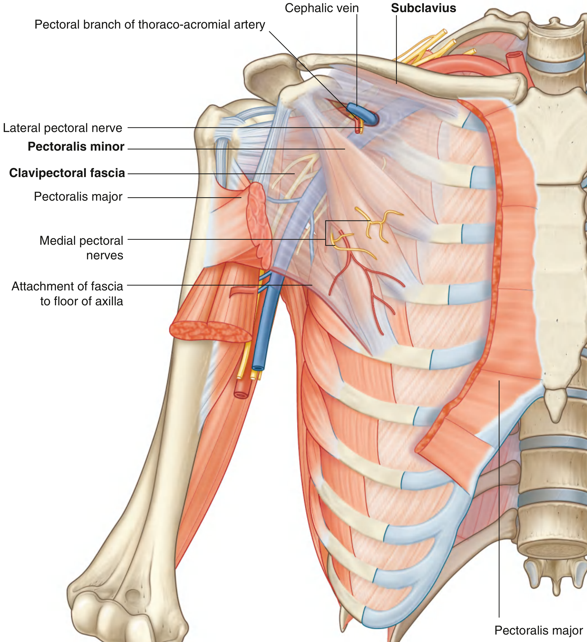
Structures Piercing the Clavipectoral Fascia
| Structure | Direction of Travel | Notes |
|---|---|---|
| Cephalic vein | Upward (superficial to deep) | Runs in the deltopectoral groove, pierces the fascia to drain into the axillary vein at the infraclavicular fossa |
| Thoraco-acromial artery | Downward (deep to superficial) | Branch of the 2nd part of axillary artery; emerges through the fascia and divides into pectoral, deltoid, clavicular, and acromial branches |
| Lateral pectoral nerve | Downward (deep to superficial) | From the lateral cord of the brachial plexus (C5, C6, C7); supplies pectoralis major |
| Lymphatics | Upward | Pass from infraclavicular nodes to the apical (level III) nodes of the axilla |
Mnemonic
"CALL" - Cephalic vein, Acromiothoracic (thoraco-acromial) artery, Lateral pectoral nerve, Lymphatics
Additional Notes
- The lateral thoracic artery exits the axilla inferior to the pectoralis minor (not between the two muscles), so it passes through the fascia at a slightly different level.
- The medial pectoral nerve does NOT pierce the clavipectoral fascia; it penetrates directly through the pectoralis minor muscle itself.
- The clavipectoral fascia also exerts traction on the subclavian vein wall (it is fused to it), helping to keep the lumen patent and facilitating venous return to the SVC - (THIEME Atlas of Anatomy, p. 401).
Create an anatomy diagram poster showing clavipectoral fascia and traversing structures
Detailed medical anatomy education poster of the clavipectoral fascia and its traversing structures. Show an anterior view of the right shoulder and upper chest with the clavicular head of pectoralis major removed to expose the deep structures. Label and illustrate: the clavipectoral fascia (shown as a semi-transparent blue sheet), subclavius muscle (superior), pectoralis minor muscle (inferior), and the four structures piercing through the fascia: (1) cephalic vein in blue traveling upward through the deltopectoral groove into the axillary vein, (2) thoraco-acromial artery in red descending from the axillary artery and branching into pectoral, deltoid, clavicular and acromial branches, (3) lateral pectoral nerve in yellow passing downward to pectoralis major, (4) lymphatic vessels shown as thin green channels passing upward to apical axillary nodes. Also show the clavicle at top, ribs and intercostal spaces medially, coracoid process laterally, pectoralis major (cut edge), deltoid muscle, axillary vein and axillary artery. Include a title "Clavipectoral Fascia & Traversing Structures" at the top, a color-coded legend at the bottom, clean white background, professional medical illustration style with clear anatomical labels and leader lines.
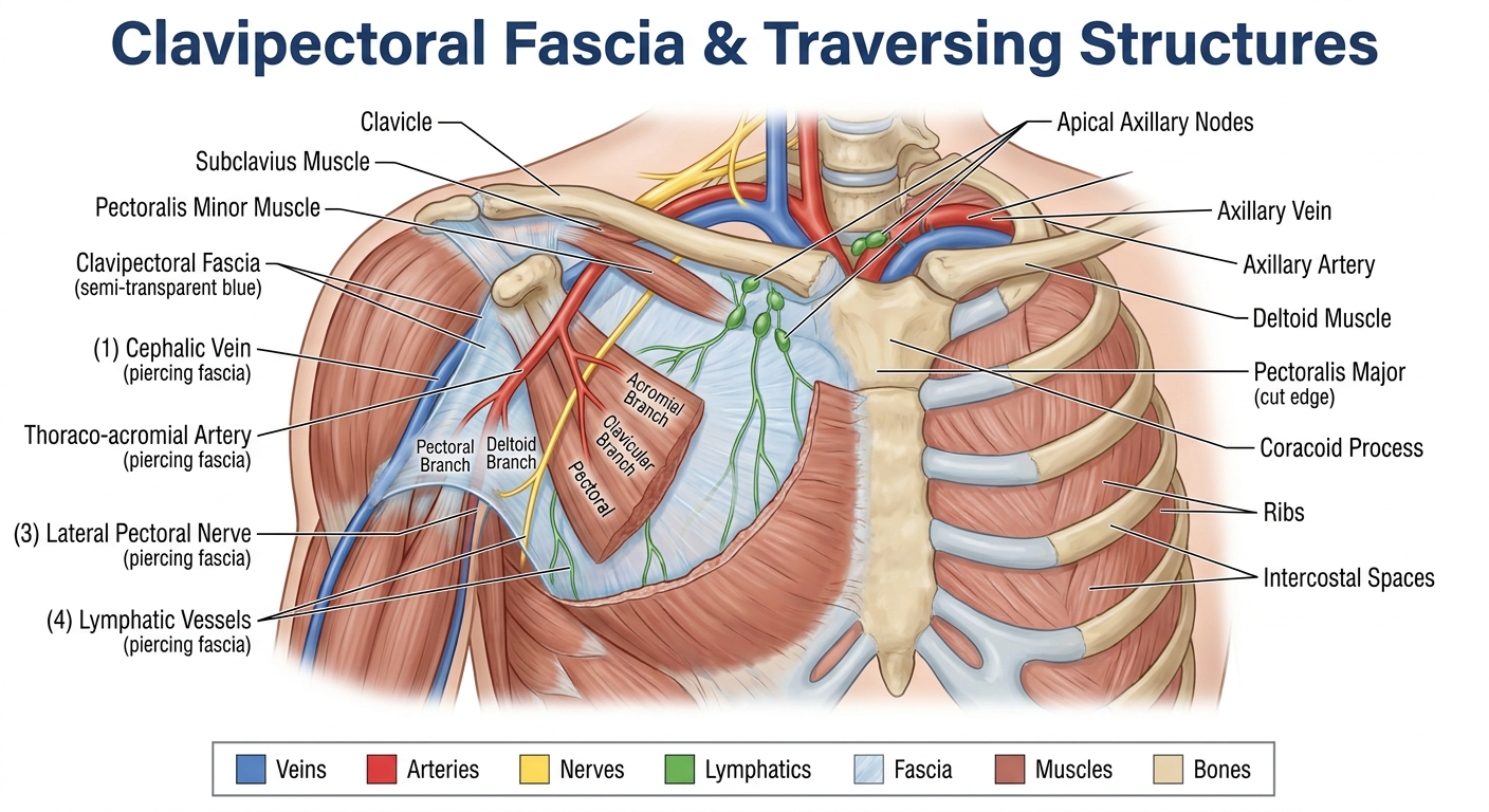
- Cephalic vein (blue) - traveling upward through the deltopectoral groove to drain into the axillary vein
- Thoraco-acromial artery (red) - descending from the axillary artery with its four branches (pectoral, deltoid, clavicular, acromial)
- Lateral pectoral nerve (yellow) - passing downward to supply pectoralis major
- Lymphatic vessels (green) - passing upward from infraclavicular to apical axillary nodes