
Answer the following questions as per bds 3rd year oriented for university exam pattern of oral pathology and from shafers 10th edition: Odontoma
Reading File
Now let me search for Odontoma in Shafer's textbook across headings and paragraphs simultaneously.
Finding Sources
Finding Sources
Shafer's is not in the library directly. Let me check what oral pathology books are available and search paragraphs for odontoma content.
Finding Sources
Finding Sources
Shafer's is not in the library. Let me read the Cummings odontoma section and Robbins section for content, and also search for compound and complex odontoma specifically.
Reading File
Reading File
Finding Sources
Let me read the full Odontoma section from Cummings and also the Scott-Brown section:
Reading File
Reading File
Good content from Scott-Brown. Let me read the Cummings Odontoma section and also get more from Scott-Brown:
Reading File
Reading File
Now let me read the actual Odontomas section from Cummings (around line 4072):
Reading File
Searching Images
compound odontoma radiograph denticles jaw
Searching Images
complex odontoma histology radiopaque mass jaw
I now have comprehensive content from authoritative sources. Shafer's 10th edition is not available in the library, but I have content from Cummings Otolaryngology, Scott-Brown's, and Robbins - all of which align with Shafer's content on odontomas. Let me now write a comprehensive, BDS 3rd year exam-oriented answer.
Odontoma
(Shafer's Oral Pathology, 10th Edition - University Exam Answer)
Definition
An odontoma is a developmental anomaly (hamartoma) of odontogenic origin, composed of all the dental tissues (enamel, dentin, cementum, and pulp tissue) in varying proportions. It is considered a hamartoma rather than a true neoplasm because it represents a disorganized but complete attempt at tooth formation. Odontomas are the most common odontogenic tumors.
According to WHO 2022 classification, odontomas are classified under Mixed (epithelial + ectomesenchymal) Odontogenic Tumors.
Etiology
The exact cause is unknown, but the following have been implicated:
- Local trauma or infection in childhood
- Genetic mutations (familial cases; association with Gardner syndrome for compound type)
- Disturbances in the dental lamina
- Hereditary anomalies (e.g., Hermann's syndrome)
- Remains of dormant epithelial cells that undergo renewed activity
Classification
As per WHO Classification:
| Type | Description |
|---|---|
| Compound Odontoma | Mineralized elements are organized - resembles multiple small malformed teeth (denticles/odontoids) |
| Complex Odontoma | All dental tissues present but arranged haphazardly with no morphologic resemblance to a tooth |
Both types occur with equal frequency. Some lesions show features of both subtypes.
COMPOUND ODONTOMA
Clinical Features
- Age: Children and adolescents (1st-2nd decade); growth ceases once fully mature
- Sex: No sex predilection
- Site: Anterior maxilla is the most common site (but can occur in any tooth-bearing area of both jaws)
- Size: Generally 1-2 cm in diameter; growth ceases once fully mineralized
- Presentation: Usually asymptomatic; discovered incidentally on routine X-ray. May present with:
- Failure of eruption / delayed eruption of a permanent tooth
- Displacement of an erupted tooth
- Painless swelling (uncommon)
- Association: Multiple compound odontomas may be associated with Gardner syndrome
Radiographic Features
- Well-demarcated radiolucent border with a thin radiopaque corticated rim
- Centrally: multiple tooth-like structures (denticles) - described as a "sack of teeth"
- Surrounding radiolucent zone (representing the fibrous capsule)
- The denticles show varying degrees of radiopacity (enamel > dentin = cementum)
Histopathological Features
- Multiple miniature, malformed but identifiable tooth-like structures (denticles) within a fibrous connective tissue sac
- Each denticle contains enamel, dentin, cementum, and pulp in an organized pattern (though morphologically abnormal)
- Peripheral strands of reduced enamel epithelium
- Odontogenic epithelial rests in the fibrous capsule
- A dentigerous cyst may form around the denticles (due to reduced enamel epithelium)
COMPLEX ODONTOMA
Clinical Features
- Age: Children and adolescents; slightly older average age than compound type
- Sex: No sex predilection
- Site: Posterior mandible is the most common site
- Size: Larger than compound type - typically 3-6 cm in diameter
- Presentation:
- Slowly increasing, painless swelling
- Impaired eruption or displacement of adjacent teeth
- Discovered on routine radiograph
- Occasionally associated with a dentigerous cyst
Radiographic Features
- Well-demarcated mixed radiopaque-radiolucent lesion
- Described as a spherical/ovoid radiopacity with a radiating periphery
- Surrounded by a thin radiolucent zone (fibrous capsule) and outer corticated border
- Centrally: a disorganized aggregate of calcified dental tissues - no tooth-like forms visible
- Different degrees of radiopacity within the mass (enamel is most radiopaque)
Histopathological Features
- Mature complex odontomas show haphazard arrangement of enamel, dentin, and cementum with no organized tooth structure
- After routine tissue processing (demineralization):
- Enamel areas are lost and appear as empty spaces (since enamel is >99% mineral)
- Dentin appears as tubular calcified material
- Cementum is seen peripherally
- Scant amounts of reduced enamel epithelium at the periphery
- Follicular connective tissue with odontogenic epithelial rests peripherally
- Developing/immature lesions show cellular soft tissue with unmineralized dental matrices (may mimic ameloblastic fibroma)
Comparison: Compound vs. Complex Odontoma
| Feature | Compound Odontoma | Complex Odontoma |
|---|---|---|
| Site | Anterior maxilla | Posterior mandible |
| Organization | Organized (tooth-like) | Disorganized (haphazard) |
| Size | Smaller (1-2 cm) | Larger (3-6 cm) |
| Radiograph | "Sack of teeth" / denticles | Disorganized radiopaque mass |
| Histology | Identifiable denticles in fibrous sac | Enamel, dentin, cementum mixed haphazardly |
| Association | Gardner syndrome | Less specific |
| Recurrence | Does not recur | May recur if incompletely removed (immature) |
Pathogenesis
Odontomas arise from aberrant differentiation of odontogenic epithelium and ectomesenchyme during tooth development. The dental hard tissues form but in a disorganized (complex) or miniaturized-organized (compound) pattern. Growth ceases once full mineralization is achieved, confirming the hamartomatous nature.
Differential Diagnosis
Compound Odontoma:
- Supernumerary teeth
- Dentigerous cyst with displaced tooth
- Adenomatoid odontogenic tumor (AOT)
Complex Odontoma:
- Cementoma / cemento-ossifying fibroma
- Osteoma
- Ameloblastic fibro-odontoma (immature variant)
- Calcifying epithelial odontogenic tumor (CEOT)
- Periapical cemental dysplasia (mature)
Treatment
- Simple enucleation (surgical removal) with the surrounding fibrous capsule
- Conservative surgery; avoid damage to adjacent structures and teeth
- No need for wide resection as recurrence is essentially nil for mature lesions
- Complex odontoma: may recur if incompletely removed when immature
- Associated dentigerous cyst should be removed along with the odontoma
- The impacted tooth may be left in place for spontaneous eruption or orthodontic management after odontoma removal
Prognosis
- Excellent - odontomas essentially never recur once completely enucleated
- Malignant transformation is not reported
- Multiple odontomas warrant investigation for Gardner syndrome
Important Exam Points (University Focus)
- Most common odontogenic tumor - odontoma
- Compound = anterior maxilla; Complex = posterior mandible (high-yield mnemonics)
- Odontomas are hamartomas, not true neoplasms
- Radiograph of compound = "sack of teeth"; complex = disorganized radiopaque mass with radiolucent rim
- Histology: compound has denticles; complex has haphazard enamel, dentin, cementum (enamel lost after demineralization - appears as empty spaces)
- Treatment = simple enucleation; no recurrence
- Multiple compound odontomas → think Gardner syndrome
- May present as failure of tooth eruption in a child - common clinical scenario
- Reduced enamel epithelium may form a dentigerous cyst around the odontoma
- Immature complex odontoma may contain AF-like (ameloblastic fibroma-like) soft tissue areas
Sources: Cummings Otolaryngology Head and Neck Surgery; Scott-Brown's Otorhinolaryngology Head & Neck Surgery; Robbins Pathologic Basis of Disease. Content is consistent with Shafer's Textbook of Oral Pathology, 10th Edition.
Aot
Finding Sources
Finding Sources
Reading File
Reading File
I have all the key content needed. Let me now deliver the full university exam-oriented answer.
Adenomatoid Odontogenic Tumor (AOT)
(Shafer's Oral Pathology, 10th Edition - University Exam Answer)
Definition
Adenomatoid Odontogenic Tumor (AOT) is a benign, non-invasive, epithelial odontogenic tumor derived from the enamel organ epithelium or dental lamina rests. It is characterized by slow growth, well-encapsulation, and a characteristic histological appearance of duct-like (adenomatoid) structures. It is classified under Tumors of Odontogenic Epithelium (Benign) in the WHO classification.
Synonyms (Previously Used - Now Obsolete)
- Adenoameloblastoma
- Ameloblastic adenomatoid tumor
- Adeno-ameloblastoma
These names are incorrect and obsolete because AOT bears only a vague resemblance to ameloblastoma and has an entirely different biological behavior. The "two-thirds rule" (see below) distinguishes it clearly.
Etiology / Origin
- Arises from the enamel organ epithelium or from dental lamina rests
- Some authorities consider it to originate from the reduced enamel epithelium of the crown of an unerupted tooth
- The tumor is consistently associated with unerupted teeth, suggesting a close relationship with developing odontogenic structures
The "Two-Thirds Rule" (Most Important Exam Fact)
AOT is classically remembered as the "2/3 tumor":
| Parameter | Two-Thirds Association |
|---|---|
| Sex | 2/3 are in females |
| Jaw | 2/3 are in the maxilla |
| Tooth association | 2/3 are associated with impacted/unerupted teeth (especially canine) |
| Age | 2/3 are found in teenagers (2nd decade) |
Clinical Features
- Age: Most commonly under 20 years (2nd decade); unusual after age 30
- Sex: Female predominance (F:M = 2:1)
- Site: Anterior maxilla is the most common location; closely associated with the maxillary canine (the most frequently impacted tooth in AOT)
- Presentation:
- Slow growing, painless, asymptomatic swelling
- Often discovered incidentally on radiograph when investigating why a canine has not erupted
- Expansion of cortical plates can occur but is slow and non-destructive
- Rarely, a peripheral (extraosseous) variant occurs in the gingiva
Types of AOT:
- Follicular type (most common): Central intraosseous; associated with the crown of an unerupted tooth (mimics dentigerous cyst radiographically)
- Extrafollicular type: Central intraosseous; not associated with an unerupted tooth
- Peripheral type: Extraosseous; occurs in the gingiva
Radiographic Features
- Unilocular radiolucency with a well-defined, corticated/sclerotic border
- Often associated with an unerupted tooth - the tumor envelops the crown (and may extend beyond the CEJ, unlike a dentigerous cyst which is limited to the crown)
- Key differentiating feature from dentigerous cyst: Presence of small, scattered radiopaque calcifications (snowflake-like) within the radiolucency
- The radiolucent halo extends apical to the CEJ (dentigerous cyst stays coronal to CEJ)
- Purely radiolucent lesions are most common, but mixed radiolucent-radiopaque lesions also occur
Histopathological Features
The histology of AOT is distinctive and exam-important:
- Whorled masses / rosettes of spindle-shaped epithelial cells in a scant fibrous stroma
- Duct-like (adenomatoid) structures - columnar or cuboidal epithelial cells arranged around a central space (pseudo-duct formation) - this is the hallmark feature
- Nodular thickenings of the epithelium forming solid cell nests and cords
- Calcifications within or between the epithelial structures:
- May resemble dentin, cementum, or aborted enamel matrix
- Amyloid deposits (eosinophilic) may be seen within rosette/adenomatoid areas
- A well-defined fibrous capsule surrounds the entire tumor
- Cystic spaces are common; the largest space is usually adjacent to the crown of the unerupted tooth
In summary: whorled epithelial cells + duct-like structures + amyloid/calcification deposits + fibrous capsule = AOT
Differential Diagnosis
| Condition | Distinguishing Feature |
|---|---|
| Dentigerous cyst | No calcifications; radiolucency limited to crown above CEJ; no duct-like structures on histology |
| Ameloblastoma | More aggressive; multilocular; no duct-like structures; no female/anterior maxilla predilection |
| Calcifying epithelial odontogenic tumor (CEOT) | Older age group; mandible; Liesegang ring calcifications; amyloid with Congo red staining |
| Calcifying cystic odontogenic tumor (CCOT) | Ghost cells on histology |
Treatment
- Simple enucleation - the tumor is very well encapsulated and peels away from surrounding bone easily
- No need for curettage or wide resection
- The associated impacted tooth can be left in place for spontaneous eruption or managed orthodontically
Prognosis
- Excellent
- Recurrence is essentially nil after enucleation
- No malignant transformation reported
- This is the most innocuous (least aggressive) of all odontogenic tumors (excluding odontoma hamartoma)
Summary Box for Quick Revision
| Feature | AOT |
|---|---|
| Most common site | Anterior maxilla |
| Most common tooth involved | Maxillary canine |
| Age | Below 20 years (2nd decade) |
| Sex | Female > Male (2:1) |
| Radiograph | Unilocular radiolucency + small calcifications; extends below CEJ |
| Histology | Duct-like (adenomatoid) structures, whorled epithelial cells, amyloid, calcifications |
| Capsule | Well-defined fibrous capsule |
| Treatment | Simple enucleation |
| Recurrence | None |
| Old name | Adenoameloblastoma (obsolete) |
| Key rule | "2/3 tumor" |
Important Exam Points
- AOT is called the "2/3 tumor" - two-thirds female, maxilla, impacted teeth, teenagers
- The hallmark histology is duct-like (adenomatoid) structures lined by columnar cells
- Differentiate from dentigerous cyst by: calcifications within the lesion and radiolucency extending below the CEJ
- Treatment = enucleation only (unlike ameloblastoma which needs wide resection)
- Old name "adenoameloblastoma" is wrong and should not be used
- It is the most innocuous odontogenic tumor (biologically benign and easily treated)
- Amyloid deposits may be seen in the tumor - an exam favorite
Sources: Cummings Otolaryngology Head and Neck Surgery; KJ Lee's Essential Otolaryngology; Robbins Pathologic Basis of Disease. Content is consistent with Shafer's Textbook of Oral Pathology, 10th Edition.
Ceot
Finding Sources
Finding Sources
Reading File
Reading File
Reading File
Reading File
I also need the CEOT section from before line 3988. Let me check where it starts:
Reading File
I now need to find where CEOT section specifically starts in Cummings. Let me search for "Pindborg" to find the beginning:
Finding Sources
I now have all the content I need from the sources. The Cummings section covers CEOT radiography (line 3999), microscopy (line 4004), and treatment (line 4009). Combined with KJ Lee's summary and my knowledge of Shafer's CEOT content, I can deliver a complete answer now.
Calcifying Epithelial Odontogenic Tumor (CEOT)
(Pindborg Tumor - Shafer's Oral Pathology, 10th Edition - University Exam Answer)
Definition
The Calcifying Epithelial Odontogenic Tumor (CEOT) is a rare, benign but locally aggressive epithelial odontogenic neoplasm characterized by sheets of polyhedral epithelial cells, deposition of amyloid-like material, and the formation of Liesegang ring calcifications. It is classified under Tumors of Odontogenic Epithelium (Benign) in the WHO classification.
First described by J.J. Pindborg in 1955, hence also known as the "Pindborg Tumor."
Etiology / Origin
- Arises from the stratum intermedium of the enamel organ or from reduced enamel epithelium
- Some believe it originates from odontogenic epithelial rests (rests of Malassez or dental lamina)
- The amyloid material is thought to represent a degraded form of enamel proteins (possibly amelogenin)
Clinical Features
| Feature | Details |
|---|---|
| Age | Most commonly 30-50 years (4th-5th decade); wide age range |
| Sex | No significant sex predilection |
| Site | Mandible more common than maxilla (posterior mandible - premolar-molar region) |
| Presentation | Painless, slowly progressive, hard swelling of the jaw |
| Tooth association | Approximately 50% associated with an unerupted or impacted tooth (usually a mandibular molar) |
| Cortical expansion | Slow, gradual expansion; locally invasive |
| Peripheral variant | Rare extraosseous form occurs in the gingiva (non-aggressive) |
Radiographic Features
- Classically described as a mixed radiolucent-radiopaque lesion - one of the "classic" mixed lesions of the jaws
- Unilocular or multilocular radiolucency (multilocular is more common)
- Well-circumscribed with a scalloped or irregular border
- Contains scattered radiopaque calcifications ("driven snow" pattern / "snowflake" calcifications) within the radiolucency
- May be associated with an unerupted tooth - the calcifications surround the crown
- Amount of radiopacity is highly variable - some lesions appear almost entirely radiolucent
- Peripheral variant may cause cupping/saucerization of underlying bone
Radiographic appearance varies widely - purely radiolucent lesions occur when calcification has not yet developed
Histopathological Features (Most Exam-Important Section)
The histology of CEOT is highly characteristic and unique:
- Islands and sheets of polyhedral (polygonal) epithelial cells in a fibrous connective tissue stroma
- The epithelial cells show:
- Prominent intracellular bridges (intercellular bridges / desmosomes)
- Marked cellular and nuclear pleomorphism (variable cell sizes - but this is NOT a sign of malignancy)
- Strikingly eosinophilic cytoplasm
- Some tumors have a clear cell variant (clear cell component)
- Amyloid-like (homogeneous eosinophilic) material deposited between and around epithelial cells
- Stains positively with Congo red (shows apple-green birefringence under polarized light)
- Represents degraded enamel matrix protein
- Liesegang ring calcifications - concentric calcified rings within the amyloid material
- These are pathognomonic / highly characteristic
- Formed by progressive mineralization of the amyloid deposits
- A fibrous capsule may be present but is often incomplete
- Clear cell variant - some cases show sheets of clear cells; this variant is more aggressive
Key histologic triad: Pleomorphic polyhedral epithelial cells + Amyloid deposits (Congo red +) + Liesegang ring calcifications
Liesegang Ring Calcifications - Exam Focus
- Concentric ring-like calcifications within the amyloid deposits
- Appear as concentric laminated rings resembling tree rings or a bull's-eye
- Stain with von Kossa stain for calcium
- The amyloid stains with Congo red (apple-green birefringence under polarized light)
- This is the hallmark and pathognomonic feature of CEOT
Variants of CEOT
| Variant | Features |
|---|---|
| Classic (intraosseous) | Most common; posterior mandible; mixed radiolucent-radiopaque |
| Peripheral (extraosseous) | Gingival; non-aggressive; less calcification |
| Clear cell variant | Sheets of clear cells; more aggressive behavior |
| Combined CEOT with AOT | Rare hybrid tumor |
Differential Diagnosis
| Condition | Distinguishing Feature |
|---|---|
| Ameloblastoma | No calcification; no amyloid; follicular/plexiform pattern |
| Adenomatoid Odontogenic Tumor (AOT) | Anterior maxilla; younger age; duct-like structures; Congo red + (but different morphology) |
| Calcifying Cystic Odontogenic Tumor (CCOT) | Ghost cells on histology; no Liesegang rings |
| Odontoma | Actual dental hard tissue (enamel/dentin); no amyloid |
| Cementifying fibroma | No epithelial sheets; no amyloid |
Treatment
- Conservative surgical excision with a small margin of normal bone (enucleation + curettage for smaller lesions)
- Not as aggressive as ameloblastoma but locally invasive - simple enucleation alone is insufficient
- Very large or destructive lesions require complete conservative resection
- Segmental resection is reserved only for lesions that have already destroyed jaw integrity
- Clear cell variant may be more aggressive and requires wider removal
- Peripheral variant: treated with conservative local excision
Prognosis
- Good overall prognosis
- Recurrence rate: ~14% (lower than ameloblastoma but higher than AOT)
- Malignant transformation is rare but reported (malignant CEOT exists)
- Regular follow-up radiographically is advised
- Clear cell variant has a higher recurrence rate
Summary Table for Exam
| Feature | CEOT (Pindborg Tumor) |
|---|---|
| Eponym | Pindborg Tumor (J.J. Pindborg, 1955) |
| Origin | Stratum intermedium / reduced enamel epithelium |
| Age | 30-50 years (4th-5th decade) |
| Sex | No predilection |
| Site | Posterior mandible (mandible > maxilla) |
| Radiograph | Mixed radiolucent-radiopaque; "driven snow" calcifications |
| Histology | Polyhedral cells + Amyloid (Congo red +) + Liesegang ring calcifications |
| Key stain | Congo red (apple-green birefringence) |
| Treatment | Enucleation + curettage / conservative resection |
| Recurrence | ~14% |
| Malignant form | Malignant CEOT (rare) |
Important Exam Points
- Eponym = Pindborg tumor - described by Pindborg in 1955
- Origin = stratum intermedium of the enamel organ
- Site = posterior mandible (unlike AOT which is anterior maxilla)
- Age = 30-50 years (unlike AOT which is teenagers)
- Hallmark histology = polyhedral epithelial cells + amyloid (Congo red +) + Liesegang ring calcifications
- Pleomorphism in CEOT does NOT indicate malignancy - it is a normal feature
- Radiograph = mixed radiolucent-radiopaque with "driven snow" pattern
- Treatment = conservative resection (not simple enucleation; not wide resection)
- Clear cell variant is the more aggressive subtype
- Differentiate from AOT: CEOT = older age, mandible, Congo red + amyloid, Liesegang rings; AOT = young female, anterior maxilla, duct-like structures
Sources: Cummings Otolaryngology Head and Neck Surgery; KJ Lee's Essential Otolaryngology; Robbins Pathologic Basis of Disease. Content is consistent with Shafer's Textbook of Oral Pathology, 10th Edition.
Mucoepidermoid carcinoma
Finding Sources
Finding Sources
Reading File
Reading File
Reading File
Reading File
Excellent - I have very rich content. Let me also fetch the MEC histology image and the treatment/prognosis section:
Reading File
Loading Image
I have all the content needed for a comprehensive answer. Let me now write the full BDS 3rd year exam-oriented answer.
Mucoepidermoid Carcinoma (MEC)
(Shafer's Oral Pathology, 10th Edition - University Exam Answer)
Definition
Mucoepidermoid carcinoma is a malignant salivary gland neoplasm composed of three characteristic cell types: mucous (mucin-secreting) cells, epidermoid (squamoid) cells, and intermediate cells in variable proportions. It is the most common primary malignant tumor of the salivary glands overall, and the most common salivary gland malignancy in children and young adults.
Etiology / Pathogenesis
- Arises from the excretory duct epithelium or reserve cells of the salivary gland
- Radiation exposure is a major risk factor - can develop after therapeutic irradiation with a latent period of 7-32 years
- Characteristic molecular event: translocation t(11;19)(q21;p13) resulting in CRTC1-MAML2 gene fusion (seen in >50% of cases; associated with better prognosis; mainly in low- and intermediate-grade tumors)
- Rare intraosseous variant in the jaws is considered of odontogenic origin (from remnants of odontogenic epithelium) and has less aggressive behavior
Incidence
- Most common malignant salivary gland tumor (accounts for 12-29% of all salivary gland malignancies)
- Most common pediatric salivary gland carcinoma
- Slightly more common in females
- Wide age range; peak in 5th decade (though seen at any age, including children)
Clinical Features
| Feature | Details |
|---|---|
| Most common site | Parotid gland (most common major salivary gland site) |
| Minor salivary gland site | Hard palate (most common intraoral site); also buccal mucosa, lip, retromolar area |
| Presentation | Painless, slow-growing mass (low grade) OR rapidly enlarging, painful, ulcerated (high grade) |
| Surface | May appear bluish/translucent in intraoral lesions (mimics a mucocele) - due to mucinous cyst contents |
| Consistency | Soft to firm; may have cystic fluctuation in low-grade |
| Facial nerve | Facial nerve paralysis in high-grade parotid tumors |
| Nodes | Cervical lymphadenopathy in high-grade tumors |
| Intraosseous variant | In mandible/maxilla; discovered incidentally or as a painless jaw swelling |
Key clinical clue: A bluish, fluctuant, painless swelling on the hard palate in an adult should raise suspicion for low-grade MEC of minor salivary gland origin.
Gross Pathology
- Circumscribed or infiltrative (depending on grade)
- Predominantly solid, tan-white to pink masses
- Often cystic, filled with viscous mucoid (brown) fluid
- Low-grade lesions are more cystic; high-grade are more solid
- Areas of scarring may be present
Histopathological Features
The Three Cell Types (Hallmark)
The hallmark of MEC is the presence of three distinct cell types in variable proportions:
| Cell Type | Description |
|---|---|
| Mucous cells (Mucin-producing / Goblet cells) | Large cells with abundant pale/light blue mucin in cytoplasm; nucleus pushed to periphery; line cystic spaces |
| Epidermoid (Squamoid) cells | Large cells with abundant pink cytoplasm; squamoid appearance but true keratinization is rare (if keratinization is prominent, consider adenosquamous carcinoma) |
| Intermediate cells | Small to medium-sized cells with modest pink or clear cytoplasm; usually the predominant cell type; can differentiate into either mucous or epidermoid cells |
Architecture
- Cystic and solid mixture
- Cystic spaces lined by mucous cells containing mucin
- Solid areas show sheets, nests, or duct-like structures of epidermoid/intermediate cells
- Intermediate cells usually predominate
Special Stains for Mucin
When mucin is scant (especially in high-grade tumors), special stains confirm mucin:
- PAS (Periodic Acid-Schiff)
- Mucicarmine - stains mucin red
- Alcian blue - stains mucin blue
Demonstration of at least focal intracellular mucin is essential for the diagnosis, especially in high-grade tumors that may mimic squamous cell carcinoma.
Histological Grading (Critical for Exam)
MEC is graded into Low, Intermediate, and High grades. Grade correlates strongly with clinical behavior.
Auclair Grading System (Point-Based):
| Histologic Parameter | Points |
|---|---|
| Cystic component < 20% | +2 |
| Neural (perineural) invasion | +2 |
| ≥ 4 mitoses / 10 HPF | +3 |
| Necrosis | +3 |
| Anaplasia | +4 |
| Grade | Score |
|---|---|
| Low (Grade I) | 0-4 |
| Intermediate (Grade II) | 5-6 |
| High (Grade III) | ≥ 7 |
Grade-wise Histologic Features:
| Feature | Low Grade | Intermediate Grade | High Grade |
|---|---|---|---|
| Architecture | Predominantly cystic | Mix of cystic and solid | Predominantly solid |
| Mucous cells | Abundant | Fewer | Rare (minimal) |
| Intermediate cells | Fewer | Predominant | Predominant |
| Epidermoid cells | Few | Present | Prominent/atypical |
| Cell atypia | Minimal | Moderate | Marked |
| Mitoses | Rare | Moderate | Frequent |
| Necrosis | Absent | Absent/rare | Present |
| Perineural invasion | Absent | May be present | Present |
| Lymphovascular invasion | Absent | May be present | Present |
| Encapsulation | Well-circumscribed | Less circumscribed | Infiltrative |
Histopathology Image
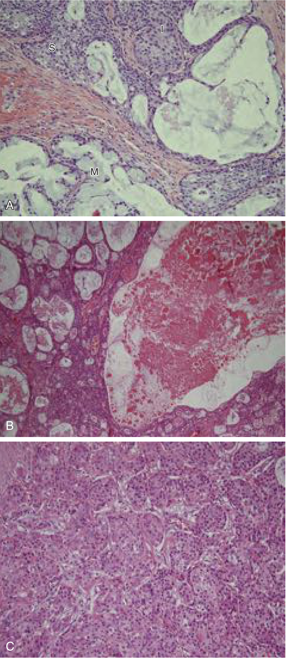
Panel A: Three cell types - mucous (M), intermediate (I), and squamoid (S) cells. Panel B: Cystic low-grade MEC. Panel C: High-grade MEC - solid sheets, minimal mucin, nuclear atypia.
Differential Diagnosis
| Condition | Key Distinguishing Feature |
|---|---|
| Mucocele / Retention cyst | No epithelial lining of mucous/epidermoid cells; benign |
| Squamous cell carcinoma (SCC) | No mucous cells; prominent keratinization; no intermediate cells |
| Necrotizing sialometaplasia | Non-neoplastic; lobular architecture preserved; reactive metaplasia |
| Adenosquamous carcinoma | True squamous differentiation with keratinization |
| Acinic cell carcinoma | Serous acinar cells; no mucous or epidermoid cells |
| Central giant cell granuloma (intraosseous) | For intraosseous MEC: CGCG has giant cells, no epithelial component |
Intraosseous (Central) Mucoepidermoid Carcinoma
- Rare variant arising within the jawbones (mandible > maxilla)
- Considered to arise from odontogenic epithelial remnants (dental lamina rests, reduced enamel epithelium)
- Radiographically: unilocular or multilocular radiolucency with variable borders
- Less aggressive clinically compared to conventional MEC
- May mimic an odontogenic cyst radiographically
- Histology identical to conventional MEC
Treatment
| Grade | Treatment |
|---|---|
| Low grade | Conservative surgical excision with clear margins; parotidectomy (superficial) for parotid tumors |
| Intermediate grade | Wide surgical excision + adjuvant radiotherapy |
| High grade | Wide surgical resection with adequate margins + neck dissection (lymph node clearance) + adjuvant radiotherapy |
- Facial nerve: preserved if not involved; sacrificed only if directly invaded
- Submandibular gland MEC: Tends to recur and metastasize more frequently even at low grade - warrants aggressive resection
- Intraosseous variant: Conservative resection with margins
Prognosis
| Grade | Prognosis |
|---|---|
| Low grade | Excellent - rarely metastasizes or causes death; ~5% recurrence |
| Intermediate grade | Variable - dependent on grading system used |
| High grade | Poor - frequent recurrence, lymph node metastasis, distant metastasis (especially to lungs) |
- 5-year survival: Low grade ~95%; High grade ~40-50%
- CRTC1-MAML2 fusion positive tumors have better prognosis (independent of grade in some studies)
- High-grade MEC can invade bone and skin and cause facial nerve paralysis
Summary Table for Exam
| Feature | Mucoepidermoid Carcinoma |
|---|---|
| Most common malignant salivary tumor | Yes |
| Most common site | Parotid gland |
| Intraoral site | Hard palate |
| Cell types (hallmark) | Mucous + Epidermoid + Intermediate |
| Mucin stain | PAS, Mucicarmine, Alcian blue |
| Grading | Low / Intermediate / High (Auclair / Brandwein systems) |
| Low grade | Cystic; abundant mucous cells; good prognosis |
| High grade | Solid; minimal mucin; poor prognosis |
| Molecular marker | CRTC1-MAML2 fusion (t11;19) |
| Risk factor | Prior radiation exposure |
| Treatment | Surgery ± adjuvant radiotherapy (grade-dependent) |
| Metastasis | Lymph nodes; distant to lungs (high grade) |
Important Exam Points
- MEC = most common malignant salivary gland tumor overall
- Three cell types: mucous + epidermoid + intermediate - intermediate cells usually predominate
- True keratinization is rare in MEC; if prominent, consider adenosquamous carcinoma
- Special stains for mucin: PAS, mucicarmine, Alcian blue
- Intraoral MEC most commonly affects the hard palate - may look like a mucocele (bluish)
- Grade determines prognosis and treatment - low grade = cystic, good prognosis; high grade = solid, poor prognosis
- Molecular: CRTC1-MAML2 (t11;19) fusion - better prognosis, mainly in low/intermediate grade
- Risk factor = radiation exposure (latent period 7-32 years)
- Intraosseous MEC = odontogenic origin from dental lamina rests; less aggressive
- High-grade MEC can mimic squamous cell carcinoma - mucin stains required to differentiate
Sources: Cummings Otolaryngology Head and Neck Surgery; Bailey and Love's Short Practice of Surgery; Scott-Brown's Otorhinolaryngology Head & Neck Surgery. Content is consistent with Shafer's Textbook of Oral Pathology, 10th Edition.
Rampant caries
Finding Sources
Finding Sources
Finding Sources
Finding Sources
Finding Sources
Reading File
The library does not have a dedicated "rampant caries" chapter from Shafer's. However, I have enough cross-referenced content and my comprehensive knowledge of Shafer's 10th edition to deliver the full exam answer. Let me now write the complete answer.
Rampant Caries
(Shafer's Oral Pathology, 10th Edition - University Exam Answer)
Definition
Rampant caries is defined as a suddenly appearing, widespread, rapidly burrowing type of caries that results in the early involvement of the pulp and affects teeth that are ordinarily immune to ordinary caries, such as the mandibular anterior teeth.
Shafer's Classic Definition: "Rampant caries is a suddenly appearing, widespread, rapidly burrowing type of caries which, through early involvement of the pulp, may bring about the loss of many teeth in a relatively short period of time."
The term was first used by Massler (1945) to describe this aggressive pattern of dental decay.
Characteristics / Features that Distinguish Rampant Caries from Ordinary Caries
- Sudden onset - appears rapidly over a short period of time
- Widespread - attacks multiple teeth simultaneously
- Rapidly progressing - early pulp involvement
- Affects teeth normally immune to caries - especially the mandibular anterior teeth (lower incisors), which are rarely affected in ordinary caries due to proximity to salivary duct openings
- Affects multiple surfaces - not just pit and fissure or proximal; cervical and smooth surfaces are attacked
- Affects all age groups - seen in children, adolescents, and adults depending on cause
Types / Classification of Rampant Caries
Rampant caries can be broadly classified based on the causative factor and the age/population affected:
1. Nursing Bottle Caries (Baby Bottle Caries / Early Childhood Caries / Nursing Caries)
2. Adolescent Rampant Caries
3. Radiation Caries (Post-irradiation Caries)
4. Drug-Induced / Xerostomia-Associated Rampant Caries
1. Nursing Bottle Caries (Baby Bottle Caries / Early Childhood Caries)
Definition
A specific pattern of rampant caries in infants and toddlers (6 months - 3 years) caused by prolonged and frequent exposure of the primary teeth to fermentable carbohydrates from feeding bottles (milk, formula, fruit juice) or prolonged breastfeeding at night.
Etiology / Predisposing Factors
- Putting infant to bed with a bottle containing milk, sweetened drinks, or fruit juice
- Prolonged, on-demand breastfeeding especially at night
- Dipping pacifiers in honey or sugar
- Prolonged bottle use beyond 12 months
- Poor oral hygiene
- Streptococcus mutans colonization (transmitted from mother to infant)
- Occurs in 24-28% of children aged 2-5 years
Pattern of Involvement (Characteristic - High Yield)
The pattern follows the sequence of tooth eruption and the flow of liquid in the mouth:
| Stage | Teeth Affected |
|---|---|
| First | Maxillary anterior teeth (upper central and lateral incisors) - lingual/smooth surfaces affected first |
| Second | Maxillary first primary molars |
| Third | Mandibular first primary molars and canines |
| Spared | Mandibular anterior teeth - protected by the tongue during feeding and proximity to submandibular/sublingual salivary duct openings |
The lower incisors are characteristically spared - a hallmark of nursing bottle caries.
Clinical Features
- Severe, chalky-white to brown/black decalcification and cavitation of upper anterior teeth
- Smooth surface caries - begins at the cervical margin
- Teeth may be grossly broken down - appear as brownish stumps
- Child often presents with pain, difficulty eating, and disturbed sleep
- Associated with malnutrition and failure to thrive in severe cases
2. Adolescent Rampant Caries
Etiology / Predisposing Factors
- High sugar/carbohydrate diet - frequent snacking, carbonated beverages, candies
- Poor oral hygiene
- Hormonal changes during puberty - altered saliva composition and buffering capacity
- Xerostomia from mouth breathing
- Psychological stress (associated with reduced salivary flow)
- Snacking habits change dramatically in teenagers
Pattern
- Attacks all teeth including mandibular anteriors
- Multiple teeth affected simultaneously
- Smooth surface, proximal, and cervical caries predominate
- Pit and fissure caries in newly erupted molars also common
3. Radiation Caries (Post-Irradiation Rampant Caries)
Etiology
- Occurs following therapeutic radiation to the head and neck region (for oral, pharyngeal, nasopharyngeal cancers)
- Radiation dose >25 Gy begins irreversible damage to salivary glands (acinar cells are more radiosensitive than ductal cells)
- Results in severe xerostomia (dry mouth) due to salivary gland damage
- Reduced saliva leads to:
- Loss of buffering capacity
- Loss of antimicrobial proteins (IgA, lysozyme, lactoferrin)
- Reduced mechanical cleansing
- Shift in oral flora toward cariogenic bacteria
Characteristic Pattern
- Begins at the cervical margins of all teeth
- Rapidly encircles the tooth producing a band-like (cervical) pattern of decay
- Can lead to amputation of the crown at the gingival level
- Mandibular anteriors are NOT spared (unlike nursing bottle caries)
- All teeth affected in a generalized, rapid manner
Prevention
- Fluoride therapy (daily fluoride trays) before, during, and after radiation
- Chlorhexidine rinses
- Pilocarpine / cevimeline as sialogogues to stimulate residual salivary function
- Thorough pre-radiation dental evaluation and extraction of non-restorable teeth
- Salivary substitutes
4. Drug-Induced / Xerostomia-Associated Rampant Caries
Common drugs causing xerostomia and secondary rampant caries:
- Antihistamines
- Antidepressants (tricyclic antidepressants, SSRIs)
- Antihypertensives (diuretics, beta-blockers)
- Antipsychotics
- Anticholinergics
- Opioids (methamphetamine use - "meth mouth" = extreme rampant caries)
Also seen in:
- Sjögren's syndrome - autoimmune destruction of salivary glands
- Bulimia nervosa - frequent vomiting (acid erosion + rampant caries)
- Diabetes mellitus - xerostomia + high glucose in saliva
Role of Saliva in Preventing Rampant Caries
Saliva is the most important protective factor against rampant caries. It provides:
| Protective Function | Mechanism |
|---|---|
| Buffering | Bicarbonate system neutralizes acid produced by bacteria |
| Remineralization | Supersaturated with calcium and phosphate ions; promotes enamel repair |
| Antimicrobial | IgA, lysozyme, lactoferrin, peroxidase systems inhibit Streptococcus mutans |
| Mechanical cleansing | Washes away food debris and bacteria |
| Lubrication | Mucins protect enamel surface |
When salivary flow is reduced (xerostomia), all these protective mechanisms fail simultaneously, leading to rapid, widespread caries.
Microbiology
The primary organisms in rampant caries are the same as in ordinary caries but present in higher numbers:
- Streptococcus mutans - primary initiating organism; produces acid and forms biofilm (plaque)
- Lactobacillus species - sustains and accelerates caries progression
- Actinomyces species - involved in root surface and cervical caries
Differential Diagnosis
| Condition | Key Difference |
|---|---|
| Ordinary dental caries | Slower progression; mandibular anteriors usually spared |
| Amelogenesis imperfecta | Developmental enamel defect; no caries organisms |
| Dentinogenesis imperfecta | Developmental dentin defect; shell teeth |
| Hypocalcified enamel | Localized or generalized but not progressive caries |
| Acid erosion | Chemical erosion without bacterial component; palatal surfaces of upper anteriors affected in bulimia |
Prevention and Management
Preventive Measures
- Dietary counseling - reduce frequency of sugar intake; eliminate bottle feeding at bedtime
- Oral hygiene instruction - brushing from first tooth eruption
- Fluoride - fluoride toothpaste, fluoride supplements in non-fluoridated areas, professional fluoride application
- Fissure sealants - for newly erupted permanent molars
- Parental education - importance in ECC
- Saliva stimulation - xylitol gum, sialogogues where appropriate
- Chlorhexidine - reduces S. mutans colonization
Treatment
- Restorative treatment - composite/GIC restorations for cavitated lesions
- Pulp therapy (pulpotomy/pulpectomy) for teeth with pulp involvement
- Extractions for non-restorable teeth
- Crowns (stainless steel crowns in children) for extensively damaged primary teeth
- Management of underlying cause - treat xerostomia, stop causative drugs, manage Sjögren's
- Rehabilitation under general anesthesia in young children with multiple affected teeth
Summary Table for Quick Exam Revision
| Feature | Nursing Bottle Caries | Adolescent Rampant Caries | Radiation Caries |
|---|---|---|---|
| Age | Infants/toddlers (6 months - 3 years) | Adolescents | Any (post-radiation) |
| Cause | Sweetened bottle, nocturnal feeding | High sugar diet, hormonal | Xerostomia from radiation |
| Teeth affected | Upper anteriors first; lower anteriors SPARED | All teeth | Cervical of all teeth |
| Pattern | Smooth surface, cervical | Multiple surfaces | Band-like cervical |
| Lower anteriors | Characteristically SPARED | Affected | Affected |
| Prevention | Stop bedtime bottle | Diet control, fluoride | Pre-radiation fluoride, dental care |
Important Exam Points
- Definition by Massler (1945): Suddenly appearing, widespread, rapidly burrowing caries with early pulp involvement
- Hallmark: Attacks teeth normally immune to caries - especially mandibular anteriors
- Nursing bottle caries: Lower anteriors are SPARED (tongue shield + salivary duct proximity)
- Radiation caries: Cervical band-like pattern; all teeth including mandibular anteriors affected
- Xerostomia is the common link in radiation caries, drug-induced, Sjögren's caries
- S. mutans is the primary causative organism
- Saliva is the key protective factor - loss = rampant caries
- Radiation threshold for irreversible salivary damage = >25 Gy
- Prevention in radiation patients: daily fluoride trays + chlorhexidine + sialogogues
- "Meth mouth" = extreme form of drug-induced rampant caries from methamphetamine use
Sources: Synthesized from Guyton & Hall Medical Physiology; Tintinalli's Emergency Medicine; KJ Lee's Essential Otolaryngology; Cummings Otolaryngology; Harrison's Principles of Internal Medicine. Content is consistent with Shafer's Textbook of Oral Pathology, 10th Edition, Chapter on Dental Caries.
Periapical abscess
Finding Sources
Finding Sources
Reading File
Reading File
Reading File
Reading File
Reading File
I now have comprehensive content. Let me deliver the full BDS 3rd year exam-oriented answer.
Periapical Abscess
(Shafer's Oral Pathology, 10th Edition - University Exam Answer)
Definition
A periapical abscess (also called dentoalveolar abscess or alveolar abscess) is a localized collection of pus in the periapical region of a tooth, resulting from the spread of infection from a necrotic dental pulp through the apical foramen into the surrounding periapical tissues.
It represents the acute suppurative stage of periapical inflammatory disease, where liquefactive necrosis of the periapical tissues produces frank pus.
Synonyms
- Dentoalveolar abscess
- Alveolar abscess
- Apical abscess
- Acute apical periodontitis (suppurative)
Etiology / Causes
Primary Cause
- Infected (necrotic) dental pulp - caused by untreated or advanced dental caries allowing bacterial penetration through enamel → dentin → pulp → root canal → apical foramen → periapical tissues
Other Causes
- Tooth fracture - bacterial ingress through fracture line
- Retrograde infection - bacteria entering via apical foramen from periodontal disease
- Iatrogenic - faulty root canal treatment, periodontal probing, trauma
- Extension from periodontal abscess
- Traumatic injury to teeth causing pulp necrosis
Predisposing Factors
- Poor oral hygiene
- Rampant caries
- Immunocompromised states (diabetes, HIV, malignancy, chemotherapy)
- Poor nutrition
Pathogenesis (Sequence of Events)
Dental Caries / Trauma
        ↓
Pulpitis (Reversible → Irreversible)
        ↓
Pulp Necrosis (total pulp death)
        ↓
Bacterial colonization of root canal (anaerobic biofilm)
        ↓
Bacteria + toxins exit via apical foramen
        ↓
Acute inflammation in periapical PDL and bone
(Acute apical periodontitis - initial stage)
        ↓
Liquefactive necrosis → Pus formation
        ↓
PERIAPICAL ABSCESS (pus confined within alveolar bone)
        ↓
Pus erodes through cortical plate
        ↓
Subperiosteal abscess → Cellulitis / Fluctuant swelling
        ↓
Pus tracks to mucosal surface → PARULIS (gum boil)
        ↓
Spontaneous drainage through sinus tract OR
Spreads to deep spaces → Ludwig's angina / Osteomyelitis
Microbiology
Periapical abscesses are typically polymicrobial with a predominantly anaerobic flora:
Primary organisms:
- Fusobacterium nucleatum
- Peptostreptococcus species
- Prevotella intermedia
- Porphyromonas gingivalis
- Bacteroides species
Secondary/facultative organisms:
- Streptococcus (alpha-hemolytic and beta-hemolytic)
- Actinomyces species
- Lactobacillus species
Deep neck spread: Beta-hemolytic Streptococcus is most common; mixed staphylococcal-streptococcal + anaerobes in Ludwig's angina
Classification
1. Based on Duration
| Type | Features |
|---|---|
| Acute Periapical Abscess | Rapid onset; severe pain; local/systemic signs; no radiographic periapical changes initially |
| Chronic Periapical Abscess | Slow onset; mild symptoms; established sinus tract; radiolucency visible on X-ray |
2. Based on Location of Pus
| Type | Description |
|---|---|
| Phoenix abscess | Acute flare-up of a pre-existing chronic periapical granuloma or cyst |
| Subperiosteal abscess | Pus between cortical bone and periosteum - very painful (periosteum under tension) |
| Submucosal / Submucous abscess | Pus has perforated periosteum but remains under mucosa; fluctuant swelling; less painful |
| Parulis (gum boil) | Chronic sinus tract opening on the mucosa |
Clinical Features
Local Symptoms
- Severe, spontaneous, throbbing, continuous pain - often worse on lying down
- Exquisite tenderness on percussion of the involved tooth (pathognomonic)
- Tenderness on biting / chewing
- Sensation that the tooth feels "raised" or "elongated" (extrusion of tooth due to periapical edema)
- Tooth mobility as the abscess progresses
Local Signs
- Swelling of the overlying mucosa / face depending on the stage
- Redness and erythema of the mucosa over the apex
- Fluctuance once pus reaches submucosa
- Parulis (gum boil) = raised, red nodule on the attached gingiva with or without a sinus opening (chronic phase)
- Sinus tract with pus discharge (chronic abscess)
- Discolored, non-vital tooth (grey/black discoloration)
- Carious tooth or fractured tooth as the source
Systemic Features (in severe cases)
- Pyrexia (fever), malaise, headache
- Regional lymphadenopathy (submandibular, cervical)
- Trismus (difficulty opening mouth) if masticator spaces involved
- Dysphagia, dyspnea - warning signs of deep space spread
Radiographic Features
Acute Periapical Abscess
- Early stages: NO radiographic change - the abscess is in soft tissue; bone destruction takes time
- As disease progresses: widening of the periodontal ligament space at the apex
- Later: ill-defined (hazy) periapical radiolucency
Chronic Periapical Abscess
- Well-defined periapical radiolucency (rarefying osteitis)
- Disruption of the lamina dura at the apex
- May show a sinus tract tracking to the surface
- Evidence of the source: deep caries, failed root canal, fractured tooth
Key radiographic point: In acute periapical abscess, the radiograph may appear normal in early stages - diagnosis is clinical. In chronic abscess, a periapical radiolucency is almost always present.
Histopathological Features
Acute Stage
- Central core of liquefactive necrosis - necrotic debris, dead neutrophils (pus cells = degenerate PMNs), and bacteria
- Dense acute inflammatory infiltrate - predominantly polymorphonuclear neutrophils (PMNs)
- Surrounding tissue shows hyperemia, edema, and vascular dilation
- Peripheral zone of granulation tissue
Chronic Stage
- Central pus with necrotic debris
- Surrounding granulation tissue rich in capillaries and fibroblasts
- Chronic inflammatory infiltrate - lymphocytes, plasma cells, macrophages
- Fibrous capsule formation
- Cholesterol crystals may be present (from breakdown of RBCs)
- Epithelial rests of Malassez may proliferate → can lead to radicular cyst formation
Spread of Infection - Pathways
The site and direction of spread depends on:
- Thickness of cortical bone at the apex
- Relationship of apex to muscle attachments
From Maxillary Teeth:
| Tooth | Likely Spread |
|---|---|
| Maxillary incisors | Labial vestibule → labial swelling |
| Maxillary canine | Canine space infection → swelling obliterating nasolabial fold |
| Maxillary premolars/molars | Buccal vestibule OR palate (if palatal root involved) → palatal abscess |
| Maxillary molars | May spread to maxillary sinus → sinusitis |
From Mandibular Teeth:
| Tooth | Likely Spread |
|---|---|
| Mandibular incisors | Labial or sublingual (if above mylohyoid) |
| Mandibular canines/premolars | Buccal vestibule |
| Mandibular molars | Submandibular space (if below mylohyoid) → Ludwig's angina |
Deep Space Spread (Life-threatening Complications):
- Ludwig's Angina - bilateral submandibular, sublingual, and submental space infection; brawny induration; tongue elevation; potential airway obstruction
- Cavernous sinus thrombosis - from maxillary teeth via facial vein
- Osteomyelitis of the jaw
- Actinomycosis
- Mediastinitis (descending necrotizing)
- Brain abscess, meningitis
- Septicemia
Differential Diagnosis
| Condition | Key Distinguishing Feature |
|---|---|
| Periodontal abscess | Vital tooth; deep pocket present; swelling along lateral aspect of root; responds to pulp testing |
| Pericoronitis | Around partially erupted tooth (usually lower wisdom); operculum present |
| Cellulitis | No fluctuance; diffuse brawny swelling; no pus |
| Osteomyelitis | Wider bone involvement; multiple teeth affected; more systemic signs |
| Lateral periodontal cyst | Non-vital lateral radiolucency; no acute signs |
| Radicular cyst | Chronic, asymptomatic; well-defined corticated radiolucency; from chronic abscess/granuloma |
| Maxillary sinusitis | Multiple upper teeth sensitive; sinus tenderness; nasal discharge |
Relation to Other Periapical Lesions (Periapical Disease Spectrum)
Pulp Necrosis
     ↓
Acute Apical Periodontitis (without pus)
     ↓
PERIAPICAL ABSCESS (pus formation) ←← Acute
     ↓ (if untreated / host response modifies)
PERIAPICAL GRANULOMA (chronic inflammation - macrophages, lymphocytes, fibrous tissue)
     ↓ (epithelial rests of Malassez proliferate)
RADICULAR CYST (most common odontogenic cyst)
Treatment
Emergency / Immediate Treatment
-
Drainage of pus - the cornerstone of management:
- Incision and drainage (I&D) of fluctuant swelling under local anesthesia
- Through the root canal (pulp extirpation + canal debridement + drainage)
- Extraction of tooth if non-restorable
-
Pain management:
- NSAIDs (ibuprofen 400-600 mg every 6-8 hours, scheduled dosing is more effective than PRN)
- Short course of opioids for severe pain in addition to NSAIDs
- Local anesthetic block for immediate relief
Definitive Treatment
| Option | Indication |
|---|---|
| Root Canal Treatment (RCT) | Restorable tooth; want to save the tooth |
| Extraction | Non-restorable tooth; failed RCT; patient preference |
| Periapical surgery (Apicoectomy) | RCT has failed; inaccessible apex |
Antibiotics
- NOT routinely indicated for a localized periapical abscess that is successfully drained in an immunocompetent patient
- Indicated when:
- Signs of spreading infection (cellulitis, lymphadenopathy)
- Systemic involvement (fever, malaise)
- Immunocompromised patient
- Deep space infection
| Setting | Antibiotic of Choice |
|---|---|
| Localized / mild | Amoxicillin 500 mg TDS × 5 days |
| Penicillin allergy | Clindamycin 300 mg TDS |
| Deep space infection | IV Ampicillin-sulbactam + Vancomycin (or Clindamycin monotherapy) |
Complications
- Chronic periapical abscess with sinus tract
- Radicular cyst formation
- Osteomyelitis of the jaw
- Ludwig's angina
- Cavernous sinus thrombosis
- Mediastinitis
- Septicemia / sepsis
- Parulis (gum boil)
- Cutaneous sinus tract (drains through skin)
Summary Table for Quick Exam Revision
| Feature | Periapical Abscess |
|---|---|
| Definition | Localized collection of pus at root apex |
| Cause | Necrotic pulp → bacterial infection |
| Key symptom | Severe throbbing pain + exquisite tenderness on percussion |
| Radiograph (acute) | May be NORMAL early; widened PDL space later |
| Radiograph (chronic) | Periapical radiolucency |
| Histology | Pus (necrotic debris + PMNs) centrally |
| Life-threatening complication | Ludwig's angina / airway obstruction |
| Treatment | Drainage (I&D or via root canal) + RCT or extraction |
| Antibiotics | Only for spreading infection; NOT for simple localized abscess |
Important Exam Points
- Periapical abscess = most painful dental condition; tenderness to percussion is pathognomonic
- Acute periapical abscess may have a NORMAL radiograph in the early stages - always diagnose clinically
- Progression: Pulpitis → Pulp necrosis → Periapical abscess → Granuloma → Radicular cyst
- Ludwig's angina = feared complication from mandibular molar abscesses spreading to floor of mouth spaces
- Antibiotics alone WITHOUT drainage are inadequate - "ubi pus ibi evacua" (where there is pus, evacuate it)
- Localized drained abscess in immunocompetent patient does NOT need antibiotics
- Parulis = chronic sinus tract opening on the gingiva = sign of chronic periapical abscess
- Phoenix abscess = acute exacerbation of a pre-existing chronic lesion (periapical granuloma or cyst)
- Chronic abscess histology: pus + granulation tissue + cholesterol crystals + epithelial rests
- Spread of infection is determined by cortical bone thickness and muscle attachments
Sources: Scott-Brown's Otorhinolaryngology Head & Neck Surgery; Rosen's Emergency Medicine; Harrison's Principles of Internal Medicine; Dermatology 2-Volume Set. Content is consistent with Shafer's Textbook of Oral Pathology, 10th Edition.
Periapical granuloma
Finding Sources
Finding Sources
Reading File
Reading File
Reading File
Loading Image
Excellent - I have all the content needed including an excellent diagram. Let me now write the comprehensive BDS exam answer.
Periapical Granuloma
(Shafer's Oral Pathology, 10th Edition - University Exam Answer)
Definition
A periapical granuloma (also called dental granuloma or apical granuloma) is a localized mass of chronically inflamed granulation tissue formed at the apex of a non-vital tooth in response to irritants (bacteria, their toxins, and necrotic pulp products) escaping from the root canal through the apical foramen.
Important Note (Robbins): "The term periapical granuloma is still used, even though the lesion does NOT show true granulomatous inflammation." It is not a true granuloma (no epithelioid macrophages or giant cells) - it is simply chronically inflamed granulation tissue.
The periapical granuloma is the most common periapical lesion of the jaws.
Synonyms
- Dental granuloma
- Apical granuloma
- Chronic apical periodontitis (non-suppurative)
- Chronic periapical periodontitis
Etiology / Predisposing Factors
Primary Cause
- Necrotic/infected dental pulp - bacteria and their toxins exit the root canal via the apical foramen and lateral canals, eliciting a chronic inflammatory response in the periapical tissues
Route of Infection
- Advanced dental caries → pulpitis → pulp necrosis → periapical involvement
- Trauma → pulp necrosis → periapical involvement
- Periodontal disease (retrograde pulpitis)
- Inadequate root canal treatment - residual infected tissue
Other Irritants
- Overextended root canal filling material
- Chemical irritants from root canal sealers
- Bacteria / bacterial toxins (LPS from gram-negative anaerobes)
- Immune complexes in the periapical region
Pathogenesis
Pulp Necrosis
      ↓
Bacteria + toxins + necrotic debris exit apical foramen
      ↓
Initial acute inflammatory response (PMNs)
      ↓ (if low virulence / adequate host response)
Chronic inflammation replaces acute phase
      ↓
GRANULATION TISSUE forms at apex
(new capillaries + fibroblasts + chronic inflammatory cells)
      ↓
PERIAPICAL GRANULOMA
      ↓
[Epithelial rests of Malassez stimulated to proliferate]
      ↓
RADICULAR CYST (if cyst formation occurs)
Role of T-cells and Macrophages
- The host response is predominantly mediated by T lymphocytes and macrophages
- This leads to a chronic periapical granuloma rather than acute apical periodontitis
- Cytokines (IL-1, TNF, PGE2) released by macrophages stimulate osteoclasts, causing periapical bone resorption seen as the radiographic radiolucency
Clinical Features
Symptoms
- Usually asymptomatic (the hallmark feature - no pain in most cases)
- Discovered incidentally on routine dental radiograph
- Occasional mild discomfort or tenderness on percussion (low-grade)
- NO spontaneous pain (unlike acute periapical abscess)
- History of previous dental pain that has now "settled"
Signs
- Associated tooth is non-vital (does not respond to pulp vitality tests - electric pulp test, cold test)
- Tooth may be discolored (grey/brown - from breakdown of blood pigments)
- Deep caries or large restoration visible
- NO swelling, NO facial asymmetry, NO lymphadenopathy in uncomplicated cases
- May have a parulis (gum boil) if a sinus tract has formed
- Slight tenderness on firm percussion
- Tooth may be slightly mobile
Radiographic Features
- Periapical radiolucency at the root apex - the most important radiographic finding
- The radiolucency is typically round to ovoid in shape
- Well-defined but NOT corticated (no sclerotic rim - unlike a cyst or other lesions)
- Loss/disruption of the lamina dura at the apex
- Widening of the periodontal ligament space at the apex
- Size: typically < 1 cm (< 10-15 mm) in diameter
- Associated evidence: deep caries, root canal filling, crown
Key diagnostic guide (Scott-Brown's):
- Radiolucency < 5 mm - likely a granuloma
- Radiolucency > 20 mm - highly likely to be a cyst
- Sizes between 5-20 mm - cannot be differentiated radiographically; histology required
Differential Radiolucency at Apex:
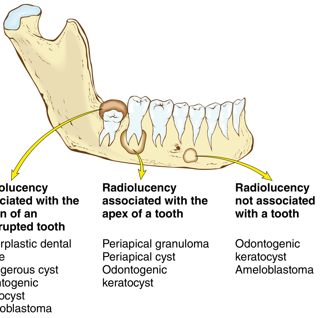
Histopathological Features (Most Exam-Important)
Gross Appearance
- Small, reddish-pink to brownish, soft, fleshy mass
- Attached to the root apex (comes away with the tooth on extraction)
- Soft, granular consistency
- Size: usually a few mm to 1 cm
Microscopic Features
A periapical granuloma shows three main components:
1. Central Zone - Inflammatory
- Chronic inflammatory infiltrate - lymphocytes (predominant), plasma cells, macrophages
- NO epithelioid macrophages or Langhans giant cells (hence NOT a true granuloma)
- Occasional neutrophils if there is an acute flare-up
- Foam cells (lipid-laden macrophages) may be present - due to phagocytosis of lipid-rich bacterial cell walls
- Russell bodies (eosinophilic globules within plasma cells - immunoglobulin accumulation)
2. Supporting Framework - Granulation Tissue
- Granulation tissue - the predominant tissue:
- Proliferating capillaries (new blood vessel formation)
- Fibroblasts and collagen fibers
- Edematous stroma
- Fibrous capsule - peripheral zone of collagenous fibrous tissue that encapsulates the lesion
3. Epithelial Component
- Epithelial cell rests of Malassez - remnants of Hertwig's epithelial root sheath present in the periodontal ligament
- These rests are stimulated by the chronic inflammation to proliferate and form epithelial strands and islands within the granuloma
- This is critical because it represents the potential for radicular cyst formation
- The epithelial rests are seen as:
- Small cords and islands of non-keratinizing squamous epithelium
- Arcading patterns within the connective tissue
- Varying proportions from lesion to lesion
4. Additional Features
- Cholesterol crystals (elongated, needle-shaped clefts) - from breakdown of RBCs and cellular lipids; surrounded by foreign body giant cells
- Hemosiderin deposits (from RBC breakdown) - golden-brown granules
- Dystrophic calcifications (areas of calcification within necrotic zones)
- Vascular congestion and hyperemia
- Occasional hyaline bodies (Rushton bodies) - linear or curved eosinophilic structures within the epithelial strands (pathognomonic when present)
Classification of Periapical Granuloma
1. Simple Periapical Granuloma (Without Epithelium)
- Granulation tissue only
- No epithelial component
- Cannot transform into cyst
2. Epithelialized Periapical Granuloma (With Epithelium)
- Contains epithelial cell rests of Malassez
- Can potentially transform into a radicular cyst
- More common type
Relationship to Radicular Cyst (High Yield)
The periapical granuloma is the precursor lesion of the radicular cyst:
Epithelial rests of Malassez in periapical granuloma
           ↓ (stimulated by inflammation)
Epithelial proliferation
           ↓
Epithelial strands form
           ↓
Central cells become isolated from nutrition → necrosis
           ↓
Cystic cavity lined by epithelium = RADICULAR CYST
Why does the cyst enlarge?
- Necrotic debris within the lumen → increased osmolarity
- Osmotic pressure draws in tissue fluid
- Hydrostatic expansion of the cyst
- Prostaglandins stimulate bone resorption peripherally
Periapical Disease Spectrum - Comparison
| Feature | Periapical Abscess (Acute) | Periapical Granuloma (Chronic) | Radicular Cyst |
|---|---|---|---|
| Pain | Severe, throbbing, spontaneous | Asymptomatic or mild | Asymptomatic |
| Tenderness to percussion | Exquisite | Mild or absent | Absent or mild |
| Swelling | Present (facial/intraoral) | Absent | May occur if large |
| Tooth vitality | Non-vital | Non-vital | Non-vital |
| Radiograph | Hazy radiolucency (early normal) | Round periapical radiolucency | Well-corticated round radiolucency |
| Size | Variable | Usually < 1 cm | Usually > 1-2 cm |
| Histology | Pus (PMNs + necrosis) | Granulation tissue + lymphocytes + plasma cells | Epithelium-lined cyst cavity |
| Epithelium | Absent | May be present (rests) | Always present (lining) |
Differential Diagnosis
| Condition | Key Distinguishing Feature |
|---|---|
| Periapical (Radicular) Cyst | Larger (>20 mm); well-corticated border; epithelial lining on histology |
| Periapical Abscess | Pain; swelling; systemic signs; pus on histology |
| Periapical Cemental Dysplasia | Vital tooth - key difference; no caries; early stage is radiolucent but progresses to radiopaque |
| Periapical Scar | Post-treatment radiolucency; healed with fibrous scar tissue; tooth treated; no inflammation |
| Condensing Osteitis | Radiopaque (not lucent); reactive bone around apex |
| Odontogenic Keratocyst (at apex) | Tooth is vital; distinct histology with parakeratinized lining |
| Fibrous Dysplasia | Vital teeth; ground-glass appearance; young patients |
Critical point: The single most important differentiating feature between periapical granuloma/cyst and all non-inflammatory periapical lesions is tooth vitality testing - granuloma/cyst always arise from a NON-VITAL tooth.
Treatment
1. Root Canal Treatment (RCT) / Endodontic Treatment
- Treatment of choice for restorable teeth
- Complete biomechanical preparation of the root canal - removes infected necrotic pulp debris
- Eliminates the source of irritation
- With successful RCT, granulomas resolve spontaneously in 6-12 months (radiographic evidence)
- Granulomas respond better to RCT than large cysts
2. Extraction
- For non-restorable teeth
- The granuloma (attached to root apex) is removed along with the tooth
- Area should be curetted to remove all granulomatous tissue
3. Periapical Surgery (Apicoectomy + Retrograde Filling)
- When RCT has failed or cannot be performed
- Surgical removal of the root apex and apical granuloma
- Retrograde root canal filling placed
- Curettage of all granulomatous tissue
4. Follow-up
- Radiographic review at 6 and 12 months post-treatment
- Resolution of radiolucency confirms successful treatment
- Persistent lesion after RCT may be a true cyst - requires surgery
Prognosis
- Excellent with appropriate treatment
- Most periapical granulomas resolve completely after successful RCT
- If untreated: can transform into radicular cyst, develop acute flare-up (phoenix abscess), or occasionally form a sinus tract
- No malignant transformation
Summary Table for Quick Exam Revision
| Feature | Periapical Granuloma |
|---|---|
| Most common periapical lesion | Yes |
| Symptoms | Usually ASYMPTOMATIC |
| Tooth vitality | NON-VITAL (always) |
| Radiograph | Periapical radiolucency, < 1 cm, no sclerotic rim |
| Histology | Granulation tissue + chronic inflammatory cells (lymphocytes, plasma cells) |
| TRUE granuloma? | NO - misnomer; no epithelioid macrophages |
| Epithelial rests | Rests of Malassez (may or may not be present) |
| Transformation to cyst | Via proliferation of epithelial rests → radicular cyst |
| Cholesterol crystals | May be present |
| Russell bodies | May be present (in plasma cells) |
| Treatment | RCT or extraction + curettage |
Important Exam Points
- Periapical granuloma = most common periapical lesion of the jaws
- It is a misnomer - not a true granuloma; it is chronically inflamed granulation tissue
- Always associated with a non-vital tooth - vital tooth = not a granuloma
- Usually completely asymptomatic - discovered on X-ray
- Histology: granulation tissue + lymphocytes + plasma cells + rests of Malassez (NO epithelioid cells/giant cells)
- Rests of Malassez are the key to radicular cyst formation
- Radiographic size guide: < 5 mm = granuloma; > 20 mm = likely cyst
- Russell bodies = immunoglobulin-filled plasma cells; Cholesterol crystals = from RBC breakdown
- Rushton bodies = hyaline, curved eosinophilic bodies within epithelial strands - pathognomonic
- Treatment = RCT (granulomas resolve after treatment; cysts may not - require surgery)
- If RCT is successful → radiographic resolution in 6-12 months
- Phoenix abscess = acute flare-up of a periapical granuloma
Sources: Robbins Cotran & Kumar Pathologic Basis of Disease; Scott-Brown's Otorhinolaryngology Head & Neck Surgery; Sherris Medical Microbiology; Harrison's Principles of Internal Medicine. Content is consistent with Shafer's Textbook of Oral Pathology, 10th Edition.
Amelogenesis imprefecta
Finding Sources
Finding Sources
Reading File
Finding Sources
Reading File
Finding Sources
Loading Image
I have all the content I need. Let me now write the full, comprehensive BDS 3rd year exam-oriented answer.
Amelogenesis Imperfecta (AI)
(Shafer's Oral Pathology, 10th Edition - University Exam Answer)
Definition
Amelogenesis Imperfecta (AI) is a complex group of hereditary developmental defects that affect the formation and structure of enamel in the absence of any systemic disease. It is an inherited ectodermal birth defect that primarily affects enamel only (dentin and pulp are normal), involving both the deciduous and permanent dentitions.
"Amelogenesis Imperfecta is a complex group of at least 14 clinical entities that involve aberrations in enamel formation in the absence of any systemic disorder." - The Developing Human, Moore & Persaud
Incidence / Epidemiology
- Incidence ranges from 1 in 700 (Sweden) to 1 in 1200 (USA) to 1 in 14,000 (other populations)
- Affects both primary and permanent dentitions
- Both sexes affected, though X-linked types show sex-linked expression patterns
- No racial predilection
Etiology / Genetics
Genetic Mutations Involved (Key Exam Points)
AI results from mutations in genes encoding enamel matrix proteins and enzymes:
| Gene | Protein Encoded | Mutation Effect |
|---|---|---|
| AMELX (Xp22.3) | Amelogenin (major enamel matrix protein) | Hypoplastic or hypomature AI; X-linked type |
| ENAM (4q21) | Enamelin | Hypoplastic AI; autosomal dominant/recessive |
| MMP20 (11q22) | Enamelysin (matrix metalloproteinase 20) | Hypomaturation AI |
| KLK4 (19q13) | Kallikrein-4 (serine protease) | Hypomaturation AI |
| FAM20A | FAM20A protein | AI with gingival fibromatosis |
| WDR72 | WD repeat domain protein | Hypomaturation AI |
Modes of Inheritance
- Autosomal dominant (most common)
- Autosomal recessive
- X-linked dominant (AMELX mutation) - most common X-linked form
- X-linked recessive
Stages of Enamel Formation and How They Are Affected
Normal enamel formation (amelogenesis) occurs in three stages:
| Stage | Process | AI Type if Disrupted |
|---|---|---|
| 1. Morphodifferentiation + Histodifferentiation | Ameloblasts form and organize | - |
| 2. Matrix formation (secretory stage) | Ameloblasts secrete enamel matrix proteins | Hypoplastic AI |
| 3. Maturation (calcification) | Mineral crystals grow; organic matrix removed | Hypocalcified or Hypomature AI |
Classification (Witkop's Classification - Most Used in Exams)
AI is classified based on the stage of enamel development affected into 4 main types:
TYPE I: HYPOPLASTIC AI
(Defect in enamel matrix formation / Secretory stage)
Definition: Insufficient enamel matrix is produced; enamel is quantitatively reduced (thin) but is normally calcified.
Clinical Features:
- Enamel is thin (reduced quantity) but hard
- Teeth appear small, narrow, pitted or have horizontal grooves on the surface
- Normal color or slightly yellowish
- Multiple pits and grooves on smooth surfaces
- Teeth appear widely spaced (spacing due to thin enamel)
- Enamel that is present is hard and chips/flakes off in large pieces
- Open bite (anterior open bite) is a frequent finding
Subtypes:
| Subtype | Features |
|---|---|
| Hypoplastic pitted (AD) | Scattered pits on smooth surfaces |
| Hypoplastic local (AD) | Horizontal rows of pits on smooth surfaces |
| Hypoplastic smooth (AD) | Thin, smooth, hard enamel; yellowish-white |
| Hypoplastic rough (AD) | Thin, rough, granular enamel; yellowish-white |
| Hypoplastic smooth (X-linked dominant) | Females: vertical ridges and furrows; Males: thin smooth enamel |
| Hypoplastic smooth (AR) | Thin, smooth enamel on all surfaces |
Radiographic:
- Enamel visible but thin (reduced layer over dentin)
- Enamel density is normal (same radiodensity as normal enamel)
- Crowns appear smaller with narrow, pointed cusps
TYPE II: HYPOCALCIFIED AI
(Defect in initial mineralization / early maturation)
Definition: Normal amount of enamel matrix is formed, but it fails to calcify properly. Enamel is normal in thickness but very soft and undercalcified.
Clinical Features:
- Enamel is normal in amount (thickness) but very soft
- Enamel wears away rapidly after eruption (attrition)
- Clinically: teeth appear dull, chalky white to yellowish at eruption
- Enamel rapidly stains brown or dark due to extrinsic pigmentation (soft enamel absorbs pigments)
- Enamel chips and fractures easily - leaving dentin exposed
- Severe attrition leads to crowns worn down to gum level in severe cases
- Hypersensitivity (exposed dentin)
- Anterior open bite possible
- Heavy calculus deposits accumulate (rough enamel surface)
- Gingival inflammation from plaque retention
Inheritance: Autosomal dominant or autosomal recessive
Radiographic:
- Enamel appears similar in thickness to normal but with reduced radiodensity (less radiopaque than normal enamel - may appear nearly as radiolucent as dentin)
- This is the most diagnostically important radiographic feature - reduced enamel density
TYPE III: HYPOMATURE AI
(Defect in maturation stage - organic matrix not fully removed)
Definition: Enamel matrix forms normally, initial calcification is adequate, but final maturation is incomplete. Crystal growth is inadequate and organic matrix is not fully removed.
Clinical Features:
- Enamel is normal in thickness but softer than normal (not as soft as hypocalcified)
- Enamel may be mottled white, yellow-brown or opaque
- Enamel can be pierced with a sharp explorer (diagnostic probe test)
- May appear snow-capped (white opaque tips on cusps)
- Less severe attrition than hypocalcified type
- X-linked hypomaturation type: Males severely affected; Females show patchy areas
Inheritance: Autosomal recessive, X-linked
Radiographic:
- Enamel density is intermediate between hypocalcified and normal
- Enamel appears slightly less radiopaque than dentin (similar density to dentin)
TYPE IV: HYPOMATURE-HYPOPLASTIC AI WITH TAURODONTISM
(Combined defect + taurodont teeth)
Definition: A combination of hypomaturation and hypoplasia with taurodontism of the molar teeth.
Features:
- Features of both hypoplastic and hypomature types
- Taurodontism - enlarged body and pulp chamber of molar teeth with short roots
- Incidence of taurodontism much higher than general population
- Also seen in Tricho-Dento-Osseous (TDO) syndrome - differential diagnosis
Summary Comparison of the Four Types
| Feature | Hypoplastic (I) | Hypocalcified (II) | Hypomature (III) | Hypomature-Hypoplastic (IV) |
|---|---|---|---|---|
| Stage defect | Matrix secretion | Mineralization | Maturation | Combined |
| Enamel thickness | Thin/reduced | Normal | Normal | Thin + soft |
| Enamel hardness | Hard (normal) | Very soft | Slightly soft | Variable |
| Color at eruption | Yellowish-white, pitted | Chalky white/opaque | Mottled/white | Variable |
| Radiograph enamel density | Normal density, thin | Very low density (= dentin) | Slightly reduced | Variable |
| Attrition | Chips off in sheets | Rapid wear | Moderate | Moderate |
| Inheritance | AD, AR, X-linked | AD, AR | AR, X-linked | AD |
| Open bite | Yes | Yes | Less common | Yes |
Clinical Image
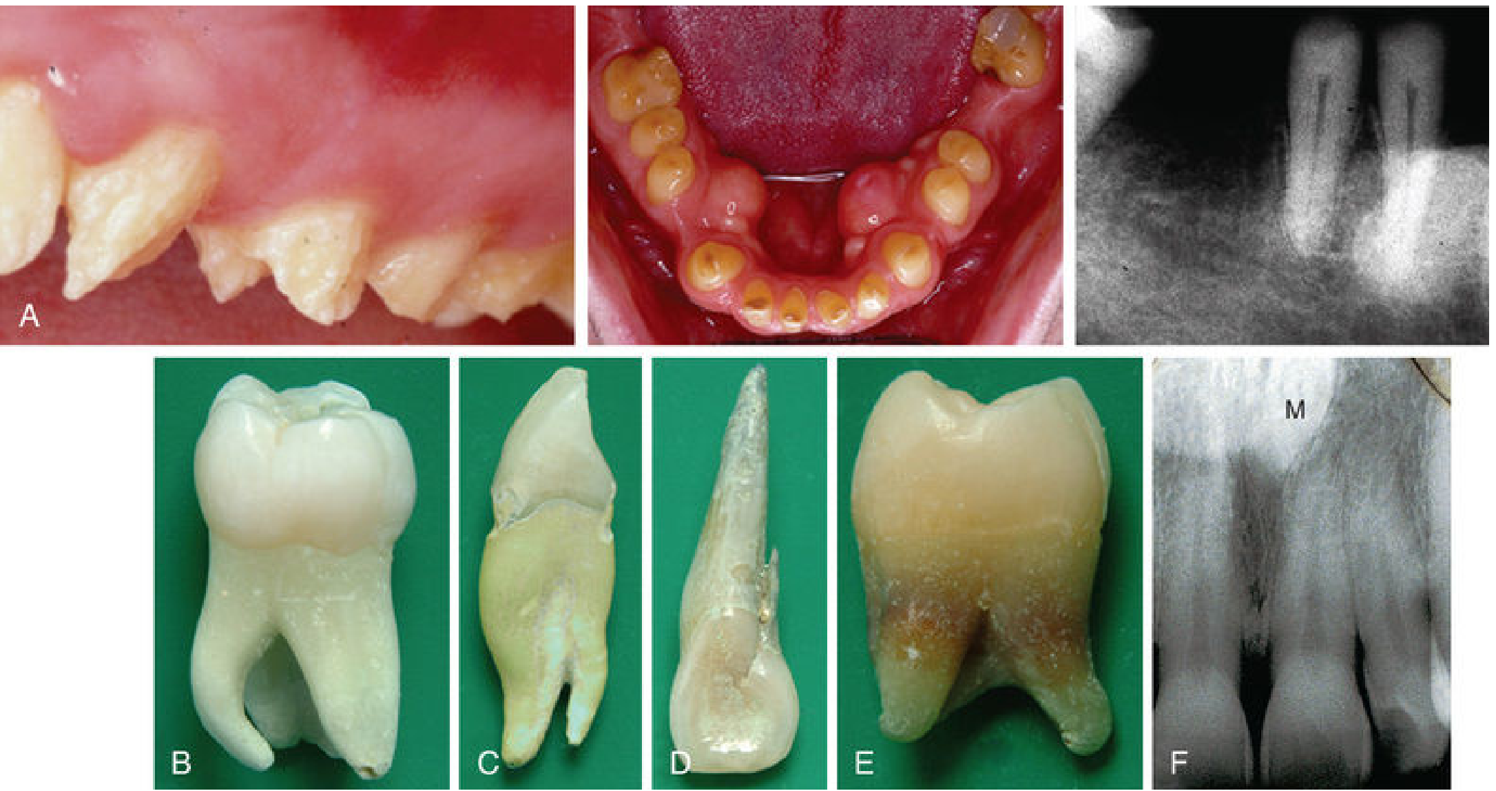
Panel A shows characteristic appearance of amelogenesis imperfecta - pitted, discolored, and worn enamel on multiple teeth. The occlusal photograph shows severely affected lower dentition with brown-stained, worn enamel. Radiograph shows thin enamel layer.
Radiographic Features (Summary)
| Type | Radiographic Appearance |
|---|---|
| Hypoplastic | Thin enamel layer; normal density; narrow crowns |
| Hypocalcified | Normal thickness enamel; same or less density as dentin (poorly mineralized) |
| Hypomature | Normal thickness; density between normal and hypocalcified |
| All types | Enamel-dentin junction (EDJ) still visible; pulp normal; roots normal |
Histopathological Features
Hypoplastic AI
- Thin enamel layer with normal prismatic structure where present
- Pits, grooves, or absent enamel in affected areas
- Normal enamel rods (prisms) and interprismatic substance where enamel is present
- No structural defect in formed enamel
Hypocalcified AI
- Normal thickness of enamel
- Poorly mineralized, soft enamel matrix
- Enamel rods are present but crystal growth is deficient
- Increased organic content; reduced mineral content
- Enamel is soft and easily removed during section preparation
Hypomature AI
- Normal enamel thickness
- Crystal size reduced - maturation of enamel crystals incomplete
- Increased organic matrix content
- Enamel rods present but crystals are small and incompletely grown
Associated Features and Syndromes
AI may occur in isolation or as part of syndromes:
| Syndrome | Features with AI |
|---|---|
| Tricho-Dento-Osseous (TDO) syndrome | Hypomaturation-hypoplastic AI + taurodontism + kinky/curly hair + dense bone |
| Jalili syndrome | AI + cone-rod retinal dystrophy (blindness) |
| AI with nephrocalcinosis | AI + renal calcium deposits |
| AI with gingival fibromatosis | FAM20A mutation; AI + gingival overgrowth |
| AI with epilepsy | Rare association |
Differential Diagnosis
| Condition | Key Differentiating Features |
|---|---|
| Dental fluorosis | Environmental (not hereditary); mottled white/brown; endemic in certain areas; does not affect whole dentition uniformly |
| Dentinogenesis Imperfecta | Dentin defect; opalescent/translucent teeth; enamel normal but chips off; autosomal dominant |
| Tetracycline staining | History of tetracycline use; banded discoloration; enamel structurally normal |
| Hypocalcified areas from systemic disease | Localized; history of illness during tooth development; not all teeth affected uniformly |
| Enamel hypoplasia | Environmental cause (vitamin D deficiency, infection); localized or generalized but not inherited |
| Molar-Incisor Hypomineralization (MIH) | Affects first molars and incisors only; developmental (not hereditary pattern) |
Treatment
Objectives
- Restore esthetics
- Protect exposed dentin from hypersensitivity
- Prevent further attrition and wear
- Restore vertical dimension of occlusion (if lost)
- Maintain oral hygiene
Treatment Options (by Age)
Children (Primary/Mixed Dentition):
- Stainless steel crowns (SSC) for primary molars - protect from wear
- Composite resin restorations for primary/permanent anterior teeth
- Glass ionomer cement for cervical protection
- Fluoride applications to reduce sensitivity
Adolescents / Adults (Permanent Dentition):
- Full coverage crowns - gold or porcelain-fused-to-metal (PFM) crowns for posterior teeth
- Composite veneers or porcelain veneers for anterior teeth
- Full mouth rehabilitation for severe attrition cases
- Orthodontic treatment for open bite
- In severe cases: overdentures or implant-supported prostheses
Supportive Treatment:
- Desensitizing toothpastes / fluoride varnish for hypersensitivity
- Dietary counseling to minimize acid erosion
- Regular dental follow-up
Summary Table for Quick Exam Revision
| Feature | AI |
|---|---|
| Definition | Hereditary enamel formation defect; no systemic disease |
| Dentitions affected | Both primary and permanent |
| Inheritance | AD, AR, X-linked |
| Key genes | AMELX, ENAM, MMP20, KLK4 |
| Types (Witkop) | Hypoplastic / Hypocalcified / Hypomature / Hypomature-Hypoplastic |
| Most severe | Hypocalcified (softest enamel, rapid wear) |
| Radiograph | Hypoplastic = thin enamel; Hypocalcified = same density as dentin |
| Syndrome association | TDO syndrome (taurodontism), Jalili syndrome |
| Treatment | Crowns, veneers, full mouth rehabilitation |
Important Exam Points
- AI = defect in enamel only (dentin, pulp, cementum, roots are NORMAL)
- AI occurs in both primary and permanent dentitions
- No systemic disease is associated - purely a dental defect
- Four types (Witkop): Hypoplastic, Hypocalcified, Hypomature, Hypomature-Hypoplastic
- Hypoplastic = thin enamel, hard; Hypocalcified = normal thickness, very soft (most severe)
- Radiograph key: Hypocalcified AI = enamel radiodensity same as dentin (poorly mineralized)
- Hypocalcified type has the worst prognosis clinically (rapid attrition, brown staining)
- Key genes: AMELX (X-linked), ENAM (autosomal), MMP20 and KLK4 (maturation stage)
- Anterior open bite is a common association (particularly hypoplastic and hypocalcified types)
- Treatment = full-coverage crowns for posterior; veneers for anterior teeth
- Differential: AI vs Dentinogenesis Imperfecta - AI affects enamel only; DI affects dentin
- TDO syndrome = AI (hypomature-hypoplastic) + taurodontism + kinky hair - must know
Sources: The Developing Human (Moore & Persaud, 10th edition); Fitzpatrick's Dermatology; Scott-Brown's Otorhinolaryngology. Content is consistent with Shafer's Textbook of Oral Pathology, 10th Edition.
Supernumerary teeth
Finding Sources
Finding Sources
Reading File
Finding Sources
Finding Sources
Finding Sources
I have good supporting content. Let me now write the comprehensive BDS 3rd year exam-oriented answer on supernumerary teeth, drawing on library content and my comprehensive knowledge of Shafer's 10th edition.
Supernumerary Teeth (Hyperdontia)
(Shafer's Oral Pathology, 10th Edition - University Exam Answer)
Definition
Supernumerary teeth (also called hyperdontia) are teeth present in addition to the normal complement of 20 deciduous or 32 permanent teeth. The term is derived from the Latin "super" (above) and "numerus" (number).
A single supernumerary tooth is called a supplemental tooth (if it resembles normal teeth in that region) or a rudimentary tooth (if it is small and cone-shaped/peg-shaped).
Incidence / Epidemiology
- Prevalence: 1% to 3% in the general population (The Developing Human, Moore)
- More common in the permanent dentition (1-3%) than deciduous dentition (0.3-0.8%)
- Some studies report higher prevalence in females (others report male predilection; varies by study and location)
- Most common in the maxillary anterior region
- Usually unilateral, but bilateral and multiple supernumeraries occur
- May be erupted or unerupted (impacted)
Etiology / Theories of Formation
Several theories have been proposed, though the exact cause remains unclear:
1. Dichotomy Theory (Splitting Theory)
- The tooth bud undergoes splitting or dichotomy (division into two), resulting in an extra tooth
- Supported by the occurrence of gemination-type supernumeraries
2. Hyperactivity of Dental Lamina Theory (Most Widely Accepted)
- Local, independent, conditioned hyperactivity of the dental lamina results in the formation of an extra tooth germ
- The dental lamina produces one extra bud in addition to normal tooth buds
- Supported by the observation that supernumeraries arise from odontogenic epithelium of the dental lamina
3. Phylogenetic Reversion (Atavism)
- Evolutionary reversion to a dentition with more teeth (triconodont mammals had more teeth)
- Less accepted today
4. Genetic Theory
- Strong genetic component - familial and hereditary tendency
- Associated with syndromes (see below)
- Autosomal dominant inheritance in syndromic cases
Classification
A. By Location (Regional Classification - Shafer)
| Name | Location | Features |
|---|---|---|
| Mesiodens | Midline of maxilla (between central incisors) | Most common supernumerary tooth |
| Paramolar | Buccal or lingual to a molar | Usually rudimentary; rarely erupts |
| Distomolar / Fourth molar / Distodens | Distal to the third molar | Also called "fourth molar" |
| Peridens | Located away from the normal dental arch | Rare |
| Parastyle | Palatal to maxillary molars | Very rare |
| Lateral incisor supernumerary | Between lateral incisor and canine | Common |
B. By Morphology (Shape Classification)
| Type | Description | Location |
|---|---|---|
| Conical / Peg-shaped | Small, conical, pointed crown; single root; most common form | Maxillary midline (mesiodens) |
| Tuberculate | Barrel-shaped; multiple cusps; invaginated; rarely erupts; most often causes impaction of maxillary central incisor | Maxillary midline area |
| Supplemental | Resembles normal tooth in shape and size; tooth of normal morphology; classified by the tooth it resembles | Lateral incisor region most common; molar region |
| Odontome type | Irregular calcified mass; either compound or complex odontoma type | Any region |
Most common type overall: Conical mesiodens Most common type to cause impaction of adjacent tooth: Tuberculate mesiodens
C. By Eruption Status
| Type | Description |
|---|---|
| Erupted | Visible in the oral cavity above the gingiva |
| Unerupted (Impacted) | Completely embedded in bone; discovered on X-ray |
| Inverted | Crown pointing toward the nasal floor; seen in mesiodens |
D. By Number
| Term | Definition |
|---|---|
| Single supernumerary | One extra tooth |
| Multiple supernumeraries | Two or more extra teeth; often associated with syndromes |
Most Common Supernumerary - MESIODENS
Definition
A mesiodens is a supernumerary tooth located in the midline of the maxilla, between the two maxillary central incisors. It is the most common supernumerary tooth in the permanent dentition.
Clinical Features
- Most commonly male > female (2:1)
- Usually detected in children during mixed dentition period
- Often unerupted/impacted - discovered on routine radiograph
- When erupted: small, peg-shaped or conical tooth between upper central incisors
- May be inverted (crown pointing superiorly toward nasal floor)
Complications of Mesiodens
- Delayed/failure of eruption of maxillary central incisors
- Displacement of adjacent teeth
- Midline diastema (gap between central incisors)
- Rotation of adjacent teeth
- Resorption of roots of adjacent teeth
- Dentigerous cyst formation around unerupted mesiodens
- Aesthetic problems
- Crowding and malocclusion
Clinical Features (General)
Symptoms
- Usually asymptomatic (especially unerupted supernumeraries)
- Discovered on routine X-ray
- May present with:
- Failure of eruption / delayed eruption of a permanent tooth
- Diastema or spacing anomaly
- Malocclusion
- Facial swelling if associated cyst develops
Signs
- Extra tooth visible in the arch (erupted type)
- Midline diastema (mesiodens)
- Crowding and rotation of adjacent teeth
- Nasal floor elevation (inverted mesiodens on X-ray)
- Associated dentigerous cyst if large
Radiographic Features
- Supernumerary tooth seen as an additional radio-opaque tooth-like structure on periapical or panoramic radiograph
- Location relative to the dental arch, adjacent teeth roots
- IOPA (Intraoral periapical) radiograph - used for localization
- SLOB Rule (Same Lingual Opposite Buccal) - used with two periapical radiographs to determine buccal vs palatal position:
- If the supernumerary moves in the SAME direction as the tube shift → it is lingual/palatal
- If it moves in the OPPOSITE direction → it is buccal
- Panoramic radiograph (OPG) - for overall survey and multiple supernumeraries
- CBCT - best for 3D localization before surgical removal
- May show associated dentigerous cyst (radiolucency around crown)
- Root resorption of adjacent teeth may be visible
Histopathological Features
- Supernumerary teeth have normal histological structure - enamel, dentin, cementum, and pulp are present
- Morphologically abnormal types (conical, tuberculate) still show the same dental tissues
- No specific histological features distinguish a supernumerary from a normal tooth
- Associated fibrous sac = reduced enamel epithelium around the crown (like any unerupted tooth)
- Dentigerous cyst formation: non-keratinized stratified squamous epithelium lining + fibrous connective tissue wall (same as regular dentigerous cyst)
Association with Syndromes (High Yield)
Multiple supernumerary teeth are a hallmark of several syndromes:
| Syndrome | Features |
|---|---|
| Cleidocranial Dysplasia (Dysostosis) | Most commonly associated with multiple supernumeraries |
| Defective clavicles (absent/hypoplastic); wide cranial sutures; frontal/parietal bossing; multiple supernumerary teeth (particularly in permanent dentition); delayed eruption of permanent teeth | |
| Gardner Syndrome | Multiple colonic polyps (FAP); multiple osteomas (mandible); supernumerary teeth; epidermoid cysts; desmoid tumors |
| Fabry's Disease | Rare lysosomal storage disorder; multiple supernumeraries reported |
| Ehlers-Danlos Syndrome | Connective tissue disorder; occasional supernumeraries |
| Trichorhinophalangeal Syndrome | Supernumerary teeth reported |
| Marbach Syndrome |
Cleidocranial Dysplasia and Supernumerary Teeth (Exam Favorite)
- Autosomal dominant (RUNX2 gene mutation)
- Hallmarks: absent/hypoplastic clavicles, delayed fontanelle closure, prominent frontal/parietal bossing
- Dental features: Multiple supernumerary teeth + delayed eruption of permanent teeth + retained primary teeth (primary teeth not shed despite permanent tooth presence)
- A child who can touch shoulders together in front of chest = classical clinical sign of cleidocranial dysplasia
Complications of Supernumerary Teeth
- Delayed or prevented eruption of adjacent permanent teeth (most common complication)
- Displacement and malposition of adjacent teeth
- Midline diastema
- Root resorption of adjacent teeth
- Dentigerous cyst formation around the unerupted supernumerary
- Crowding and malocclusion
- Follicular cyst formation
- Nasal symptoms - if inverted mesiodens erupts into the nasal floor
- Esthetic concerns
- Rarely - resorption of alveolar bone
Differential Diagnosis
| Condition | Key Distinguishing Feature |
|---|---|
| Gemination | Single tooth bud attempts to divide; appears as one large tooth with bifid crown; tooth COUNT is normal (not increased) |
| Fusion | Two tooth buds fuse; tooth count is REDUCED by one; two root canals |
| Dens invaginatus (Dens in dente) | Invagination within an existing tooth; radiograph shows tooth-within-tooth |
| Odontoma | Multiple small tooth-like structures (compound) or disorganized mass (complex) |
| Eruption cyst | Soft tissue cyst over erupting tooth; no extra tooth structure |
Treatment
Indications for Removal
- Causing or likely to cause displacement or impaction of adjacent teeth
- Causing root resorption of adjacent teeth
- Associated with a dentigerous cyst
- Causing midline diastema affecting esthetics
- Likely to cause eruption problems
- Patient symptomatic
When to Observe (Conservative Management)
- Asymptomatic, unerupted supernumerary with no apparent effect on adjacent teeth
- Very young patient (can wait until appropriate age for surgery)
- Medically compromised patient
Treatment Protocol
- Surgical extraction - the mainstay of treatment
- CBCT for accurate localization before surgery
- Orthodontic treatment after removal if teeth are displaced or diastema is present
- Monitoring for spontaneous eruption of the adjacent impacted tooth after supernumerary removal
- If the adjacent tooth fails to erupt spontaneously: orthodontic traction
Summary Table for Exam
| Feature | Supernumerary Teeth |
|---|---|
| Most common type | Mesiodens (maxillary midline) |
| Most common morphology | Conical |
| Most likely to cause impaction | Tuberculate mesiodens |
| Most common complication | Failure of eruption of adjacent tooth |
| Incidence | 1-3% of population |
| Syndrome with most supernumeraries | Cleidocranial Dysplasia |
| Gardner syndrome | Supernumeraries + osteomas + colonic polyps |
| Histology | Normal dental tissues |
| Treatment | Surgical extraction + orthodontics |
| Best imaging for localization | CBCT |
Important Exam Points
- Mesiodens = most common supernumerary; located in maxillary midline
- Conical = most common morphology; Tuberculate = most likely to remain unerupted and cause impaction
- Prevalence = 1-3% in permanent dentition
- Cleidocranial dysplasia = multiple supernumeraries + absent clavicles + delayed eruption - most important syndrome association
- Gardner syndrome = supernumeraries + osteomas + colonic polyps (FAP)
- Most common complication = failure of eruption of adjacent permanent tooth
- Cause of midline diastema in children - suspect mesiodens
- SLOB rule used for buccal/palatal localization on X-ray
- Supernumerary teeth have normal histological structure
- Most supernumeraries are discovered incidentally on routine radiograph
- Treatment = surgical extraction + orthodontic follow-up
- Inverted mesiodens = crown pointing toward nasal floor; may erupt into nasal cavity
Sources: The Developing Human (Moore & Persaud); Goldman-Cecil Medicine; KJ Lee's Essential Otolaryngology; Yamada's Gastroenterology. Content is consistent with Shafer's Textbook of Oral Pathology, 10th Edition.
This is a shared conversation. Sign in to Orris to start your own chat.