
PE
What does PE refer to in your question?
What aspect of Pulmonary Embolism would you like to cover?
"pulmonary embolism"[MeSH Terms] AND "diagnosis"[MeSH Terms]
pulmonary embolism CT pulmonary angiography saddle embolus
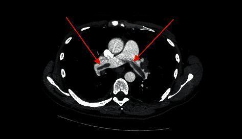
This diagnostic axial CT angiography (CTA) scan of the chest reveals a classic saddle pulmonary embolism. The primary visual finding is a large, low-attenuation filling defect centrally located at the bifurcation of the main pulmonary artery. This thrombus straddles the carina and extends into both the right and left pulmonary arteries. Two red arrows indicate the specific sites where the hypodense embolus obstructs the contrast-enhanced pulmonary vasculature. The surrounding mediastinal structures, including the ascending aorta and the superior vena cava, are visible and show normal contrast enhancement. This imaging modality is the gold standard for diagnosing acute pulmonary embolism, and the 'saddle' configuration demonstrated here indicates a massive or submassive embolic event with high clinical significance for hemodynamic instability.
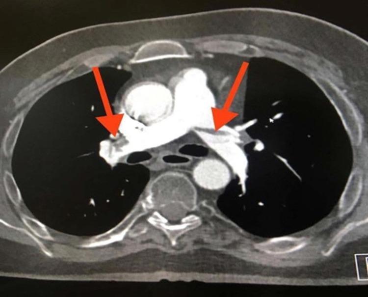
This diagnostic image is an axial cut from a CT Pulmonary Angiography (CTPA) demonstrating extensive pulmonary embolism. The primary visual finding is a large, low-attenuation filling defect appearing as a 'saddle embolus' at the bifurcation of the main pulmonary artery, extending into both the right and left main pulmonary arteries (indicated by red arrows). The central filling defects are characterized by absent or significantly reduced contrast enhancement within the vessel lumens, which are otherwise surrounded by hyperdense contrast material. Additional thromboembolic material is visible in the lobar and segmental branches bilaterally. The main pulmonary artery shows evidence of distension secondary to the large obstructive clot burden. This image serves as a classic clinical example of acute, high-burden pulmonary embolism in a patient with risk factors such as prolonged immobility, illustrating the critical role of CTPA in diagnosing life-threatening vascular obstructions.

**Imaging Modality:** Contrast-enhanced Computed Tomography (CT) of the chest, specifically a CT Pulmonary Angiography (CTPA) protocol. **Anatomical Region:** Axial section of the thorax at the level of the pulmonary artery bifurcation. **Observed Pathology:** Large, bilateral pulmonary embolism. A prominent "saddle" embolus is visible, characterized by low-attenuation (hypodense) filling defects within the lumen of the main pulmonary artery, extending into both the right and left main pulmonary arteries. **Characteristic Visual Features:** * **Filling Defects:** Central non-opacified areas surrounded by radiopaque contrast media, creating the classic "Polo Mint" sign in cross-section or "railway track" sign in longitudinal views. * **Vascular Anatomy:** The pulmonary trunk appears dilated. * **Surrounding Structures:** The ascending and descending aorta, superior vena cava, and vertebral body are visible and unremarkable in this single slice. Metal streak artifacts are present anteriorly from sternal wires, suggesting prior cardiac surgery. **Key Diagnostic Features:** The presence of a large, centrally located thrombus straddling the bifurcation of the pulmonary trunk (saddle embolus) is the pathognomonic finding for acute major pulmonary embolism.
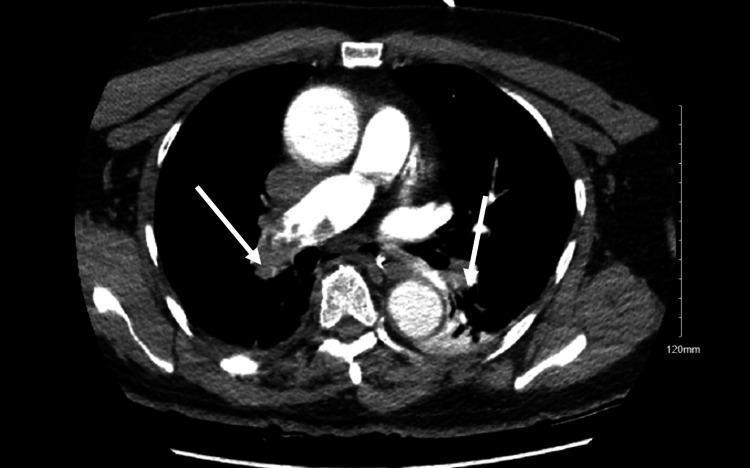
This diagnostic image is an axial contrast-enhanced CT Pulmonary Angiography (CTPA) of the chest. It demonstrates significant bilateral acute pulmonary emboli within the pulmonary arterial tree. Key findings include prominent, low-attenuation filling defects (darker regions) contrasted against the bright, opacified blood in the pulmonary arteries. Specifically, white arrows highlight large clots obstructing the main right and left pulmonary arteries, consistent with a saddle embolus configuration. Smaller peripheral filling defects are also visible in the branching segments of the arterial tree. Other visible thoracic structures include the ascending and descending aorta, the heart, and the superior vena cava, which show normal contrast enhancement. The image serves as a clinical illustration of massive pulmonary embolism causing significant vascular obstruction, often associated with hemodynamic instability and right ventricular strain in an acute clinical setting.
Pulmonary Embolism (PE) - Comprehensive Overview
Definition & Epidemiology
Pathophysiology (Virchow's Triad)
- Stasis (immobility, prolonged bed rest, long-haul travel)
- Vessel wall injury (surgery, trauma, indwelling catheters)
- Hypercoagulability (malignancy, oral contraceptives, pregnancy, Factor V Leiden, protein C/S deficiency, antiphospholipid syndrome)
- Respiratory compromise - the obstructed lung segment is ventilated but not perfused (V/Q mismatch), creating dead space and hypoxemia
- Hemodynamic compromise - increased pulmonary vascular resistance leads to acute RV pressure overload, RV failure, and reduced LV preload; sudden death results from blockage of pulmonary outflow
Clinical Presentation
Symptoms
- Dyspnea (most common; 75-80% of patients, but absent in ~25%)
- Chest pain (second most common; pleuritic in only ~20%)
- Cough, hemoptysis (when pulmonary infarction occurs)
- Palpitations, fatigue, vague malaise
- Unexplained unilateral leg swelling (DVT source; <30% of PE patients)
- Syncope (<5% of presentations, but raises suspicion when unexplained)
A patient with fever >38.6°C or productive sputum cough is more likely to have pneumonia than PE.
Signs
- Tachycardia (most common ECG finding)
- Tachypnea, hypoxia
- Hypotension / cardiogenic shock (high-risk/massive PE)
- Neck vein distension, RV S3 (overt right heart failure)
- Leg swelling / tenderness
ECG Findings
- Tachycardia (most common)
- S1Q3T3 pattern (McGinn-White sign)
- Symmetric T-wave inversions in V1-V4
- Incomplete or complete right bundle branch block
Diagnosis
Step 1 - Pretest Probability
| Wells Score Variable | Points |
|---|---|
| Clinical signs/symptoms of DVT | 3 |
| PE more likely than alternative diagnosis | 3 |
| Heart rate >100 bpm | 1.5 |
| Immobilization/surgery in last 4 weeks | 1.5 |
| Prior DVT or PE | 1.5 |
| Hemoptysis | 1 |
| Active malignancy | 1 |
Step 2 - D-Dimer
- High pretest probability patients
- Hospitalized/postoperative patients (many comorbidities elevate baseline levels)
- Patients >80 years (specificity drops to ~10%)
- Second/third trimester pregnancy
Step 3 - Imaging
Risk Stratification
| Category | Old Term | Definition |
|---|---|---|
| High-risk | Massive | SBP ≤90 mmHg for ≥15 min OR cardiogenic shock |
| Intermediate-risk | Submassive | Normotensive + RV dysfunction (echo or CTPA) and/or elevated troponin/BNP |
| Low-risk | - | No hemodynamic compromise, no RV strain, low sPESI score |
Management
Anticoagulation (all PE categories)
- Rivaroxaban or apixaban - can be started directly without parenteral bridging (after initial loading doses for 1-3 weeks)
- Dabigatran or edoxaban - require 5-10 days of parenteral anticoagulation first
- DOACs are noninferior to LMWH/warfarin for efficacy and have significantly less major bleeding (especially intracranial hemorrhage)
Duration of Anticoagulation
- Provoked PE (reversible trigger): 3 months minimum
- Unprovoked PE: at least 3-6 months; consider indefinite based on recurrence risk and bleeding profile
- Cancer-associated PE: LMWH or DOAC (edoxaban/rivaroxaban) indefinitely while cancer active
Thrombolysis (High-Risk/Massive PE)
IVC Filters
PE Response Team (PERT)
Special Populations
Complications
- CTEPH (Chronic Thromboembolic Pulmonary Hypertension): Occurs in ~1-3.8% after first PE, ~13% after recurrent VTE. Progressive exertional dyspnea, RV failure. Diagnosed by V/Q mismatch + right heart catheterization. Treated with pulmonary endarterectomy (definitive), riociguat, or balloon pulmonary angioplasty.
- Post-PE syndrome: Persistent dyspnea and functional limitation after PE, even without CTEPH.
- Recurrent VTE: Risk is highest in unprovoked PE and cancer-associated VTE.
Summary Algorithm
- Assess pretest probability (Wells or Geneva score)
- Low/moderate probability → D-dimer: if negative, PE excluded; if positive → CTPA
- High probability → CTPA directly (skip D-dimer)
- Hemodynamically unstable → Bedside echo; if RV dysfunction confirmed → systemic thrombolysis or embolectomy without delay
- Confirmed PE → Risk-stratify → anticoagulate; escalate therapy if high/intermediate-risk
Recent evidence note: A 2025 meta-analysis (PMID: 40269600, Catheter Cardiovasc Interv) compared mechanical thrombectomy versus catheter-directed thrombolysis in PE, with results supporting growing use of catheter-based approaches for intermediate/high-risk cases. A 2025 meta-analysis (PMID: 40412084) evaluated AI-based deep learning models for PE detection on CTPA, finding high sensitivity but variable performance. Neither fundamentally changes current guideline management but they may influence future practice.
Create a comparison poster of DOAC vs. warfarin for PE anticoagulation
A clean, professional medical comparison poster titled "DOAC vs. Warfarin for PE Anticoagulation" with two columns side by side. Left column in deep blue labeled "DOACs (Direct Oral Anticoagulants)" listing: Agents: Rivaroxaban, Apixaban, Dabigatran, Edoxaban. Fixed dosing, no routine monitoring. Rapid onset (1-4 hours). Most can start without parenteral bridging (rivaroxaban, apixaban). Noninferior efficacy vs warfarin. Significantly less intracranial bleeding. Predictable pharmacokinetics. Preferred in renal function >30 mL/min. Limited reversal agents (andexanet alfa for Xa inhibitors, idarucizumab for dabigatran). Avoid in pregnancy and breastfeeding. Cost: higher. Right column in deep red labeled "Warfarin (Vitamin K Antagonist)" listing: Variable dosing, requires INR monitoring (target 2.0-3.0). Slow onset - requires 5-10 day heparin bridge. Many drug and food interactions. Teratogenic (weeks 6-12) - avoid in pregnancy. Reversal: Vitamin K, FFP, PCC. Lower cost, widely available. Preferred in: mechanical heart valves, antiphospholipid syndrome, severe renal failure, pregnancy. At the bottom, a highlighted box: "DOACs are now first-line for most PE patients per current guidelines." Include a small molecular/blood clot icon at the top. Medical infographic style, white background, clear typography.
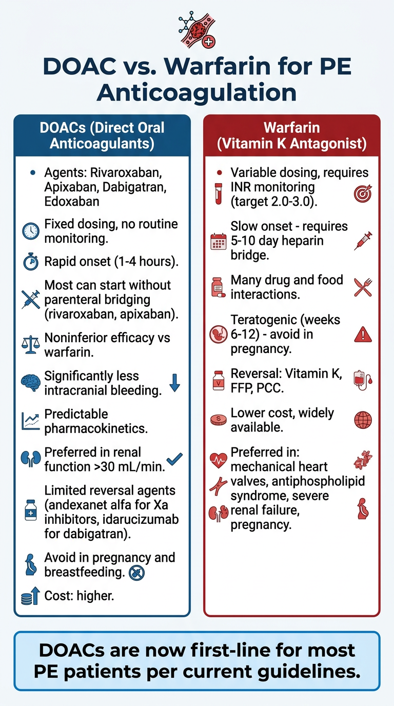
- Convenience (fixed dosing, no INR checks)
- Safety (less intracranial/major bleeding)
- Speed (rapid onset, most skip the heparin bridge)
- Predictability
- Mechanical heart valves (DOACs contraindicated)
- Antiphospholipid antibody syndrome (trials showed DOACs inferior)
- Severe renal failure (CrCl <15-30 mL/min depending on agent)
- Pregnancy/breastfeeding (LMWH is actually the go-to here, not warfarin either)
- Cost is a hard barrier