
spondylosis, spondylolisthesis, spondylitis, spondylolysis, define describe all of them clinical characteristics and presentations signs symptoms investigation treatment physiotherapy rehabilitation give more diagrams to explain them appropriately
Reading File
Checking Date & Time
Finding Sources
Searching Images
spondylosis spondylolisthesis spondylolysis spondylitis spine diagram
Now let me read the most comprehensive sections in parallel:
Reading File
Reading File
Searching Images
ankylosing spondylitis bamboo spine sacroiliitis radiograph
Reading File
Finding Sources
Searching Images
cervical lumbar spondylosis osteophyte disc degeneration MRI
Reading File
Reading File
Reading File
Reading File
Reading File
Searching Images
spondylolysis pars interarticularis scotty dog sign oblique radiograph
Reading File
Searching Images
spondylolisthesis grading Meyerding classification slip vertebra
Reading File
Searching Images
physiotherapy rehabilitation exercises spine spondylolisthesis spondylosis core strengthening
I now have comprehensive data to synthesize a thorough response. Let me also quickly get the AS treatment section.
Finding Sources
Reading File
Finding Sources
Reading File
Here is a comprehensive, structured guide to all four conditions:
The "Spondylo-" Conditions: A Complete Clinical Guide
Quick Mnemonic: Think of them by what is broken, slipped, inflamed, or degenerated:
- Spondyl-osis = degenerative arthrosis (wear & tear)
- Spondyl-olysis = lysis (break/fracture) of the pars interarticularis
- Spondyl-olisthesis = olisthesis (Greek: to slip) — one vertebra slides forward
- Spondyl-itis = itis (inflammation) — usually ankylosing
1. SPONDYLOSIS
Definition
Spondylosis is a non-specific, progressive degenerative condition of the spine involving the vertebral bodies, intervertebral discs, facet joints, and ligaments. It is essentially spinal osteoarthritis. It typically affects patients over 60 years of age and worsens with advancing age.
"Spondylosis is a term for non-specific progressive degenerative changes of the vertebral bodies, facet joints, and central canal which typically affects patients greater than 60 years of age." — ROSEN's Emergency Medicine
Pathophysiology
- Intervertebral discs lose water content and height → disc space narrowing
- Osteophyte (bone spur) formation at vertebral endplates
- Ligamentum flavum hypertrophies and calcifies
- Facet joint deterioration and subluxation
- Foraminal and canal narrowing → nerve root or spinal cord compression
- Can affect cervical, thoracic, or lumbar spine
Clinical Presentation
| Region | Syndrome | Features |
|---|---|---|
| Cervical | Radiculopathy | Neck pain radiating down arm ± paresthesias, weakness; aggravated by Spurling maneuver (extension + rotation + axial load) |
| Cervical | Myelopathy | Spastic gait, upper motor neuron signs, hand clumsiness, bladder dysfunction |
| Lumbar | Radiculopathy | Low back pain ± leg pain in dermatomal distribution |
| Lumbar | Neurogenic claudication | Buttock/leg pain with standing/walking (worsens with extension); relieved by sitting/bending forward — "Shopping cart sign" |
| All | Axial pain | Local stiffness, aching, worse in the morning |
Key Signs:
- Spurling test positive (cervical radiculopathy)
- Decreased deep tendon reflexes (radiculopathy)
- Hyperreflexia, clonus, Hoffman's sign (myelopathy)
- Positive straight leg raise (lumbar)
Investigations
| Investigation | Finding |
|---|---|
| X-ray | Disc space narrowing, osteophytes, endplate sclerosis, foraminal narrowing |
| MRI (gold standard) | Disc degeneration (T2 hypointense), disc herniation, canal/foraminal stenosis, cord compression, myelomalacia (T2 hyperintense cord signal) |
| CT | Bony detail — osteophyte extent, foraminal stenosis |
| EMG/NCS | Localizes radiculopathy, differentiates from peripheral neuropathy |
Cervical Spondylosis — MRI:
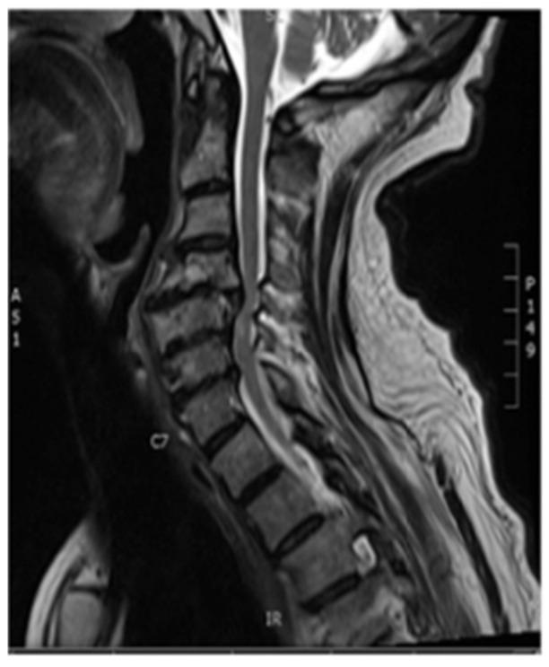
T2-weighted sagittal MRI: multilevel cervical spondylosis with disc degeneration, posterior osteophyte complex, canal stenosis, and cord compression with myelomalacia signal at C5–C6.
Lumbar Spondylosis — Lateral X-ray:
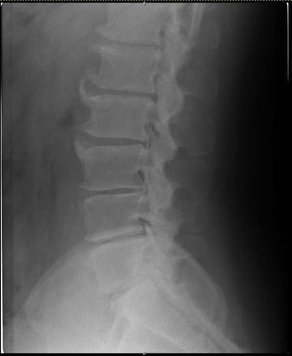
Lateral radiograph: prominent anterior osteophyte formation at multiple lumbar levels, disc space narrowing, endplate sclerosis — classic lumbar spondylosis.
Cervical Spondylosis lateral X-ray:
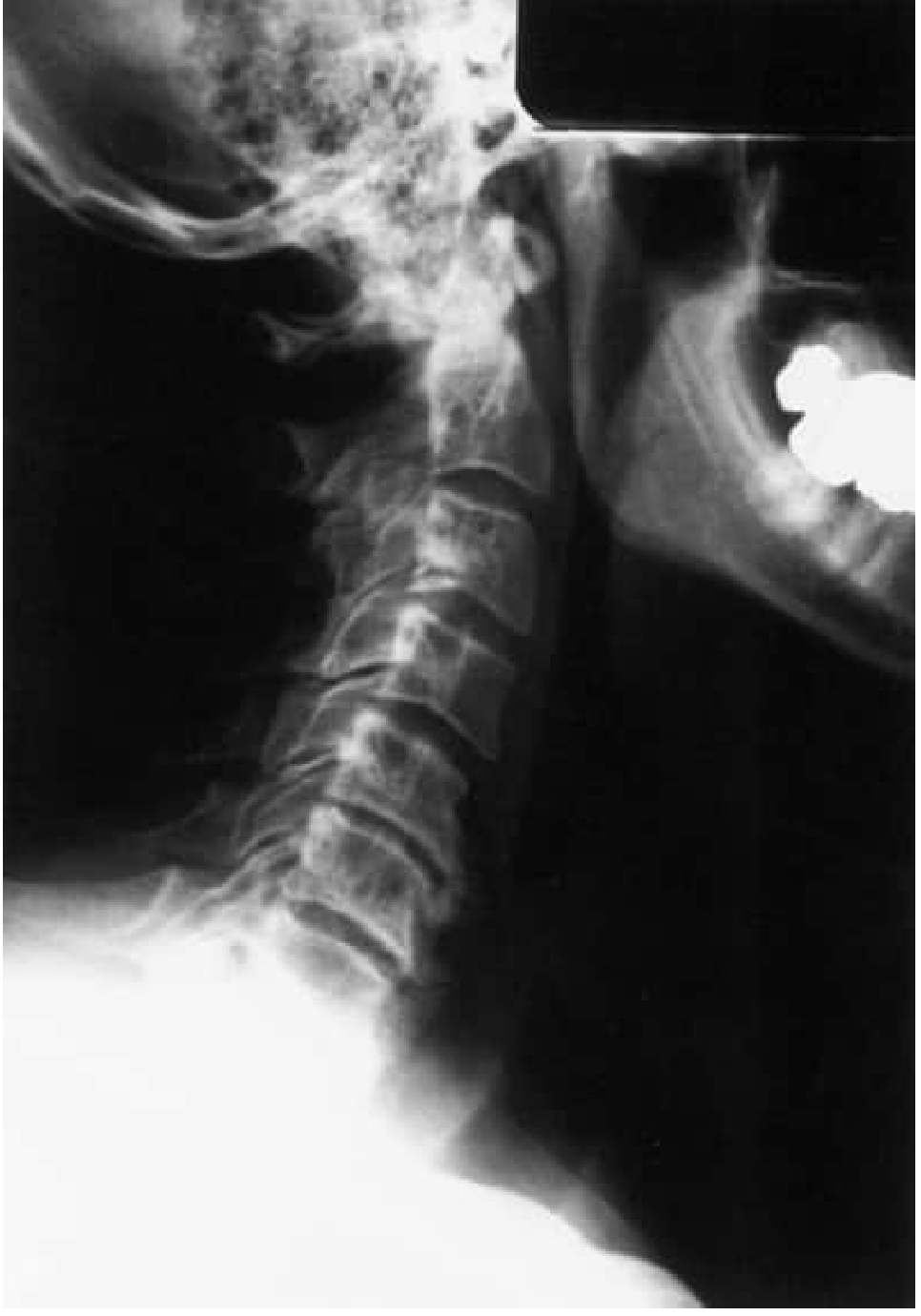
Lateral cervical radiograph: typical spondylosis and osteoarthritis changes. (Bradley and Daroff's Neurology)
Treatment
Conservative (first-line):
- NSAIDs / analgesics for pain
- Soft cervical collar (short-term cervical)
- Physiotherapy and exercise
Physiotherapy & Rehabilitation:
- Cervical: cervical traction, isometric neck strengthening, postural correction, chin tucks, deep cervical flexor strengthening, hot/cold therapy
- Lumbar: core stabilization (McGill Big 3: bird-dog, side plank, curl-up), flexion-based exercises for neurogenic claudication, McKenzie extension protocol for disc-dominant pain, aquatic therapy
- Activity modification; avoid prolonged static postures
- Ergonomic adjustments
Interventional:
- Epidural steroid injections for radicular pain
- Facet joint injections
Surgical indications:
- Progressive neurological deficit
- Myelopathy
- Intractable radiculopathy failing 6 weeks conservative management
- Procedures: ACDF (anterior cervical discectomy and fusion), laminectomy, laminoplasty, TLIF/PLIF
2. SPONDYLOLYSIS
Definition
A stress fracture or defect of the pars interarticularis (the bony bridge connecting the superior and inferior articular processes of a vertebra). It is one of the most common causes of low back pain in children and adolescents.
"Spondylolysis — defect in the pars interarticularis. One of the most common causes of low back pain in children and adolescents." — Miller's Review of Orthopaedics
Epidemiology
- Most common at L5 (85–95%), followed by L4
- Prevalence ~6% in general population
- Common in gymnasts, football linemen, weightlifters (repetitive hyperextension)
- Probable hereditary predisposition
Pathophysiology
- Fatigue fracture from repetitive hyperextension + rotation stresses on the pars interarticularis
- The pars is the weakest point of the posterior neural arch
- Can be unilateral (rarely progresses) or bilateral (risk of spondylolisthesis)
- Fibrocartilaginous reparative tissue at the defect site = Gill nodule (can compress nerve root)
Clinical Presentation
Symptoms:
- Low back pain, aggravated by extension (hyperextension activities)
- Relieved by flexion
- May radiate to buttocks
- Occasionally: radicular symptoms from nerve root irritation (Gill nodule)
- Often found incidentally
Signs:
- Paraspinal muscle spasm
- Tenderness over lower lumbar spinous processes
- Positive "one-leg hyperextension test" (Stork test) — standing on the ipsilateral leg and extending the spine reproduces pain
- Usually no neurological deficit in isolated spondylolysis
Investigations
| Investigation | Finding |
|---|---|
| Lateral X-ray | Detects 80% of defects — radiolucent break in pars |
| Oblique X-ray | "Scotty dog" sign — defect appears as a collar around the dog's neck (15% additional detection) |
| CT scan | Confirms defect, characterizes healing potential |
| SPECT bone scan | Increased uptake = active/acute lesion with healing potential; guides treatment |
| MRI | Identifies pars stress reaction (bone marrow edema) without visible fracture; shows Gill nodule |
Scotty Dog Sign on oblique X-ray:
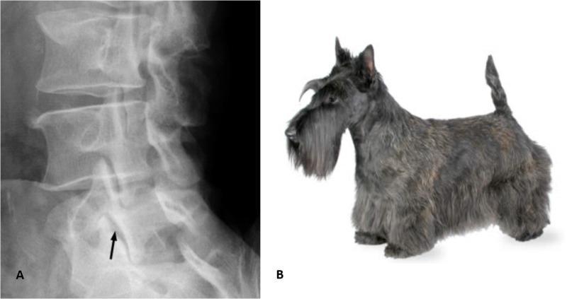
Classic Scotty dog sign: the transverse process = nose, pedicle = eye, superior articular process = ear, inferior articular process = front leg. A pars defect appears as a "collar" around the neck.
Multi-modal imaging of spondylolysis:
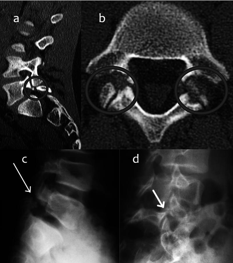
Four-modality comparison: (a) sagittal CT — clear lytic pars defect; (b) axial CT — bilateral defects (double facet sign); (c) lateral X-ray — radiolucent line; (d) oblique X-ray — classic Scotty dog collar sign.
Treatment
Conservative (first-line — most cases):
- Activity restriction (avoid hyperextension sports)
- Flexion-based exercises (flexion unloads the pars)
- Rigid thoracolumbar-sacral orthosis (TLSO) brace for 3–6 months (especially acute lesions with SPECT uptake)
- NSAIDs / analgesics
- Unilateral defects rarely progress; nonunion is common but often asymptomatic
Physiotherapy & Rehabilitation:
- Hamstring stretching (tight hamstrings increase posterior pelvic tilt and pars stress)
- Core stabilization in neutral/flexion
- Avoid extension-dominant loading (McKenzie extension is contraindicated)
- Progressive return to sport once pain-free
- Proprioceptive and neuromuscular training
Surgical:
- Rare; for persistent pain after 6 months conservative treatment
- Buck's direct repair — lag screw across pars defect with bone graft (for young patients, <10% slip, L4 or above)
- Bradford/Scott tension band wiring — alternative direct repair
- In situ posterolateral fusion (L5–S1) if associated with spondylolisthesis
3. SPONDYLOLISTHESIS
Definition
Forward slippage (anterolisthesis) of one vertebral body on the one below it. The suffix "-olisthesis" comes from the Greek word for "slipping." It most commonly occurs at L5–S1 (isthmic type) and L4–L5 (degenerative type).
Classification (Newman–Wiltse–McNab — 6 Types)
| Type | Age | Pathology |
|---|---|---|
| I — Dysplastic | Child | Congenital dysplasia of S1 superior facet |
| II — Isthmic (most common) | 5–50 yrs | Pars fracture/elongation → L5 slips on S1 |
| III — Degenerative | >40 yrs | Facet arthrosis → subluxation (L4–L5) |
| IV — Traumatic | Any age | Acute fracture other than pars |
| V — Pathologic | Any age | Bony incompetence (tumor, Paget's, etc.) |
| VI — Postsurgical | Adult | Excessive facet/lamina resection |
Grading — Meyerding Classification (% of slip)
| Grade | Slip % |
|---|---|
| I | 0–25% |
| II | 25–50% |
| III | 50–75% |
| IV | 75–100% |
| V | >100% = Spondyloptosis |
Meyerding grading — CT series:
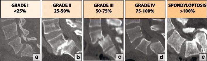
Sagittal CT series demonstrating Meyerding Grades I–V. Grade V (spondyloptosis) = complete anterior displacement of L5 off the sacrum.
Spondylolysis → Spondylolisthesis progression:
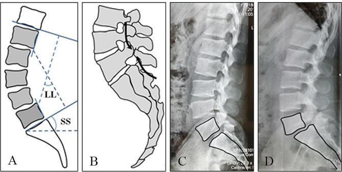
Panel A: sagittal balance parameters (lumbar lordosis, sacral slope). Panel B: stress/shear forces at L5 pars. Panels C–D: progression from spondylolysis (pars defect, no slip) to isthmic spondylolisthesis (anterior L5 displacement).
Clinical Presentation
Pediatric/Adolescent (Isthmic):
- Low back pain (instability-type)
- Hamstring tightness (classic feature — reflex splinting)
- Palpable step-off at lumbosacral junction
- "Pelvic waddle" gait
- Severe slips: L5 radiculopathy, cauda equina dysfunction, kyphosis at L5–S1, "heart-shaped buttocks"
Adult Isthmic (L5–S1):
- Low back pain aggravated by extension
- L5 radiculopathy (exiting nerve root) — pain/paresthesias down lateral leg to dorsum of foot
- Note: isthmic L5–S1 spondylolisthesis → L5 root compression (not S1, unlike disc herniation at L5–S1)
Degenerative (L4–L5, adults >40 years):
- More common in: women, Black patients, diabetics, patients with sagittally-oriented facets
- Central stenosis → neurogenic claudication (leg heaviness, cramping, shopping cart sign)
- L4–L5 degenerative spondylolisthesis → L5 traversing nerve root compression
- Bowel/bladder dysfunction (uncommon)
Key Signs:
- Palpable step-off at L5–S1 (high-grade)
- Restricted lumbar range of motion
- Positive SLR (if radiculopathy)
- Neurological deficits (in high-grade or degenerative)
Investigations
| Investigation | Finding |
|---|---|
| Lateral X-ray | Visualises the slip — Meyerding grading; pars defect may be visible |
| Oblique X-ray | Shows pars defect (Scotty dog collar) |
| MRI | Disc degeneration, foraminal stenosis, nerve compression, cord/cauda equina compression |
| CT scan | Better bony detail, confirms pars defect |
| Standing/dynamic X-rays | Assess instability and slip progression |
MRI + CT — spondylolysis with Grade I spondylolisthesis:
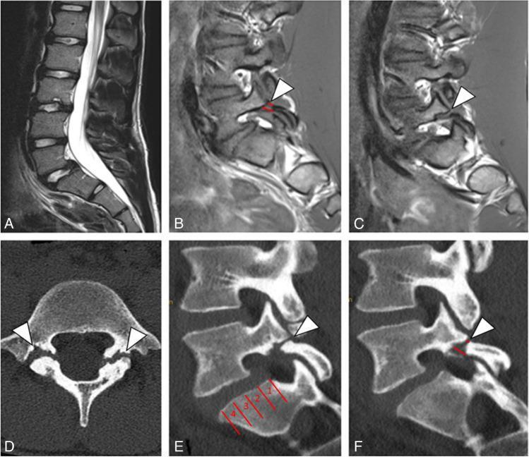
Figure A: sagittal T2 MRI — Grade I L5 anterolisthesis (~6 mm). Figures B–C: T1 sagittal MRI showing lysis gaps (arrowheads). Figure D: axial CT — bilateral pars defects. Figures E–F: sagittal CT confirming persistent lysis with Meyerding division of S1 endplate.
Treatment
Conservative:
- Activity modification
- NSAIDs
- Flexion-based exercises; avoid extension loading
- Hamstring stretching
- Adolescents with Grade I: may return to sports once asymptomatic
- Asymptomatic Grade II: restrict gymnastics/football; observe for progression
Risk factors for progression: young age, female sex, slip angle >10°, high-grade slip, dysplastic morphology
Physiotherapy & Rehabilitation:
- Hamstring stretching (critical — reduces posterior pelvic stress)
- Core stabilization (transversus abdominis, multifidus) in lumbar flexion/neutral
- Pelvic floor activation
- Postural retraining to reduce lumbar hyperlordosis
- McKenzie extension avoided in spondylolisthesis
- Aquatic therapy for pain-limited patients
- Proprioception and balance training
Surgical indications:
- Progression of slip
- Intractable pain after conservative measures
- Neurological weakness or radiculopathy
Surgical options:
- Low-grade (I–II): posterolateral in situ fusion (L5–S1) ± instrumentation; rarely decompression needed in children
- High-grade (III–V): bilateral L4–S1 posterolateral fusion; nerve root decompression if radiculopathy; reduction controversial (20–30% risk of L5 root injury)
- Degenerative: decompressive laminectomy + fusion (SPORT trial: operative outcomes significantly better than non-operative at 4-year follow-up)
- Pars repair (Buck's screw): for young patients <10% slip, defect at L4 or above
4. SPONDYLITIS (Ankylosing Spondylitis)
Definition
Ankylosing spondylitis (AS) — now termed radiographic axial spondyloarthritis (r-axSpA) — is the most common inflammatory disorder of the axial skeleton. It is a chronic, progressive, seronegative spondyloarthropathy characterized by inflammation of the sacroiliac joints and spine, leading ultimately to ankylosis (fusion).
Epidemiology
- Prevalence ~0.2% general population; ~2% in HLA-B27 positive individuals; ~20% in B27-positive relatives of affected patients
- Male:female ratio historically 3–5:1 (more recent data suggests lower ratio)
- Onset typically in young adulthood (teens to 30s); rarely after 45
- Strong association with HLA-B27 (>90% of AS patients)
Pathophysiology
- HLA-B27 misfolds in the endoplasmic reticulum → unfolded protein response → pro-inflammatory cascade
- Gut microbiome–joint axis: B27 transgenic rats raised germ-free have reduced joint disease
- Enthesitis (inflammation at tendon/ligament insertion sites) is the primary pathological process
- Progresses from sacroiliitis → lumbar → thoracic → cervical spine
- New bone formation (syndesmophytes) bridging vertebral bodies → bamboo spine
Clinical Presentation
Cardinal symptoms:
- Chronic inflammatory low back pain (>3 months, onset <45 years)
- Morning stiffness lasting >30 minutes, improving with exercise (not rest)
- Pain in sacroiliac region ± radiation to buttocks
- Night pain (wakes patient, especially in the second half of night)
- Fatigue
Axial involvement:
- Progressive loss of lumbar, thoracic, and cervical mobility
- Loss of lumbar lordosis → thoracic kyphosis → stooped "question mark" posture
- Chest expansion reduced (<5 cm)
Extra-axial manifestations:
- Peripheral oligoarthritis (asymmetrical, predominantly lower limb) — 30%
- Enthesitis (heel pain, plantar fasciitis, Achilles insertion)
- Uveitis/iritis — 40% (acute anterior uveitis; unilateral, recurrent)
- Inflammatory bowel disease (Crohn's/UC) — ~10%
- Psoriasis — ~10%
- Aortic insufficiency, cardiac conduction defects (rare)
- Apical pulmonary fibrosis (rare)
- Osteoporosis → vertebral fractures
Key Clinical Tests
| Test | Technique | Significance |
|---|---|---|
| Schober test | Mark 10 cm above L5; re-measure on full forward flexion. Normal: ≥15 cm (increment ≥5 cm) | Lumbar flexion restriction |
| Occiput-to-wall | Patient back against wall; measure occiput–wall distance | Cervical kyphosis severity |
| Chest expansion | Difference in chest circumference at max inspiration vs. expiration | Normal >5 cm |
| FABERE / Patrick test | Flexion, Abduction, External Rotation, Extension of hip — stress on SI joint | SI joint involvement |
| Gaenslen test | One hip extended off table edge | SI joint involvement |
| BASRI, BASDAI, BASFI | Disease activity and functional indices | Monitoring disease |
Modified New York Criteria for AS
Radiologic criterion:
- Sacroiliitis: grade ≥2 bilateral OR grade 3–4 unilateral
Clinical criteria:
- Low back pain/stiffness >3 months, improves with exercise but not rest
- Limitation of lumbar motion in sagittal AND frontal planes
- Limitation of chest expansion
Definite AS: radiologic criterion + ≥1 clinical criterion
Probable AS: all 3 clinical criteria present, OR radiologic criterion without clinical criteria
Investigations
| Investigation | Finding |
|---|---|
| Pelvic X-ray (AP) | Bilateral sacroiliitis — erosions, pseudowidening, sclerosis, ankylosis |
| Spinal X-ray | Squaring of vertebral bodies, "shiny corners" (Romanus lesion), syndesmophytes, bamboo spine |
| MRI (sacroiliac joints) | Active sacroiliitis (bone marrow edema) before radiographic changes — critical for early diagnosis |
| HLA-B27 | Positive in ~90%; not diagnostic alone |
| ESR, CRP | Elevated (non-specific); may be normal |
| ASAS criteria | Classification for non-radiographic axial SpA (MRI active sacroiliitis + 1 criterion, or B27 + 2 criteria) |
Bamboo spine + bilateral sacroiliitis (AS):
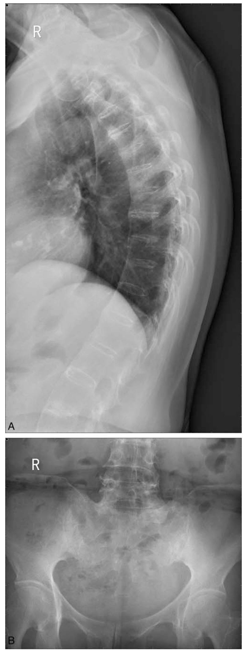
Left: lateral thoracic radiograph — "bamboo spine" with extensive syndesmophyte formation fusing thoracic vertebrae, preserved disc heights. Right: AP pelvic radiograph — Grade III bilateral sacroiliitis with subchondral sclerosis, narrowing, and partial ankylosis.
Advanced AS — complete SI fusion and bamboo spine:
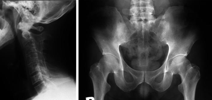
Left: cervical spine — bamboo spine with complete ankylosis, loss of cervical lordosis. Right: pelvis — Grade IV bilateral sacroiliitis (total fusion, obliterated joint spaces).
Lumbar spondylitis — marginal syndesmophytes (AS) vs. reactive arthritis:
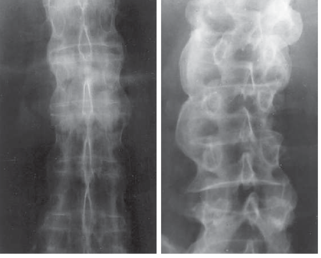
Left: AS — symmetric, marginal, "pencil-thin" syndesmophytes with spinal ligament calcification. Right: Reactive arthritis — bulky, non-marginal, asymmetric syndesmophytes.
Differential Diagnosis of AS
| Condition | Key differentiating features |
|---|---|
| DISH (Diffuse Idiopathic Skeletal Hyperostosis) | Flowing anterior osteophytes, disc heights preserved, SI joints normal |
| Degenerative disc disease | Horizontal bulky osteophytes, disc space narrowing, no sacroiliitis |
| Reactive arthritis | Asymmetrical sacroiliitis, precipitating infection, bulky non-marginal syndesmophytes |
| Psoriatic arthritis | Asymmetrical sacroiliitis, skin/nail changes, coarse syndesmophytes |
| Rheumatoid arthritis | Symmetrical peripheral joints, RF positive, no sacroiliitis |
| Osteitis condensans ilii | Sclerosis on iliac side of SI joint only, no erosions, females |
Treatment
General measures:
- Patient education and psychosocial support
- Avoid high-impact sports; swimming and water therapy ideal
- Sleep position: straight back (avoid curling)
- Deep breathing exercises; avoid smoking
- Periodic stretching during long travel
NSAIDs (first-line pharmacotherapy):
- Indomethacin, diclofenac (up to 150 mg/day), naproxen (up to 1000 mg/day)
- Continuous vs. on-demand NSAID use may slow radiographic progression
- Caution in enteropathic arthritis (risk of exacerbating IBD)
Biologic agents (second-line — when NSAIDs fail):
| Drug class | Agents |
|---|---|
| Anti-TNF-α (monoclonal Ab) | Infliximab 5 mg/kg IV q8w; Adalimumab 40 mg SC q2w; Golimumab 50 mg SC q4w; Certolizumab 200 mg SC q2w |
| Soluble TNF receptor | Etanercept 50 mg SC weekly |
| IL-17A inhibitors | Secukinumab 150 mg SC monthly; Ixekizumab |
| IL-17 receptor blocker | Brodalumab |
| JAK inhibitors | Tofacitinib, upadacitinib (newer) |
⚠️ Screen for latent TB before initiating anti-TNF therapy.
Note: Methotrexate and sulfasalazine have no efficacy for axial disease; sulfasalazine may help peripheral joint involvement.
Physiotherapy & Rehabilitation (cornerstone of management):
- Daily stretching to maintain spinal mobility (most important long-term intervention)
- Extension exercises to counteract kyphotic deformity (opposite of spondylolysis/spondylolisthesis where extension is avoided)
- Postural training — encourage erect posture, prone lying
- Chest expansion exercises and deep breathing (prevents thoracic cage restriction)
- Swimming/hydrotherapy — ideal aerobic activity (buoyancy reduces axial load)
- Group exercise programs (ASAS recommends supervised physiotherapy)
- Cervical range-of-motion exercises
- BASDAI and BASFI used to monitor response
Surgical:
- Rare; for severe deformity
- Spinal osteotomy (pedicle subtraction or Smith-Petersen) for fixed kyphotic deformity
- Joint replacement for hip involvement
- Spinal fractures in AS carry high risk of neurological injury — even minor trauma can cause devastating fracture through the ankylosed rigid spine
Summary Comparison Table
| Feature | Spondylosis | Spondylolysis | Spondylolisthesis | Spondylitis (AS) |
|---|---|---|---|---|
| Nature | Degenerative | Stress fracture | Vertebral slip | Inflammatory |
| Age | >40–60 yrs | Children/adolescents | Any (type-dependent) | Young adults (teens–30s) |
| Location | Cervical > lumbar | L5 (85–95%) | L5–S1 (isthmic); L4–L5 (degen.) | Sacroiliac → lumbar → thoracic → cervical |
| Key symptom | Pain + neurogenic claudication | Extension-aggravated back pain | Extension back pain ± radiculopathy | Inflammatory back pain, morning stiffness, improves with exercise |
| Hallmark sign | Spurling / shopping cart | Stork test / Scotty dog | Palpable step-off / pelvic waddle | Reduced Schober / bamboo spine |
| Key imaging | MRI (disc + cord) | Oblique X-ray (Scotty dog) | Lateral X-ray (Meyerding grade) | AP pelvis (sacroiliitis), MRI SI joints |
| Physio focus | Core strength, flexion for claudication | Flexion exercises, hamstring stretch | Flexion/neutral core, hamstring stretch | Extension/posture, swimming, breathing |
| Avoid in physio | — | Extension exercises | Extension/McKenzie | Kyphotic postures, immobility |
| Surgery trigger | Myelopathy / progressive neuro deficit | Chronic pain after 6 months | Grade progression / radiculopathy | Deformity / fracture |
Rehabilitation exercises — cervical and lumbar spine:
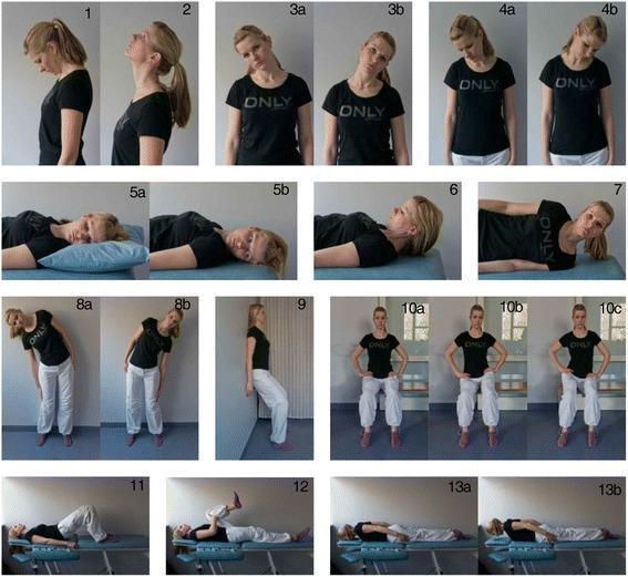
20-panel home exercise program: cervical ROM exercises (flexion, extension, rotation), lumbar mobilization, pelvic bridging, single knee-to-chest stretch, prone extension — relevant to all four spondylo-conditions.
Core stabilization exercises:

Advanced core stabilization protocol: partial curl, supine bridge, prone plank, side bridge, and quadruped (bird-dog) — essential for spondylosis, spondylolysis, and spondylolisthesis rehabilitation.
Sources: ROSEN's Emergency Medicine; Miller's Review of Orthopaedics 9th Edition; Goldman-Cecil Medicine 2 Volume Set; Bradley and Daroff's Neurology in Clinical Practice; Harrison's Principles of Internal Medicine 22E (2025).The guide above is a complete, clinically structured reference for all four conditions. Here is a brief orientation to what was covered:
What Was Covered
All four conditions were addressed with full clinical depth:
Key Distinctions to Remember
| Spondylosis | Degenerative wear-and-tear — osteophytes, disc loss, stenosis. Treat with core strengthening, flexion exercises for claudication |
| Spondylolysis | Stress fracture of the pars interarticularis — Scotty dog sign on oblique X-ray. Extension aggravates; treat with flexion exercises, bracing, hamstring stretching |
| Spondylolisthesis | Vertebral slip (forward) — Meyerding I–V grades. Palpable step-off, pelvic waddle. Treat conservatively with core/hamstring rehab; fusion for progression |
| Spondylitis (AS) | HLA-B27+ inflammatory disease — morning stiffness, sacroiliitis, bamboo spine. NSAIDs first, anti-TNF/IL-17 biologics second. Physiotherapy (extension, posture, swimming) is the cornerstone of rehab |
Physiotherapy Contrast (Critical!)
- Spondylosis/AS → Extension exercises (counteract kyphosis/degeneration)
- Spondylolysis/Spondylolisthesis → Flexion exercises + core in neutral (extension is harmful — loads the pars/slip)
This is a shared conversation. Sign in to Orris to start your own chat.