development of anal canal and recturm total detailed from singh
Reading File
Finding Sources
Finding Sources
Finding Sources
Finding Sources
Finding Sources
Finding Sources
Finding Sources
Finding Sources
Finding Sources
Reading File
Reading File
Reading File
Reading File
Reading File
Loading Image
Loading Image
Loading Image
I now have comprehensive information to provide a detailed answer. Note that "Singh" likely refers to Vishram Singh's Textbook of Clinical Embryology or Singh's Human Embryology, which is not directly in the library, but the content below is drawn from the two authoritative embryology textbooks in the library - The Developing Human (Moore & Persaud) and Langman's Medical Embryology - which cover the same material as Singh, often chapter-by-chapter.
Development of the Anal Canal and Rectum
Note: Singh's Embryology (Vishram Singh) is not directly available in this library. The following is a comprehensive account drawn from The Developing Human (Moore, Persaud & Torchia) and Langman's Medical Embryology, which are the primary sources Singh's text draws upon. The content maps precisely to what Singh covers.
1. Origin - The Hindgut
The rectum and anal canal are derivatives of the hindgut, the most caudal segment of the primitive gut tube. The hindgut gives rise to:
- Distal one-third to one-half of the transverse colon
- Descending colon
- Sigmoid colon
- Rectum
- Superior two-thirds (upper part) of the anal canal
All hindgut derivatives are supplied by the inferior mesenteric artery.
2. The Cloaca
At the caudal end of the embryo (around week 4), the cloaca is a common endoderm-lined chamber into which the hindgut opens dorsally and the allantois opens ventrally. It is closed externally by the cloacal membrane, a bilaminar structure composed of:
- Endoderm (from the cloaca)
- Ectoderm (from the anal pit / proctodeum)
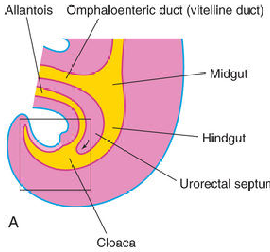
3. Partitioning of the Cloaca by the Urorectal Septum
Between weeks 4 and 7, a wedge of mesenchyme called the urorectal septum develops in the angle between the allantois and hindgut. This septum:
- Grows caudally toward the cloacal membrane
- Develops fork-like extensions that produce infoldings of the lateral cloacal walls
- These folds grow toward each other and fuse, forming a partition
This partition divides the cloaca into three parts:
- Rectum (dorsal)
- Cranial part of the anal canal (dorsal)
- Urogenital sinus (ventral - gives rise to bladder and urethra)
Current evidence indicates the urorectal septum does not actually fuse with the cloacal membrane (contrary to older teaching). The cloacal membrane ruptures by apoptosis (programmed cell death), and the anorectal lumen is temporarily closed by an epithelial anal plug. Mesenchymal proliferations then produce surface ectoderm elevations (the anal tubercles) around this plug. The canal is reopened by apoptotic cell death of the epithelial anal plug, forming the anal pit (proctodeum).
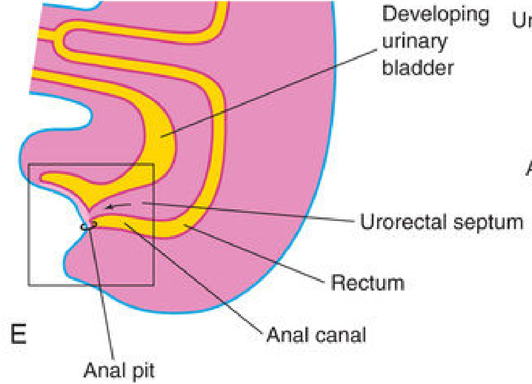
4. Development of the Anal Canal
The adult anal canal has a dual embryologic origin, which is the most clinically important concept in its development:
| Part | Origin | Tissue type |
|---|---|---|
| Superior 2/3 | Hindgut endoderm | Columnar epithelium |
| Inferior 1/3 | Anal pit (proctodeum) ectoderm | Stratified squamous epithelium |
The pectinate (dentate) line marks the approximate junction between these two parts - it lies at the inferior limit of the anal valves.
About 2 cm above the anus is the anocutaneous line (white line / Hilton's line) where the epithelium transitions from columnar to stratified squamous. At the anus itself, the epithelium is keratinized and continuous with the perianal skin.
The muscular wall (internal and external sphincters, longitudinal muscle) and connective tissue are all derived from splanchnic mesenchyme. The anal sphincter complex appears to be under Hox D gene control.
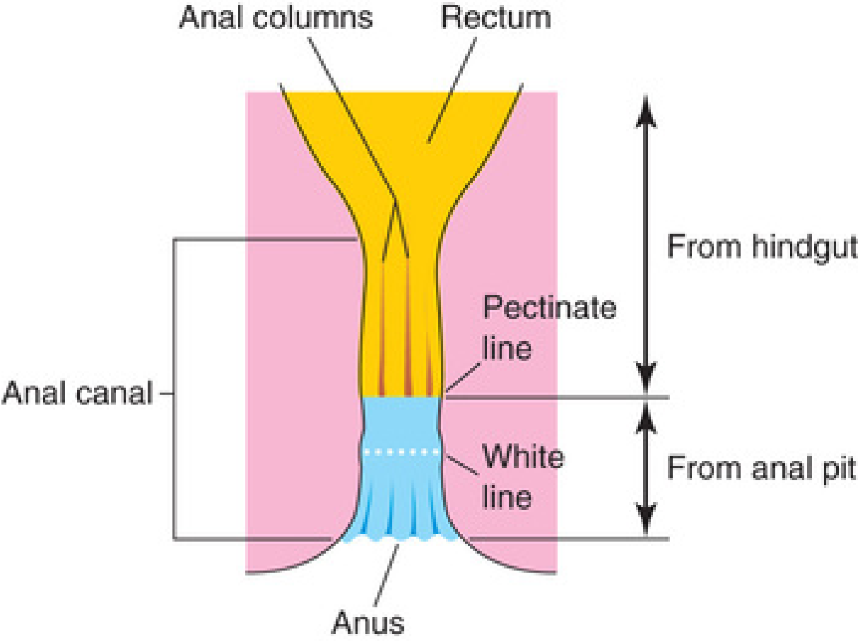
5. Clinical Significance of Dual Origin - Blood Supply, Nerves, Lymphatics
The different embryologic origins result in completely different neurovascular territories, which is clinically critical:
Superior 2/3 (Hindgut origin)
- Arterial supply: Superior rectal artery (continuation of inferior mesenteric artery)
- Venous drainage: Superior rectal vein → inferior mesenteric vein → portal system
- Lymphatics: Inferior mesenteric lymph nodes (internal iliac nodes)
- Nerve supply: Autonomic (visceral) nerves - insensitive to pain; tumors are painless, arise from columnar epithelium
Inferior 1/3 (Anal pit / ectodermal origin)
- Arterial supply: Inferior rectal arteries (branches of internal pudendal artery)
- Venous drainage: Inferior rectal vein → internal pudendal vein → internal iliac vein → systemic circulation
- Lymphatics: Superficial inguinal lymph nodes
- Nerve supply: Inferior rectal nerve (somatic) - sensitive to pain, touch, temperature, pressure; tumors are painful, arise from stratified squamous epithelium
6. Congenital Anomalies
Imperforate Anus
Failure of the anal membrane to break down (or failure of apoptosis of the epithelial anal plug). Incidence: ~1/5,000 births.
Anorectal Fistulas and Atresias
- Rectourethral fistula (male) / Rectovaginal fistula (female): The cloaca is too small, or the urorectal septum fails to extend sufficiently caudally, causing the hindgut opening to shift anteriorly into the urethra or vagina.
- Rectoperineal fistula: Likely due to misexpression of genes during epithelial-mesenchymal signaling.
- These vary in severity from a narrow tube to a fibrous remnant at the perineal surface.
Congenital Aganglionic Megacolon (Hirschsprung Disease)
- Absence of parasympathetic ganglion cells in the myenteric (Auerbach) and submucosal (Meissner) plexuses of the bowel wall, most commonly the rectum and sigmoid colon.
- Due to failure of neural crest cells to migrate into the wall of the colon during weeks 5-7.
- The RET proto-oncogene (tyrosine kinase receptor for neural crest cell migration) is the major susceptibility gene.
- Incidence: 1/5,000 newborns; M:F ratio = 4:1.
- In most cases, only rectum and sigmoid are involved; in 10-20%, more proximal colon is affected; in 3%, the entire colon.
- The aganglionic segment fails to relax, causing obstruction and dilation of the normal (proximal) bowel.
Summary Table
| Feature | Rectum & Upper 2/3 Anal Canal | Lower 1/3 Anal Canal |
|---|---|---|
| Embryologic origin | Hindgut (endoderm) | Anal pit / proctodeum (ectoderm) |
| Epithelium | Columnar | Stratified squamous (keratinized at anus) |
| Artery | Superior rectal (IMA) | Inferior rectal (internal pudendal) |
| Vein | Superior rectal → portal | Inferior rectal → systemic |
| Lymph nodes | Inferior mesenteric / internal iliac | Superficial inguinal |
| Nerves | Autonomic (visceral) | Somatic (inferior rectal nerve) |
| Pain sensitivity | None | Present |
| Landmark | Above pectinate line | Below pectinate line |
Sources: The Developing Human: Clinically Oriented Embryology (Moore, Persaud & Torchia) and Langman's Medical Embryology. These are the primary reference sources that Singh's Embryology is based upon.
acute watery diarrhoea in pediatrcs nelson based answer detailed
Reading File
Finding Sources
Finding Sources
Finding Sources
Finding Sources
Finding Sources
Finding Sources
Reading File
Reading File
Reading File
Finding Sources
Reading File
Finding Sources
Reading File
Reading File
Finding Sources
Reading File
Finding Sources
Reading File
Reading File
Reading File
I now have comprehensive information from authoritative pediatric emergency/clinical sources. Let me now compile the complete Nelson-style answer:
Acute Watery Diarrhoea in Pediatrics
Note: Nelson's Textbook of Pediatrics is not directly available in this library. This answer follows Nelson's framework precisely, drawing from Tintinalli's Emergency Medicine (Pediatric section), Rosen's Emergency Medicine, Harriet Lane Handbook, and Red Book 2021 - which all cite and align with Nelson's content.
Definition
Diarrhoea is defined as loose or liquid stools and/or an increase in the frequency of evacuations with at least 3 stools in 24 hours. Acute diarrhoea lasts <14 days (Nelson: acute <7 days, persistent 7-14 days, chronic >14 days). The child's age and diet affect stool consistency - in the first month of life, change in consistency is more specific than absolute stool number. Breastfed infants normally have several poorly formed, yellow-green stools per day.
Watery diarrhoea implies loss of large volumes of fluid without blood or mucus - the hallmark of secretory/osmotic mechanisms.
Epidemiology
- Acute viral gastroenteritis is the most common cause of diarrhoea in children
- Accounts for >500,000 deaths globally each year in children <5 years
- In the USA/Europe, viruses cause >90% of acute infectious diarrhoea; bacteria cause only 7-10% of cases in children
- Rotavirus previously caused the majority of severe cases; since introduction of oral rotavirus vaccines (RotaTeq, Rotarix), there has been an ~80% reduction in rotavirus-related hospitalizations
- Norovirus is now the pathogen responsible for the greatest burden of medically attended gastroenteritis
- Incidence peaks in winter months
- Hand washing reduces diarrhoeal disease incidence by ~30%
Pathophysiology
Three mechanisms produce acute watery diarrhoea:
1. Secretory Diarrhoea
- Increased intestinal secretion of water into the gut lumen, or inhibition of absorption
- Classic example: Vibrio cholerae produces enterotoxin → increased Cl⁻ and HCO₃⁻ secretion
- Other enterotoxin producers: Salmonella, Shigella, E. coli (ETEC), Clostridioides difficile
- Characteristics:
- Stool volume does not decrease with fasting
- Stool pH above 6
- Absence of reducing substances in stool
2. Osmotic Diarrhoea
- Poorly absorbed solutes create osmotic gradient → intraluminal movement of water and electrolytes
- Typical acute viral gastroenteritis injures small bowel epithelium → disruption of microvilli → decreased absorptive area → impaired fluid, electrolyte, and nutrient absorption
- Characteristics:
- Diarrhoea decreases or stops with fasting
- Stool pH below 6
- Presence of reducing substances in stool
3. Inflammatory / Invasive Diarrhoea
- Destruction of villous cells, dysfunction of cellular transporters
- Enteroinvasive organisms (Salmonella, Shigella, Campylobacter) → neutrophil infiltration → increased secretion + decreased absorption
- Associated with blood, mucus (dysentery)
- More relevant to bloody diarrhoea, but can initially present as watery
Why Children Are More Vulnerable
- Larger extracellular fluid compartments relative to body size - proportionately more fluid lost
- Fluid turnover in infants/young children is 3x that of adults
- Higher metabolic rates, increased body surface area to mass ratio, higher body water content
- Limited stores of metabolic substrates (fat, glycogen)
- Limited ability to access fluids when ill
- Limited renal water-conserving ability compared to adults
- Result: rapid development of hypoglycaemia, electrolyte abnormalities, dehydration, and shock
Aetiology
Viral (Majority of Cases)
| Virus | Key Features |
|---|---|
| Rotavirus | Peak age 6 months-2 years; winter; watery stool, fever, vomiting 1-3 days before diarrhoea; 5-7 day illness; vaccine-preventable |
| Norovirus | All ages; year-round; projectile vomiting prominent; 24-72 hr illness; highly contagious; foodborne/person-to-person |
| Adenovirus (serotypes 40,41) | Enteric adenovirus; year-round; prolonged diarrhoea (up to 14 days) |
| Astrovirus | Young children; mild watery diarrhoea |
| Sapovirus | Similar to norovirus |
Bacterial (7-10% in Developed Countries)
| Organism | Mechanism | Features |
|---|---|---|
| E. coli (ETEC) | Enterotoxin (LT/ST) | Traveller's diarrhoea; profuse watery stools |
| Vibrio cholerae | Cholera toxin | Rice-water stools; massive fluid loss |
| Salmonella spp. | Invasion + toxin | Fever, watery/bloody; foodborne |
| Shigella spp. | Invasion | Watery → dysentery; seizures; hemolytic-uremic syndrome |
| Campylobacter jejuni | Invasion | Bloody or watery; abdominal cramps |
| C. difficile | Cytotoxin | Post-antibiotic; pseudomembranous colitis |
Parasitic
- Giardia lamblia - watery/greasy stools, bloating, prolonged; common in childcare settings
- Cryptosporidium - watery diarrhoea, especially in immunocompromised
- Entamoeba histolytica - travel/endemic areas
High-Risk Groups for Severe Complications
- Premature infants / very low birth weight infants (up to 1 year of age)
- Infants <3 months
- Immunosuppressed or malnourished children
- Children with chronic underlying conditions (hemoglobinopathy, HIV, neoplasm)
- Recent hospitalization, broad-spectrum antibiotic use, travel to developing countries
Clinical Features
Onset: 12-72 hours after exposure. Viral AGE typically resolves within 1 week.
History (Key Points to Gather)
- Duration, frequency, amount, and consistency of stools (watery, mucusy, bloody)
- Vomiting: duration, amount, time since last vomited
- Fever, chills, myalgia, rash
- Level of alertness, activity, lethargy, irritability
- Frequency of wet diapers / urine output
- Food history, recent travel, contact with sick individuals
- Similar episodes in household or daycare
Physical Examination
- Begin with general appearance from across the room - responsiveness, activity, work of breathing
- Assess vital signs relative to age norms
- Focus on signs of dehydration (see below)
- Look for alternative diagnoses: otitis media, pyelonephritis, appendicitis, DKA
- Signs of extraintestinal/systemic spread: bone pain (osteomyelitis), altered mental status (meningitis), petechiae (HUS)
Red Flags (Requiring Immediate Evaluation)
- Bilious or bloody vomitus
- Blood in stool / hematochezia
- Significant or localised abdominal pain (peritoneal signs - consider appendicitis)
- Fever in child <3 months
- Very ill appearance / altered mental status
- Inability to maintain oral intake / signs of severe dehydration
Assessment of Dehydration
WHO/Nelson Classification
| Parameter | None to Minimal (<3% body weight) | Some / Mild-Moderate (3-9%) | Severe (>9%) |
|---|---|---|---|
| Mental status | Well, alert | Fatigued, restless, irritable | Drowsy, limp, unresponsive |
| Eyes | Normal | Mildly sunken | Very sunken |
| Tears | Normal | Decreased | Absent |
| Oral mucosa | Moist | Sticky/dry | Very dry |
| Pulse | Normal | Normal to increased | Tachycardic, weak/thready |
| BP | Normal | Normal | Low / hypotensive |
| Skin turgor | Normal | Reduced | Poor (>2 sec return) |
| Urine output | Normal | Decreased | Minimal / absent |
| Fontanelle (infants) | Normal | Sunken | Very sunken |
| Thirst | Normal | Increased | Drinks poorly/unable |
| Capillary refill | <2 sec | 2-3 sec | >3 sec |
Clinical Dehydration Score (CDS)
| Score | General Appearance | Eyes | Oral Mucosa | Tears |
|---|---|---|---|---|
| 0 | Normal | Normal | Moist | Normal |
| 1 | Thirsty, restless, lethargic but irritable | Mildly sunken | Sticky | Decreased |
| 2 | Drowsy/non-responsive, limp, cold, diaphoretic | Very sunken | Dry | None |
- Score >0 = some dehydration
- Score >5 = moderate-severe dehydration
Laboratory Investigations
- Not routinely required for acute watery diarrhoea in a well-appearing child
- CBC: only if child is ill-appearing or has bloody diarrhoea (to identify bacterial enterocolitis or HUS)
- WBC count and CRP are not reliable for distinguishing viral from bacterial gastroenteritis
- Serum glucose: measure in infants and young children (hypoglycaemia may occur in up to 9%)
- Serum electrolytes, BUN, creatinine, calcium: only in severe dehydration or specific clinical indications
- Stool culture: for bloody diarrhoea, severely ill children, immunocompromised, suspected outbreak, or recent travel
- Stool microscopy (ova & parasites): suspected Giardia, Cryptosporidium, or travel history
Management
Goals
- Prevent or treat dehydration
- Replace ongoing fluid losses
- Meet nutritional needs
- Identify and treat specific pathogens where necessary
Step 1: Oral Rehydration Therapy (ORT) - First-Line Treatment
The physiologic basis of ORT is coupled cotransport of sodium and glucose at the brush border of intestinal epithelial cells, creating a gradient for passive water absorption. This mechanism remains relatively intact even in severe diarrhoeal disease and works optimally when the Na⁺:glucose ratio is 1:1.
ORS Composition (WHO Reduced-Osmolarity, 2002)
| Component | Amount |
|---|---|
| Glucose | 75 mmol/L |
| Sodium | 75 mmol/L |
| Potassium | 20 mmol/L |
| Chloride | 65 mmol/L |
| Citrate (base) | 10 mmol/L |
| Total osmolarity | 245 mOsm/L |
Commercial solutions (Pedialyte, Enfalyte) contain 45-60 mmol/L sodium (appropriate for developed countries).
Avoid: Sports drinks, fruit juices, tea, plain water, soda - these are deficient in sodium and/or have excessive sugar, worsening fluid losses.
ORT Protocol by Dehydration Severity
| Severity | Route | Volume |
|---|---|---|
| Minimal/None | Oral (preferred fluids or dilute apple juice) | Maintenance + 10 mL/kg per stool + 2 mL/kg per emesis |
| Mild-Moderate | Oral ORS (or NG if vomiting) | 50-100 mL/kg over 3-4 hours + ongoing losses |
| Severe | IV or IO immediately | 20 mL/kg NS or Lactated Ringer's over 5-30 min; aim 60-100 mL/kg in first hour |
For vomiting patients: Start with 5 mL (1 tsp) every 2-3 minutes, increase gradually as tolerated. Nasogastric ORS at 10-20 mL/kg/h is effective even in vomiting patients and is more cost-effective than IV treatment.
Cochrane review: ORT vs IV therapy - no difference in rehydration failure, weight gain, or total fluid intake; ORT associated with shorter hospital stay. For every 25 children treated with ORT, 1 requires IV rescue.
IV Rehydration (Indications)
- Severe dehydration / haemodynamic compromise
- Altered mental status precluding safe oral administration
- Persistent vomiting failing NG route
- IV NS 20 mL/kg over 20-30 min, repeat as needed
Step 2: Nutritional Management
- Continue breastfeeding throughout illness (do not stop)
- Formula-fed infants: continue usual formula (no need to dilute)
- Early refeeding as soon as rehydration is achieved - do not fast the child
- Return to normal age-appropriate diet as soon as possible
- Special "BRAT diet" (bananas, rice, applesauce, toast) is not recommended by Nelson/AAP - it is nutritionally incomplete
- Avoid high-sugar fluids (fruit juices) as they worsen osmotic diarrhoea
Step 3: Pharmacological Treatment
Zinc Supplementation
- WHO recommendation: Zinc 20 mg/day for 10-14 days in children with diarrhoea in developing countries
- Reduces duration and severity of diarrhoea; reduces recurrence for 2-3 months
- Infants <6 months: 10 mg/day
Probiotics
- Evidence supports Lactobacillus rhamnosus GG and Saccharomyces boulardii in reducing duration of acute watery diarrhoea by ~1 day
- Most benefit in viral (rotavirus) diarrhoea
- Routine use is reasonable but not mandatory
Ondansetron (Antiemetic)
- Significantly reduces vomiting, improves ability to complete ORT
- Reduces IV fluid requirement and hospitalisation rate
- Dose: 0.15 mg/kg PO/IV (max 4 mg/dose)
- Use when vomiting is the main barrier to ORT
Antimotility Agents
- Loperamide: NOT recommended in children - risk of ileus, respiratory depression, death in infants
- Bismuth subsalicylate: limited role; avoid in children due to Reye syndrome risk
Antibiotics
- Not indicated for routine acute watery diarrhoea (viral in most cases)
- Indicated in specific situations:
| Pathogen | Indication for Antibiotics | Drug |
|---|---|---|
| Shigella | All cases (shortens illness, eradicates organism) | Azithromycin 20 mg/kg/day × 3-5 days; Ceftriaxone 50 mg/kg for severe |
| Salmonella typhi | All (enteric fever) | Ceftriaxone; Azithromycin for uncomplicated |
| Salmonella non-typhi | Only high-risk: <3 months, immunocompromised, hemoglobinopathy, HIV, bacteremia | Amoxicillin or TMP-SMX; Ceftriaxone if invasive |
| Giardia | Symptomatic | Metronidazole 15 mg/kg/day × 5-7 days |
| C. difficile | Symptomatic (discontinue causative antibiotic) | Oral metronidazole (mild-moderate); vancomycin (severe) |
| V. cholerae | All | Doxycycline (>8 yrs) or Azithromycin |
| ETEC (traveller's) | Moderate-severe | Azithromycin |
Complications
| Complication | Features |
|---|---|
| Dehydration / Hypovolaemic shock | Most common life-threatening complication |
| Electrolyte abnormalities | Hypo/hypernatraemia; hypokalaemia; metabolic acidosis (bicarbonate loss in stool) |
| Hypoglycaemia | Up to 9% incidence; especially in infants |
| Haemolytic-Uraemic Syndrome (HUS) | Post-STEC (E. coli O157:H7); microangiopathic haemolytic anaemia + thrombocytopenia + acute renal failure; do NOT treat with antibiotics |
| Febrile seizures | Associated with Shigella (Shigella toxin / febrile response) |
| Bacteraemia / Sepsis | High-risk groups |
| Reactive arthritis (Reiter's) | Post-Shigella, Salmonella, Campylobacter |
| Intussusception | Post-rotavirus vaccine (attributable risk ~5.3/100,000 vaccinated) |
| Persistent / Chronic diarrhoea | Lactase deficiency post-viral; cow's milk protein intolerance |
| Congenital Megacolon (Hirschsprung) | Differential diagnosis in neonates with obstruction |
Indications for Hospital Admission
- Severe dehydration or haemodynamic compromise
- Failed oral/NG rehydration
- Age <3 months with fever or ill appearance
- Persistent vomiting with inability to maintain ORS
- Suspected surgical diagnosis (appendicitis, intussusception, volvulus)
- Bloody diarrhoea with toxicity
- Altered mental status
- Concern for HUS
- Unreliable caregivers or unsafe home environment
Prevention
- Rotavirus vaccination (RotaTeq: 3 doses at 2, 4, 6 months; Rotarix: 2 doses at 2, 4 months) - ~80% reduction in severe rotavirus disease
- Hand hygiene - reduces diarrhoeal disease by ~30%
- Safe water, sanitation, and hygiene (WASH)
- Breastfeeding (passive immunity via secretory IgA)
- Proper food handling and storage
- Alcohol-based hand sanitizer in daycare/school settings
Key Nelson Pearls
- ORT is the gold standard - always preferred over IV in mild-moderate dehydration; IV is not superior and is associated with a longer hospital stay
- Continue feeding - early refeeding reduces illness duration and promotes mucosal recovery
- Breastfeeding must not be stopped during acute diarrhoea
- Routine stool cultures and CBC are not needed in well-appearing children with acute watery diarrhoea
- Hypoglycaemia must be checked in infants and young children
- Do not use antibiotics routinely - most acute watery diarrhoea is viral and self-limited
- Loperamide is contraindicated in children
- Bilious vomiting in an infant = malrotation/volvulus until proven otherwise
- HUS triad = microangiopathic haemolytic anaemia + thrombocytopenia + acute kidney injury; do NOT give antibiotics for STEC (worsens HUS risk)
- Ondansetron facilitates ORT by controlling vomiting - reduces hospitalisation
Sources: Tintinalli's Emergency Medicine (Pediatric Gastroenteritis chapter); Rosen's Emergency Medicine (Pediatric Diarrhoea chapter); Harriet Lane Handbook 23e; Red Book 2021 (AAP). All aligned with Nelson's Textbook of Pediatrics guidelines.
actue watery diarrhoea in pediatrics
You just asked this same question and I provided a comprehensive, detailed answer above. Here's a quick reference summary of everything covered:
What Was Covered in the Previous Answer
Definition - loose/liquid stools ≥3/day, acute <14 days
Epidemiology - viral cause in >90% of pediatric cases; norovirus now #1 burden; rotavirus vaccine reduced hospitalizations by ~80%
Pathophysiology - three mechanisms:
- Secretory (cholera toxin, ETEC) - stool persists with fasting, pH >6
- Osmotic (viral - rotavirus) - stool stops with fasting, pH <6, reducing substances present
- Inflammatory/invasive - neutrophil-mediated
Aetiology - Rotavirus, Norovirus, Adenovirus 40/41; bacteria (Salmonella, Shigella, ETEC, Vibrio cholerae); parasites (Giardia, Cryptosporidium)
Dehydration Assessment - WHO 3-tier (none <3%, mild-moderate 3-9%, severe >9%); Clinical Dehydration Score
Management:
- ORT first-line (WHO ORS: Na 75, Glucose 75, K 20, osmolarity 245 mOsm/L)
- 50-100 mL/kg over 3-4 hrs for mild-moderate; 20 mL/kg IV NS bolus for severe
- Continue breastfeeding; early refeeding; no BRAT diet
- Zinc 20 mg/day × 10-14 days (WHO countries)
- Ondansetron to facilitate ORT
- No loperamide in children
- Antibiotics only for specific pathogens (Shigella, typhoid, cholera, Giardia)
Complications - dehydration/shock, hyponatraemia, hypoglycaemia, HUS, febrile seizures
Prevention - rotavirus vaccine, hand hygiene, breastfeeding, WASH
Is there a specific aspect you want me to expand on - such as ORS preparation, a particular pathogen, dehydration scoring in detail, or the WHO IMCI algorithm? Let me know and I will go deeper on that section.
Create a clinical management flowchart (decision tree) for acute watery diarrhea
Running Command
ls -la /home/daytona/workspace/ && python3 --version && python3 -c "import PIL; print('PIL OK')" 2>/dev/null || echo "PIL not found"
Running Command
mkdir -p /home/daytona/workspace/awd-flowchart && python3 -c "import matplotlib; print(matplotlib.__version__)"
Writing File
~/awd-flowchart/create_flowchart.py
import matplotlib
matplotlib.use('Agg')
import matplotlib.pyplot as plt
import matplotlib.patches as mpatches
from matplotlib.patches import FancyBboxPatch, FancyArrowPatch
import matplotlib.patheffects as pe

# ── Canvas ────────────────────────────────────────────────────────────────────
fig, ax = plt.subplots(figsize=(22, 34))
ax.set_xlim(0, 22)
ax.set_ylim(0, 34)
ax.axis('off')
fig.patch.set_facecolor('#F8FAFC')

# ── Colour palette ─────────────────────────────────────────────────────────
C = {
    'title':    '#1A237E',
    'start':    '#0D47A1',
    'diamond':  '#1565C0',
    'green':    '#1B5E20',
    'orange':   '#E65100',
    'red':      '#B71C1C',
    'purple':   '#4A148C',
    'teal':     '#004D40',
    'grey':     '#37474F',
    'action':   '#E3F2FD',
    'action2':  '#E8F5E9',
    'action3':  '#FFF3E0',
    'action4':  '#FCE4EC',
    'warn':     '#FFF8E1',
    'arrow':    '#37474F',
    'text_dk':  '#0D0D0D',
    'white':    '#FFFFFF',
    'line':     '#90A4AE',
}

# ── Helper functions ───────────────────────────────────────────────────────

def box(ax, x, y, w, h, text, facecolor, edgecolor, fontsize=8.5,
        textcolor='#0D0D0D', bold=False, style='round,pad=0.1', wrap=True,
        radius=0.15):
    """Draw a rounded rectangle with centred, wrapped text."""
    rect = FancyBboxPatch((x - w/2, y - h/2), w, h,
                          boxstyle=f'round,pad={radius}',
                          facecolor=facecolor, edgecolor=edgecolor,
                          linewidth=1.8, zorder=3)
    ax.add_patch(rect)
    weight = 'bold' if bold else 'normal'
    ax.text(x, y, text, ha='center', va='center',
            fontsize=fontsize, color=textcolor, weight=weight,
            wrap=True, zorder=4,
            multialignment='center',
            linespacing=1.35)

def diamond(ax, x, y, w, h, text, facecolor, edgecolor, fontsize=8.5,
            textcolor='white'):
    """Draw a diamond shape."""
    dx, dy = w/2, h/2
    coords = [[x, y+dy], [x+dx, y], [x, y-dy], [x-dx, y]]
    patch = plt.Polygon(coords, closed=True,
                        facecolor=facecolor, edgecolor=edgecolor,
                        linewidth=2, zorder=3)
    ax.add_patch(patch)
    ax.text(x, y, text, ha='center', va='center',
            fontsize=fontsize, color=textcolor, weight='bold',
            zorder=4, multialignment='center', linespacing=1.3)

def arrow(ax, x1, y1, x2, y2, label='', color='#37474F', lw=1.6,
          label_side='right', fontsize=7.5):
    ax.annotate('', xy=(x2, y2), xytext=(x1, y1),
                arrowprops=dict(arrowstyle='->', color=color,
                                lw=lw, mutation_scale=16),
                zorder=2)
    if label:
        mx = (x1+x2)/2
        my = (y1+y2)/2
        offset = 0.22 if label_side == 'right' else -0.22
        ax.text(mx + offset, my, label, fontsize=fontsize,
                color=color, weight='bold', ha='center', va='center',
                zorder=5)

def hline(ax, x1, x2, y, color='#37474F', lw=1.4):
    ax.plot([x1, x2], [y, y], color=color, lw=lw, zorder=2)

def vline(ax, x, y1, y2, color='#37474F', lw=1.4):
    ax.plot([x, x], [y1, y2], color=color, lw=lw, zorder=2)


# ══════════════════════════════════════════════════════════════════════════════
# TITLE
# ══════════════════════════════════════════════════════════════════════════════
ax.add_patch(FancyBboxPatch((0.4, 32.8), 21.2, 1.0,
             boxstyle='round,pad=0.15', facecolor=C['title'],
             edgecolor='none', zorder=3))
ax.text(11, 33.32, 'CLINICAL MANAGEMENT FLOWCHART', ha='center', va='center',
        fontsize=15, color='white', weight='bold', zorder=4)
ax.text(11, 32.98, 'Acute Watery Diarrhoea in Paediatrics  |  WHO / Nelson Guidelines',
        ha='center', va='center', fontsize=9.5, color='#90CAF9', zorder=4)

# ══════════════════════════════════════════════════════════════════════════════
# STEP 1 – PRESENTATION
# ══════════════════════════════════════════════════════════════════════════════
box(ax, 11, 32.1, 7.5, 0.75,
    'CHILD PRESENTS WITH ACUTE WATERY DIARRHOEA\n(≥3 loose/liquid stools in 24 h, duration <14 days)',
    '#0D47A1', '#0D47A1', fontsize=9, textcolor='white', bold=True)

arrow(ax, 11, 31.72, 11, 31.2)

# ══════════════════════════════════════════════════════════════════════════════
# STEP 2 – RED FLAGS
# ══════════════════════════════════════════════════════════════════════════════
diamond(ax, 11, 30.75, 8, 0.9,
        'RED FLAGS PRESENT?\n(Blood in stool / bilious vomit / peritoneal signs\n/ age <3 months / altered mental status)',
        C['red'], C['red'], fontsize=8.2)

# YES → right
arrow(ax, 15, 30.75, 17.5, 30.75, label='YES', color=C['red'], label_side='right')
box(ax, 19.5, 30.75, 4.2, 0.9,
    'URGENT EVALUATION\n• Surgical consult (appendicitis, volvulus)\n• Consider HUS, sepsis, DKA\n• Admit & IV access',
    '#FCE4EC', C['red'], fontsize=7.8, textcolor=C['red'], bold=False)

# NO → down
arrow(ax, 11, 30.3, 11, 29.75, label='NO', color=C['green'], label_side='right')

# ══════════════════════════════════════════════════════════════════════════════
# STEP 3 – HISTORY & EXAM
# ══════════════════════════════════════════════════════════════════════════════
box(ax, 11, 29.3, 9.5, 0.82,
    'HISTORY & PHYSICAL EXAMINATION\nDuration • Stool frequency/consistency • Vomiting • Fever • Urine output\nTravel history • Food history • Contact with sick persons • Immunisation status',
    C['action'], C['grey'], fontsize=8, textcolor=C['text_dk'])

arrow(ax, 11, 28.89, 11, 28.35)

# ══════════════════════════════════════════════════════════════════════════════
# STEP 4 – DEHYDRATION ASSESSMENT
# ══════════════════════════════════════════════════════════════════════════════
box(ax, 11, 27.95, 10.5, 0.75,
    'ASSESS DEGREE OF DEHYDRATION  (WHO Classification)',
    '#1565C0', '#1565C0', fontsize=9, textcolor='white', bold=True)

arrow(ax, 11, 27.57, 11, 27.05)

# ── Dehydration table ─────────────────────────────────────────────────────
# Three columns: None, Mild-Mod, Severe
col_x = [4.2, 11, 17.8]
col_fc = [C['action2'], C['action3'], C['action4']]
col_ec = [C['green'], C['orange'], C['red']]
col_titles = ['NONE / MINIMAL\n(<3% body weight)',
              'MILD–MODERATE\n(3–9% body weight)',
              'SEVERE\n(>9% body weight)']
col_content = [
    'Alert, well\nNormal eyes/tears\nMoist mucosa\nNormal pulse/BP\nNormal skin turgor\nNormal urine output',
    'Restless/irritable\nSunken eyes, ↓tears\nDry/sticky mucosa\nNormal–↑HR\nReduced skin turgor\nDecreased urine',
    'Lethargic/unconscious\nVery sunken eyes, no tears\nVery dry mucosa\nTachycardia, weak pulse\nVery poor turgor (>2s)\nMinimal/no urine',
]

for i, (cx, fc, ec, title, content) in enumerate(
        zip(col_x, col_fc, col_ec, col_titles, col_content)):
    # header
    box(ax, cx, 26.65, 5.0, 0.65, title, ec, ec,
        fontsize=8, textcolor='white', bold=True)
    # body
    box(ax, cx, 25.5, 5.0, 1.55, content, fc, ec, fontsize=7.5, textcolor=C['text_dk'])

# Arrows from dehydration box down to each column header
for cx in col_x:
    arrow(ax, 11, 27.05, cx, 26.98, color=C['arrow'])

# ══════════════════════════════════════════════════════════════════════════════
# STEP 5 – MANAGEMENT BRANCHES
# ══════════════════════════════════════════════════════════════════════════════
mgmt_y_start = 24.6   # top of management boxes

# ── None/Minimal ──────────────────────────────────────────────────────────
arrow(ax, col_x[0], 24.72, col_x[0], mgmt_y_start)
box(ax, col_x[0], 23.85, 5.0, 1.45,
    'PLAN A  –  HOME MANAGEMENT\n\n'
    '• Continue breastfeeding\n'
    '• Extra fluids after each stool:\n'
    '  < 2 yrs: 50–100 mL ORS\n'
    '  ≥ 2 yrs: 100–200 mL ORS\n'
    '• Continue normal diet\n'
    '• Zinc: 10 mg/day (<6 m)\n'
    '  20 mg/day (≥6 m) × 10–14 days\n'
    '• Return if worsens',
    C['action2'], C['green'], fontsize=7.5, textcolor=C['text_dk'])

# ── Mild–Moderate ─────────────────────────────────────────────────────────
arrow(ax, col_x[1], 24.72, col_x[1], mgmt_y_start)
box(ax, col_x[1], 23.85, 5.0, 1.45,
    'PLAN B  –  ORT IN FACILITY\n\n'
    '• ORS 75 mL/kg over 3–4 hours\n'
    '• Reassess every 1–2 hours\n'
    '• Replace ongoing losses:\n'
    '  10 mL/kg per stool\n'
    '  2 mL/kg per vomit\n'
    '• If vomiting: 5 mL q2–3 min\n'
    '• NG-ORS if repeated vomiting\n'
    '• Ondansetron 0.15 mg/kg PO\n'
    '• Zinc supplementation',
    C['action3'], C['orange'], fontsize=7.5, textcolor=C['text_dk'])

# ── Severe ────────────────────────────────────────────────────────────────
arrow(ax, col_x[2], 24.72, col_x[2], mgmt_y_start)
box(ax, col_x[2], 23.85, 5.0, 1.45,
    'PLAN C  –  IV REHYDRATION\n\n'
    '• IV access immediately\n'
    '• NS or Lactated Ringer\'s:\n'
    '  20 mL/kg bolus over 15–30 min\n'
    '  Repeat until haemodynamically stable\n'
    '• Check: glucose, electrolytes,\n'
    '  BUN, creatinine, Ca²⁺\n'
    '• Treat hypoglycaemia if present\n'
    '• Switch to ORS when tolerated\n'
    '• ADMIT',
    C['action4'], C['red'], fontsize=7.5, textcolor=C['text_dk'])

# ══════════════════════════════════════════════════════════════════════════════
# ORS COMPOSITION BOX (inset, right side)
# ══════════════════════════════════════════════════════════════════════════════
box(ax, 19.2, 22.2, 4.6, 1.3,
    'WHO REDUCED-OSMOLARITY ORS\n'
    '─────────────────────────────\n'
    'Glucose       75 mmol/L\n'
    'Sodium        75 mmol/L\n'
    'Potassium     20 mmol/L\n'
    'Chloride      65 mmol/L\n'
    'Citrate       10 mmol/L\n'
    'Osmolarity   245 mOsm/L',
    '#EDE7F6', '#4A148C', fontsize=7.5, textcolor='#1A0050')

# ══════════════════════════════════════════════════════════════════════════════
# STEP 6 – REASSESSMENT AFTER PLAN B
# ══════════════════════════════════════════════════════════════════════════════
# Arrow down from Plan B
arrow(ax, col_x[1], 23.07, col_x[1], 22.45)

diamond(ax, col_x[1], 22.0, 5.2, 0.82,
        'REASSESS AFTER ORT\n(Improving?)',
        '#1565C0', '#1565C0', fontsize=8.2)

# Yes → left (Plan A)
arrow(ax, 8.4, 22.0, 6.8, 22.0, label='YES', color=C['green'], label_side='right')
box(ax, 5.1, 22.0, 3.0, 0.62,
    'STEP DOWN TO PLAN A\n+ Discharge with instructions',
    C['action2'], C['green'], fontsize=7.5, textcolor=C['text_dk'])

# No → right (Plan C)
arrow(ax, 13.6, 22.0, 15.0, 22.0, label='NO', color=C['red'], label_side='right')
box(ax, 16.5, 22.0, 3.0, 0.62,
    'ESCALATE TO PLAN C\n+ Admit for IV fluids',
    C['action4'], C['red'], fontsize=7.5, textcolor=C['text_dk'])

# ══════════════════════════════════════════════════════════════════════════════
# STEP 7 – NUTRITIONAL MANAGEMENT
# ══════════════════════════════════════════════════════════════════════════════
arrow(ax, col_x[1], 21.59, col_x[1], 21.15)
box(ax, 11, 20.75, 10.5, 0.68,
    'NUTRITIONAL MANAGEMENT  (All Plans)',
    C['teal'], C['teal'], fontsize=9, textcolor='white', bold=True)

arrow(ax, 11, 20.41, 11, 20.0)
box(ax, 11, 19.5, 10.5, 0.85,
    '• Continue breastfeeding throughout illness  •  Formula-fed: continue usual formula (do NOT dilute)\n'
    '• Restart age-appropriate diet EARLY (do not fast)  •  No BRAT diet — nutritionally incomplete\n'
    '• Avoid fruit juices, sports drinks, plain water in infants  •  Lactase-containing foods if post-viral lactose intolerance',
    '#E0F2F1', C['teal'], fontsize=7.6, textcolor=C['text_dk'])

# ══════════════════════════════════════════════════════════════════════════════
# STEP 8 – SPECIFIC AETIOLOGY / ANTIBIOTICS
# ══════════════════════════════════════════════════════════════════════════════
arrow(ax, 11, 19.07, 11, 18.55)

diamond(ax, 11, 18.1, 7.5, 0.82,
        'BLOODY STOOL / SUSPECTED BACTERIAL CAUSE\n/ TRAVEL HISTORY / IMMUNOCOMPROMISED?',
        '#4A148C', '#4A148C', fontsize=8)

# YES – right
arrow(ax, 14.75, 18.1, 16.4, 18.1, label='YES', color='#4A148C', label_side='right')
box(ax, 18.8, 18.1, 4.8, 0.88,
    'STOOL CULTURE + TARGETED ANTIBIOTICS\n'
    'Shigella → Azithromycin 20 mg/kg/day × 3–5 d\n'
    'Cholera → Azithromycin (children)\n'
    'Salmonella typhi → Ceftriaxone\n'
    'Giardia → Metronidazole 15 mg/kg/day × 5–7 d\n'
    'C. diff → Stop causative abx; Metronidazole/Vancomycin\n'
    'ETEC (traveller\'s) → Azithromycin',
    '#F3E5F5', '#4A148C', fontsize=7.2, textcolor='#1A0050')

# NO – down
arrow(ax, 11, 17.69, 11, 17.2, label='NO', color=C['green'], label_side='right')
box(ax, 11, 16.85, 7.5, 0.62,
    'VIRAL AETIOLOGY LIKELY  –  SUPPORTIVE CARE ONLY\n'
    'No antibiotics  •  Probiotics optional (L. rhamnosus GG or S. boulardii)',
    C['action2'], C['green'], fontsize=7.8, textcolor=C['text_dk'])

# ══════════════════════════════════════════════════════════════════════════════
# STEP 9 – MONITOR FOR COMPLICATIONS
# ══════════════════════════════════════════════════════════════════════════════
arrow(ax, 11, 16.54, 11, 16.05)
box(ax, 11, 15.65, 10.5, 0.68,
    'MONITOR FOR COMPLICATIONS',
    '#B71C1C', '#B71C1C', fontsize=9, textcolor='white', bold=True)

arrow(ax, 11, 15.31, 11, 14.9)
box(ax, 11, 14.3, 10.5, 1.05,
    'HUS: ↓Hb + ↓platelets + ↑creatinine → STOP antibiotics, nephrology consult\n'
    'Hyponatraemia / Hypernatraemia: correct slowly (Na change ≤10–12 mEq/L/day)\n'
    'Hypoglycaemia: dextrose 0.5–1 g/kg IV bolus (D10W: 5–10 mL/kg)\n'
    'Hypokalaemia: add KCl to IV fluids (K⁺ 20–40 mEq/L)\n'
    'Seizures (Shigella): benzodiazepine + antibiotic cover\n'
    'Persistent diarrhoea (>14 days): assess for secondary lactose intolerance, post-infectious enteropathy',
    '#FFF5F5', '#B71C1C', fontsize=7.5, textcolor=C['text_dk'])

# ══════════════════════════════════════════════════════════════════════════════
# STEP 10 – DISCHARGE CRITERIA
# ══════════════════════════════════════════════════════════════════════════════
arrow(ax, 11, 13.77, 11, 13.3)
box(ax, 11, 12.88, 10.5, 0.68,
    'DISCHARGE CRITERIA',
    C['teal'], C['teal'], fontsize=9, textcolor='white', bold=True)

arrow(ax, 11, 12.54, 11, 12.1)
box(ax, 11, 11.6, 10.5, 0.85,
    '✔ Clinically well, alert, and oriented  ✔ Vital signs within normal limits for age\n'
    '✔ Tolerating oral feeds adequately  ✔ Urine output adequate during hydration period\n'
    '✔ Intake ≥ ongoing losses  ✔ Caregiver educated on ORS, diet, warning signs',
    C['action2'], C['teal'], fontsize=7.8, textcolor=C['text_dk'])

# ══════════════════════════════════════════════════════════════════════════════
# STEP 11 – PREVENTION
# ══════════════════════════════════════════════════════════════════════════════
arrow(ax, 11, 11.17, 11, 10.7)
box(ax, 11, 10.28, 10.5, 0.68,
    'PREVENTION',
    '#1B5E20', '#1B5E20', fontsize=9, textcolor='white', bold=True)

arrow(ax, 11, 9.94, 11, 9.55)
box(ax, 11, 9.1, 10.5, 0.8,
    'Rotavirus vaccine (RotaTeq ×3 or Rotarix ×2)  •  Hand hygiene (↓ incidence by ~30%)  •  Exclusive breastfeeding\n'
    'Safe water & sanitation (WASH)  •  Proper food handling  •  Alcohol-based hand sanitiser in schools/daycare',
    '#E8F5E9', '#1B5E20', fontsize=7.8, textcolor=C['text_dk'])

# ══════════════════════════════════════════════════════════════════════════════
# RETURN SIGNS box (bottom-left)
# ══════════════════════════════════════════════════════════════════════════════
box(ax, 3.8, 9.1, 6.2, 0.68,
    'RETURN SIGNS (Advise caregivers)', '#E65100', '#E65100',
    fontsize=8.5, textcolor='white', bold=True)
arrow(ax, 3.8, 8.76, 3.8, 8.3)
box(ax, 3.8, 7.75, 6.2, 0.95,
    '• Many watery stools or worsening diarrhoea\n'
    '• Repeated vomiting / unable to drink\n'
    '• Blood in stool\n'
    '• Fever >38°C (infant <3 months: any fever)\n'
    '• Marked decrease in activity / lethargy\n'
    '• Not improved after 3–5 days',
    '#FFF3E0', '#E65100', fontsize=7.5, textcolor=C['text_dk'])

# ══════════════════════════════════════════════════════════════════════════════
# KEY REMINDERS box (bottom-right)
# ══════════════════════════════════════════════════════════════════════════════
box(ax, 18.2, 9.1, 6.2, 0.68,
    'KEY CLINICAL REMINDERS', '#1A237E', '#1A237E',
    fontsize=8.5, textcolor='white', bold=True)
arrow(ax, 18.2, 8.76, 18.2, 8.3)
box(ax, 18.2, 7.6, 6.2, 1.15,
    '⚠ Loperamide CONTRAINDICATED in children\n'
    '⚠ NO antibiotics for routine viral diarrhoea\n'
    '⚠ Do NOT stop antibiotics in STEC/HUS\n'
    '   → nephrology consult urgently\n'
    '⚠ Bilious vomiting = malrotation until\n'
    '   proven otherwise\n'
    '⚠ Always check glucose in infants\n'
    '✔ ORT is always preferred over IV\n'
    '   (no difference in efficacy; shorter LOS)',
    '#E8EAF6', '#1A237E', fontsize=7.4, textcolor='#1A0050')

# ══════════════════════════════════════════════════════════════════════════════
# Footer
# ══════════════════════════════════════════════════════════════════════════════
ax.add_patch(FancyBboxPatch((0.4, 0.15), 21.2, 0.45,
             boxstyle='round,pad=0.1', facecolor='#ECEFF1',
             edgecolor='#B0BEC5', zorder=3))
ax.text(11, 0.38,
        'Based on WHO IMCI Guidelines · Nelson\'s Textbook of Pediatrics · AAP/ESPGHAN Recommendations',
        ha='center', va='center', fontsize=7.5, color='#546E7A', zorder=4)

# ══════════════════════════════════════════════════════════════════════════════
# Save
# ══════════════════════════════════════════════════════════════════════════════
out = '/home/daytona/workspace/awd-flowchart/AWD_Pediatrics_Flowchart.png'
plt.tight_layout(pad=0)
plt.savefig(out, dpi=180, bbox_inches='tight', facecolor=fig.get_facecolor())
plt.close()
print(f'Saved: {out}')
Running Command
cd /home/daytona/workspace/awd-flowchart && python3 create_flowchart.py 2>&1
Running Command
ls -lh /home/daytona/workspace/awd-flowchart/AWD_Pediatrics_Flowchart.png
Your flowchart is ready. Download it here:
AWD Pediatrics Flowchart
Image · PNG
What the flowchart covers (top to bottom):
- Entry point - Child presents with acute watery diarrhoea
- Red flag screening - Diamond decision node: bilious/bloody vomit, peritoneal signs, age <3 months, altered mental status → urgent surgical/sepsis pathway
- History & Physical - Key assessment points
- Dehydration Assessment - WHO 3-tier table (None/Minimal, Mild-Moderate, Severe) with clinical signs in each column
- Three Management Branches:
- Plan A (None) - Home ORS + Zinc
- Plan B (Mild-Moderate) - ORT in facility + Ondansetron + Zinc
- Plan C (Severe) - IV fluids + Admit
- Reassessment node after Plan B - step up or step down
- Nutritional management - continue breastfeeding, early refeeding, no BRAT
- Antibiotic decision node - bloody stool/bacterial cause suspected → targeted therapy table
- Complications monitoring - HUS, electrolyte disorders, hypoglycaemia, seizures
- Discharge criteria
- Prevention - rotavirus vaccine, WASH, hand hygiene
- Return signs and Key clinical reminders side boxes (including loperamide contraindication, HUS warning)
This is a shared conversation. Sign in to Orris to start your own chat.