
Glp 1 receptor agoinst in OSA in detail according to standard ENT recent articles in simplified manner
GLP-1 receptor agonist obstructive sleep apnea semaglutide tirzepatide
GLP-1 receptor agonists obstructive sleep apnea ENT 2024 2025 clinical evidence semaglutide
PMID: 39621418
PMID: 40590655
https://pmc.ncbi.nlm.nih.gov/articles/PMC12538296
tirzepatide SURMOUNT-OSA obstructive sleep apnea AHI reduction
GLP-1 receptor agonist obstructive sleep apnea upper airway mechanism diagram
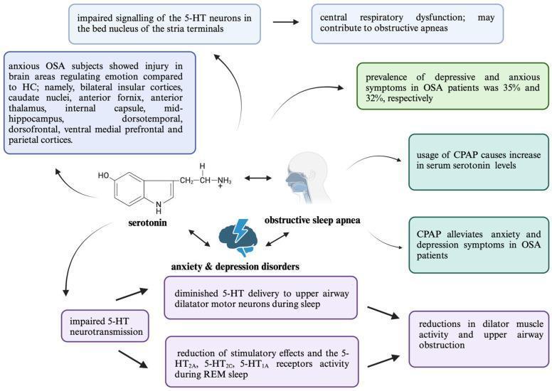
A medical infographic and pathophysiology diagram illustrating the bidirectional relationship between Obstructive Sleep Apnea (OSA), psychiatric comorbidities (anxiety and depression), and serotonin (5-HT) neurotransmission. The central visual elements include a chemical structure of serotonin and a sagittal head icon showing the upper airway. Key pathways include: 1) Pathophysiological mechanism where impaired 5-HT neurotransmission and reduced stimulatory effects on 5-HT2A, 5-HT2C, and 5-HT1A receptors during REM sleep lead to diminished upper airway dilator muscle activity and obstruction. 2) Neural injury patterns in anxious OSA subjects, highlighting damage in the bilateral insular cortices, caudate nuclei, anterior fornix, thalamus, internal capsule, hippocampus, and prefrontal/parietal cortices. 3) Clinical data indicating a prevalence of depressive and anxious symptoms at 35% and 32% respectively in OSA patients. 4) Therapeutic impact, showing that Continuous Positive Airway Pressure (CPAP) usage increases serum serotonin levels, subsequently alleviating comorbid psychiatric symptoms. This diagram serves as an educational summary of neurobiological changes and treatment responses in the context of sleep-disordered breathing and mood disorders.
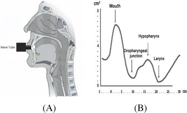
This educational composite visual illustrates acoustic pharyngometry, a non-invasive diagnostic method used to evaluate the upper airway anatomy, particularly in the context of Obstructive Sleep Apnea (OSA). Figure A is a sagittal anatomical diagram showing a wave tube inserted into the oral cavity. Visible landmarks include the nasal cavity, hard and soft palates, tongue, oropharynx, hypopharynx, larynx, and trachea, with the cervical vertebrae providing a posterior border. Figure B is a corresponding line graph plotting the airway cross-sectional area (cm²) against the distance from the mouth (cm). The graph identifies key diagnostic landmarks: a peak at the mouth (~7 cm²), a primary narrowing at the oropharyngeal junction (approx. 10 cm), a secondary widening at the hypopharynx, and a final constriction at the level of the larynx (~21 cm). This tool is clinically relevant for screening anatomical predispositions to airway collapse during wakefulness by identifying specific regions of reduced diameter or increased compliance within the respiratory tract.
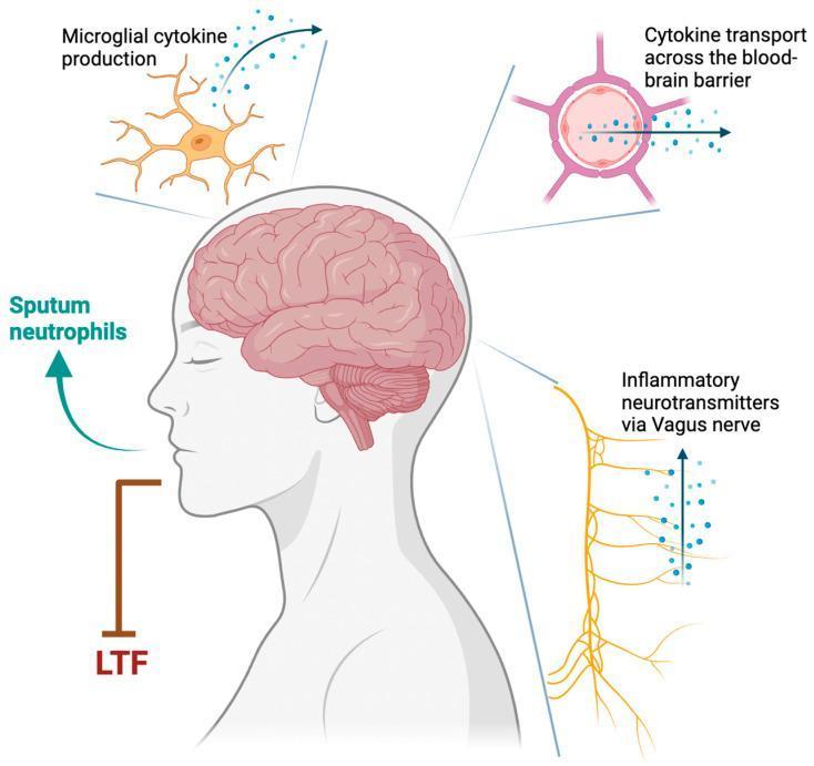
This pathophysiology diagram illustrates the mechanisms by which Type 1 inflammation in asthma may destabilize respiratory control, potentially contributing to obstructive sleep apnea (OSA). A sagittal profile of a human head and neck highlights the brain, brainstem, and central nervous system as the primary regulatory sites. Three key neural-immune pathways are detailed via callouts: 1) Microglial cytokine production, depicting an activated microglial cell with branching processes releasing signaling molecules; 2) Cytokine transport across the blood-brain barrier (BBB), shown as molecules moving from a peripheral blood vessel into the central nervous system parenchyma; and 3) Inflammatory neurotransmitters via the Vagus nerve, represented by an ascending neural pathway from the lungs/airways carrying afferent signals. Additional elements include 'Sputum neutrophils' indicating airway inflammation and 'LTF' (Long-Term Facilitation), representing a compensatory mechanism for respiratory stability that becomes compromised. The diagram serves as an educational tool for pulmonologists and neurologists to understand the neuro-inflammatory cross-talk involving central chemoreceptors and peripheral lung injury.
PMID: 41135142
GLP-1 Receptor Agonists in Obstructive Sleep Apnea (OSA)
A Simplified, Up-to-Date ENT Perspective
1. What Are GLP-1 Receptor Agonists?
| Drug | Brand Name | Type |
|---|---|---|
| Semaglutide | Ozempic / Wegovy | GLP-1 agonist |
| Liraglutide | Saxenda / Victoza | GLP-1 agonist |
| Tirzepatide | Zepbound / Mounjaro | Dual GIP + GLP-1 agonist |
- Reducing appetite (acting on the brain's hunger centers)
- Slowing gastric emptying (you feel full longer)
- Lowering blood sugar
- Promoting significant, sustained weight loss
2. Why Does OSA Relate to Obesity?
- Fat deposits around the neck and throat narrow the airway
- Tongue fat is a major contributor - weight loss directly reduces tongue fat volume, strongly linked to AHI (Apnea-Hypopnea Index) reduction
- Visceral fat presses on the diaphragm, reducing lung volumes and making airway collapse more likely
- Upper airway muscle tone is reduced in obese individuals
- Systemic inflammation from obesity destabilizes respiratory control
3. How Do GLP-1 Drugs Help OSA? (Mechanisms)
A. Primary Mechanism - Weight Loss
- Reduces adipose tissue around the pharynx and tongue
- Reduces neck circumference (a key OSA risk factor)
- Lowers pressure on the upper airway from peripharyngeal fat
- The AHI improvement tracks closely with the degree of weight loss achieved
B. Anti-inflammatory Effects
- Reduce circulating CRP (C-reactive protein) and other inflammatory markers
- Systemic inflammation disrupts upper airway muscle function; reducing it stabilizes the airway
- In SURMOUNT-OSA, high-sensitivity CRP levels fell significantly with tirzepatide
C. Central Nervous System Effects
- GLP-1 receptors exist in the brainstem and hypothalamus
- May improve ventilatory control stability (reduces loop gain - a key OSA endotype)
- May improve upper airway dilator muscle neuromuscular control, possibly independent of weight
D. Possible Weight-Independent Effects
- Early AHI improvements in SURMOUNT-OSA (Malhotra et al., Sleep Med 2025, PMID: 41135142) appeared as early as Week 4, before maximal weight loss - suggesting some direct non-weight mechanisms
- Research is still ongoing to separate weight vs. non-weight effects
4. The Clinical Evidence - Key Trials
🔬 SCALE Sleep Apnea Trial (Liraglutide) - 2016 Foundational Evidence
- Liraglutide 3.0 mg vs. placebo in obese patients with moderate-severe OSA
- Result: Greater AHI reduction than placebo; reduced body weight
- Set the stage for the GLP-1 and OSA story
🔬 SURMOUNT-OSA Trials (Tirzepatide) - The Game Changer
- 52 weeks, double-blind, placebo-controlled
- Published in NEJM (Malhotra et al., 2024)
- AHI reduced by ~29 events/hour (Study 1) and ~25 events/hour (Study 2) vs. placebo
- Average body weight loss: 18-20%
- Many participants moved from severe OSA to mild or near-remission
- AHI improvements detectable from Week 4 (Malhotra et al., Sleep Med 2025, PMID: 41135142)
- Significant reduction in hs-CRP (inflammation marker)
- Improvements in sleep-related quality of life and daytime sleepiness scores
5. Comparative Data (Real-World Evidence)
- PAP + semaglutide or tirzepatide had significantly lower rates of pulmonary hypertension and all-cause mortality vs. PAP alone at 1 and 3 years
- Tirzepatide outperformed semaglutide on these outcomes
- Tirzepatide reduced major adverse cardiovascular events (MACE) vs. liraglutide (HR 0.58) and vs. semaglutide (HR 0.86) in OSA + T2DM patients
- Tirzepatide also reduced incident OSA compared to liraglutide (HR 0.89)
6. From an ENT Perspective - What This Means Clinically
Who benefits most?
- Patients with obesity-driven OSA (BMI ≥30, large neck circumference, significant upper airway fat)
- Patients who are CPAP-intolerant or looking for adjunctive treatment
- Patients with comorbid T2DM, hypertension, or cardiovascular disease
Who may NOT benefit as much?
- Patients with OSA due to craniofacial anatomy (retrognathia, narrow jaw) - the airway is anatomically small regardless of weight
- Patients with enlarged tonsils or adenoids as the primary cause
- Patients with non-obese OSA (lean OSA patients driven by neuromuscular or anatomical factors)
- The AASM noted: GLP-1 drugs are expected to help only obesity-related OSA, not all OSA
Important ENT Perioperative Consideration
- GLP-1 drugs slow gastric emptying - patients on these medications are at increased aspiration risk under anesthesia
- Multisociety perioperative guidelines (2024) recommend holding GLP-1 drugs before elective ENT surgery
- ENT surgeons must ask about GLP-1 drug use during pre-op assessment
7. Does GLP-1 Replace CPAP?
| Parameter | CPAP | GLP-1 RA |
|---|---|---|
| Immediate AHI reduction | Yes (complete control) | Gradual (over months) |
| Disease modification | No | Yes (reduces fat load) |
| Systemic metabolic benefit | Limited | Significant |
| Works in non-obese OSA | Yes | No |
| Adherence challenge | High (device) | Injection/cost |
| FDA approved for OSA | No | Yes (tirzepatide only) |
8. Future Directions in ENT & Sleep Medicine
- Ongoing trials of semaglutide specifically for OSA (results pending)
- Research into weight-independent mechanisms (brainstem GLP-1 receptors, loop gain stabilization)
- Exploring GLP-1 drugs in patients undergoing OSA surgery to optimize body weight pre- and post-procedure
- Studying impact on upper airway anatomy (tongue fat, lateral wall fat) using MRI
- Whether GLP-1 drugs can reduce need for surgical intervention (tonsillectomy, UPPP, mandibular advancement)
Quick Summary
GLP-1 receptor agonists - especially tirzepatide (Zepbound) - represent a major step forward in OSA management. They work primarily through weight loss-mediated reduction of upper airway fat, but likely have additional anti-inflammatory and central effects. Tirzepatide is the first FDA-approved medication for OSA (December 2024). From an ENT standpoint, these drugs are most useful in obese patients with OSA, are not a replacement for CPAP, and require special consideration perioperatively due to aspiration risk. The evidence from SURMOUNT-OSA (RCT) and multiple real-world studies confirms clinically meaningful AHI reduction with good safety profiles.
- El-Solh et al., Expert Opin Pharmacother 2025 - narrative review of GLP-1 RAs in obesity-related OSA
- Malhotra et al., NEJM 2024 - SURMOUNT-OSA pivotal RCT
- Malhotra et al., Sleep Med 2025 (PMID: 41135142) - time course of tirzepatide effects in SURMOUNT-OSA
- Henney et al., Ann Am Thorac Soc 2025 (PMID: 40590655) - comparative real-world cardiovascular outcomes
- theentdr.com ENT-specific perspective by A/Prof Julius Goh, International Sleep Disorder Specialist