
Give dermatology images
Which dermatology topic would you like images for?
skin lesions tumors dermatology
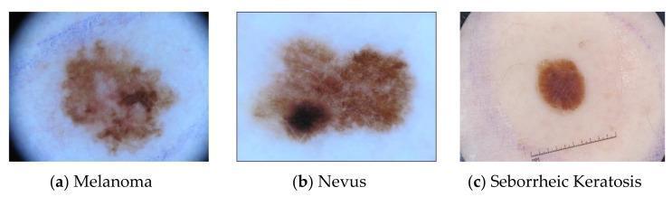
This set of three clinical dermoscopy images provides a comparative visual analysis of common skin lesions: (a) melanoma, (b) nevus, and (c) seborrheic keratosis. The melanoma image demonstrates classic ABCDE criteria, including asymmetry and irregular, notched borders with variegated pigmentation ranging from light tan to dark brown. The nevus displays a somewhat irregular but more uniform brown pigment network, though it contains a concentrated area of darker melanocytic activity. In contrast, the seborrheic keratosis presents as a well-circumscribed, more symmetrical, and uniformly dark brown plaque-like lesion with a characteristic 'stuck-on' appearance common in non-melanocytic epidermal tumors. All three images are captured under high-magnification dermoscopy to highlight surface patterns and pigment distribution. A measuring scale is visible in the seborrheic keratosis frame for size estimation. These images serve as educational benchmarks for differentiating malignant melanoma from benign melanocytic nevi and non-melanocytic keratinocytic lesions in dermatology and oncology curricula.
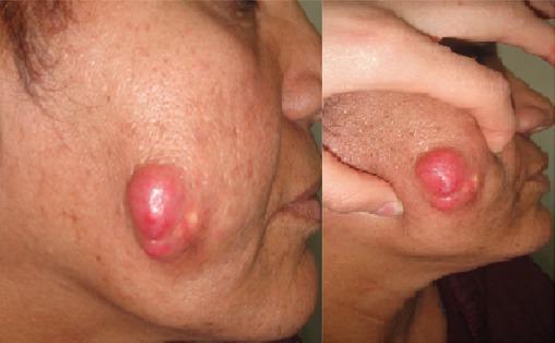
This clinical photograph consists of two views (profile and close-up) showing a prominent skin lesion on the left cheek of an adult patient. The lesion is a well-circumscribed, exophytic, polylobed nodular tumor with a distinctly erythematous or reddish-pink hue. The surface appears smooth, glistening, and tense, suggesting significant underlying pressure or fullness. There is visible evidence of subcutaneous infiltration, as the surrounding skin appears distorted and stretched. Adjacent to the primary mass, smaller pale-yellowish papular elevations are noted, which may represent secondary skin changes or satellite lesions. The medical specialty involved is dermatology or oncology, and the image illustrates the morphology of a firm, rapidly growing or infiltrative cutaneous tumor, requiring differential diagnosis between malignant processes (such as squamous cell carcinoma or Merkel cell carcinoma) and benign but aggressive entities. The educational focus is on identifying visual markers of cutaneous malignancy and the clinical presentation of infiltrative facial tumors.
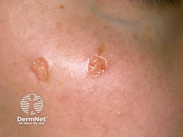
Clinical photograph of two small annular lesions on the zygomatic cheek. The imaging modality is standard color photography used in dermatology to document cutaneous tumors. The primary subject is annular trichoepithelioma, a benign adnexal neoplasm with hair follicle differentiation. The lesions appear as well-circumscribed circular plaques with slightly raised borders and a pale or atrophic center. Skin tone is light to medium; the surface is smooth with minimal scale; surrounding skin is non-inflammatory. The location corresponds to the zygomatic region of the face, within the aesthetic facial unit. The lesions are non-ulcerated, nonpalpable, and asymptomatic with no pigmentary change. Differential diagnosis includes basal cell carcinoma presenting as pearly annular rings, desmoplastic trichoepithelioma, and other adnexal tumors. Clinical significance lies in distinguishing benign trichoepitheliomas from malignant skin cancers to avoid overtreatment; biopsy or dermoscopic assessment may be considered if diagnostic uncertainty persists. This image is valuable for educational purposes, illustrating classic annular morphology and facial localization. Potential clinical use cases include dermatology training, teledermatology, lesion characterization in cutaneous tumor registries, and preoperative planning for excision with cosmetic preservation. No signs of invasion or systemic disease are apparent. Clinical notes recommended. Appropriate for patient counseling, documentation, and surgical planning in clinical practice.
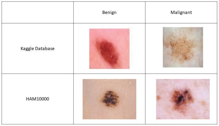
This comparison chart displays four dermoscopic images of skin lesions, organized into a 2x2 grid to illustrate the visual differences between benign and malignant tumors across two different medical datasets: Kaggle and HAM10000. In the Kaggle dataset row, the benign lesion shows a relatively symmetric, dark red-brown oval with regular borders, while the malignant lesion displays an irregular, pale tan-to-brown appearance with asymmetric pigmentation and peripheral white circular structures. In the HAM10000 row, the benign lesion is characterized by a well-demarcated, dark brown pigmentation with a fairly uniform texture. The corresponding malignant lesion exhibits hallmark features of melanoma, including asymmetry, irregular borders, and a variegated color pattern featuring a dark central focus with radial streaming or 'star-like' extensions. The image serves as an educational tool for clinical dermatology, highlighting key diagnostic morphological features such as pigment distribution, border regularity, and architectural patterns used in the differential diagnosis of skin cancer.
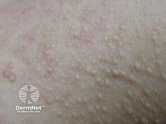
This is a dermatology clinical photograph showing multiple small, dermal papules on the facial skin, particularly on the cheeks and perioral region. The lesions are 1–3 mm, dome-shaped, skin-colored to slightly pale, with a smooth surface and firm consistency. Perifollicular localization is evident, with subtle shading around each papule reflecting superficial dermal involvement. The photo is a close-up macro view, suitable for documenting cutaneous follicular lesions. The pattern is often bilateral and symmetrical and may extend to the neck or trunk in some cases. These lesions are characteristic of fibrofolliculomas, benign hamartomas of facial hair follicles. If numerous or associated with renal tumors or pulmonary cysts, consider Birt-Hogg-Dubé syndrome. Differential diagnosis includes milia, syringomas, sebaceous hyperplasia, trichoepithelioma, and folliculitis. Clinically, the presence of multiple lesions warrants assessment for systemic associations and family history. In practice, management includes observation for cosmetic concerns, laser or surgical removal for selected lesions, and genetic counseling if a hereditary syndrome is suspected. For diagnostic confirmation, dermatopathology can reveal the classic epithelial strands in fibrous stroma. Imaging-wise, this photo supports a dermatology diagnosis and should be correlated with patient history and potential renal/pulmonary imaging if there is systemic suspicion. Consult dermatology and genetics teams for evaluation.
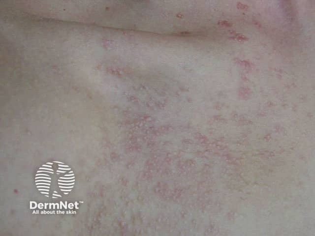
Clinical photograph (DermNet watermark) of the skin showing multiple small follicular papules with mild erythema. The image depicts a close-up, macro view of the integumentary surface, suitable for dermatologic morphology assessment. Primary subject: fibrofolliculomas, benign hair-follicle–origin hamartomas often appearing as dome-shaped, flesh-colored papules; in some individuals they may appear erythematous or coalescent in plaques. Here, the lesions are discrete to slightly grouped, with perifollicular localization and a surface that is smooth or gently papular. The distribution is trunk/torso region with no obvious mucosal involvement; there is no scale, crust, or central keratin plug evident in this frame. The image emphasizes follicular localization, hinting at adnexal origin. The technique is noninvasive clinical photography with standard lighting; no contrast or staining is used. This depiction is intended for documentation, pattern recognition, and differential diagnosis of follicular tumors in dermatology. Diagnostic significance: when multiple fibrofolliculomas are present, consider inherited syndromes such as Birt-Hogg-Dubé; correlate with patient history, lung imaging, renal surveillance. Differential includes trichofolliculoma, sebaceous hyperplasia, syringoma, trichoepithelioma, and other adnexal tumors. Clinical correlation and, if indicated, histopathology confirm diagnosis. Clinical utility includes educational use for students and trainees, aiding pattern recognition, differential narrowing, and patient counseling about benign prognosis and potential systemic associations.
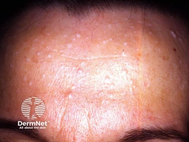
Clinical photograph of facial skin (dermatology) showing multiple fibrofolliculomas on the forehead. This frontal close-up capture highlights numerous small, dome-shaped, skin-colored papules arranged in a diffuse distribution over the frontal region and upper cheeks. The lesions are firm to touch, non-tender, and primarily atop pilosebaceous units with perifollicular accentuation. The overlying epidermis appears intact with minimal erythema; there is no evident ulceration or crusting. In life, such lesions are benign adnexal neoplasms composed of epithelial strands arising from hair follicles within a fibrous stroma; histology typically reveals anastomosing cords of basaloid cells embedded in a fibrous matrix, often with horn cysts. Clinically, the presence of multiple fibrofolliculomas, especially in conjunction with other facial papules, may suggest Birt-Hogg-Dubé syndrome, mandating assessment for renal imaging and pulmonary cysts and familial counseling. Differential considerations include trichofolliculoma, perifollicular fibroma, sebaceous hyperplasia, and milia. This image is valuable for dermatology education, clinic documentation, cosmetic planning, and teaching differential diagnosis of follicular papules. It supports correlation with genetic testing and multidisciplinary management when systemic associations are suspected. Useful for medical students, residents, and fellows refining pattern recognition of facial adnexal tumors. This description emphasizes clinical morphology, correlates with histology, and supports educational annotation for imaging repositories.
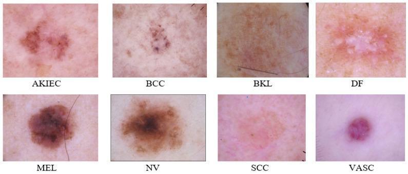
This clinical comparison chart presents eight high-resolution dermoscopic images representing common benign and malignant skin lesions from the ISIC 2019 dataset. The visual compilation serves as a diagnostic reference for dermatology. (1) AKIEC (Actinic Keratosis/Intraepithelial Carcinoma): shows brownish-red, scaly patches with irregular borders. (2) BCC (Basal Cell Carcinoma): displays dark, translucent spots with telangiectasia. (3) BKL (Benign Keratosis-like Lesion): exhibits a uniform light brown pigment and smooth texture. (4) DF (Dermatofibroma): characterized by a central white scar-like area with a peripheral reddish-brown halo. (5) MEL (Melanoma): demonstrates classic asymmetry, irregular borders, and variegated dark brown to black pigmentation. (6) NV (Melanocytic Nevus): shows a symmetric, well-defined brown lesion with uniform pattern. (7) SCC (Squamous Cell Carcinoma): presents as an erythematous, scaly plaque with poorly defined margins. (8) VASC (Vascular Lesion): appears as a well-circumscribed, cherry-red or purple papule. This comparison aids in the differential diagnosis of pigmented and non-pigmented skin tumors based on morphology, color, and border characteristics.
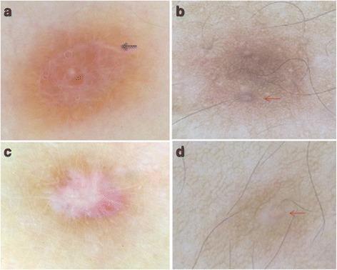
This composite diagnostic image displays four dermoscopic views (labeled a-d) of skin lesions, specifically common fibrous dermatofibromas (CFDF), showcasing atypical dermoscopic patterns. Frame 'a' demonstrates total homogeneous light-brown pigmentation with distinct white structures forming a ring-like configuration (indicated by a black arrow). Frames 'b' and 'd' show a peripheral pigment network that incorporates unusual white rings encircling follicular openings (indicated by red arrows), a pattern deviating from classic dermatofibroma presentation. Frame 'c' illustrates a discreet peripheral pigment network surrounding a central, star-shaped white patch. These images provide critical educational value for dermatology and oncology by documenting rare morphological variants of dermatofibromas, such as the white ring around follicles or total homogeneous patterns, which must be differentiated from other melanocytic and non-melanocytic skin tumors.
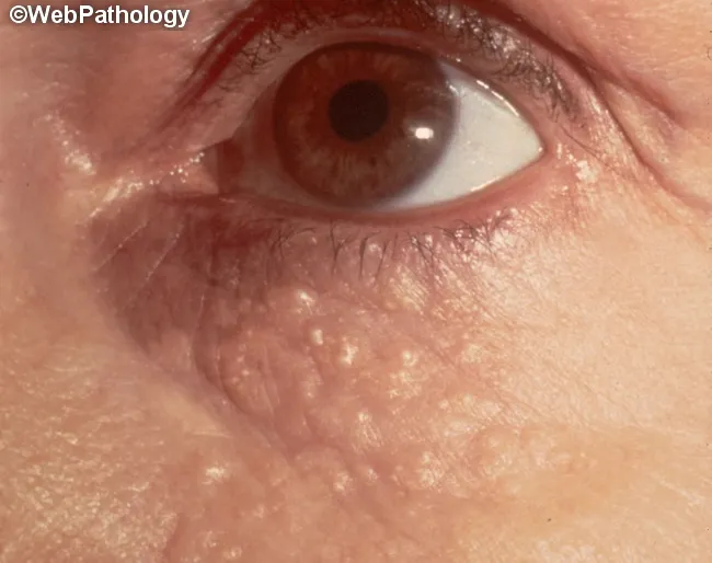
Clinical photography of the periorbital skin demonstrates multiple small, firm, yellow to skin‑colored papules clustered around the eyelids. Each lesion measures approximately 1–3 mm in diameter and lacks ulceration or crusting. The distribution is predominantly periorbital, with preference for the periorbital eyelid skin and adjacent malar area, consistent with syringomas—benign eccrine sweat duct tumors. The image emphasizes a tangential, close‑up view suitable for surface dermatology assessment, with good color fidelity to reveal the characteristic papule texture. In this condition, lesions represent benign proliferations of eccrine ducts within the dermis and are non-inflammatory, usually asymptomatic, though they may be cosmetically distressing. Differential diagnosis includes milia, xanthelasma, sebaceous hyperplasia, and other adnexal tumors; however, syringomas typically present as small, skin‑colored or yellow papules in clusters around the eyelids, whereas milia are white and more superficial. The clinical significance lies in distinguishing benign syringomas from other eyelid lesions to guide management, which is often cosmetic (laser ablation, electrodessication, or topical therapies) rather than urgent. This image is valuable for dermatology and pathology education, clinic photo atlases, and differential diagnosis training in eyelid lesions and periorbital dermatology. Correlates with onset in adolescence or early adulthood and a female predilection; may accompany genetic syndromes in rare cases.
1. Melanoma vs. Nevus vs. Seborrheic Keratosis (Dermoscopy)
2. Infiltrative Facial Tumor
3. Annular Trichoepithelioma (Benign Adnexal Tumor)
4. ISIC 2019 Dermoscopy Reference Panel
5. Benign vs. Malignant Dermoscopy (Kaggle / HAM10000)
6. Dermatofibroma - Atypical Dermoscopic Patterns
7. Fibrofolliculomas - Facial (Birt-Hogg-Dubé Associations)
8. Syringomas - Periorbital (Benign Eccrine Tumors)