
Types of mi and Omi vs nomi
Reading File
Now let me search the medical library for MI types and STEMI/NSTEMI comparisons in parallel.
Finding Sources
Finding Sources
Reading File
Reading File
Reading File
Reading File
Reading File
Finding Sources
Finding Sources
Searching the Web
OMI vs NOMI occlusion myocardial infarction paradigm STEMI NSTEMI
I now have comprehensive data from the textbooks and the web. Let me fetch the ECG diagram images and compile the full answer.
Loading Image
Loading Image
Loading Image
Here is a complete breakdown of MI types and the OMI vs. NOMI paradigm.
Types of Myocardial Infarction
Part 1 - The Universal Classification (Types 1-5)
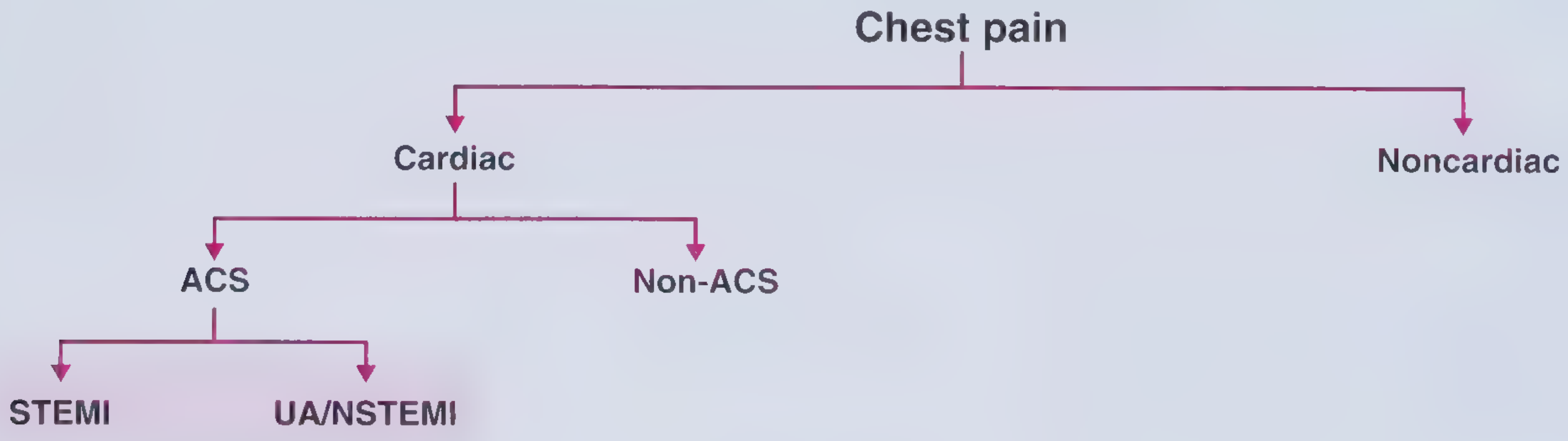
The Fourth Universal Definition of MI (Thygesen et al., 2018) classifies MI into 5 mechanistic types:
| Type | Name | Mechanism |
|---|---|---|
| 1 | Spontaneous MI | Atherosclerotic plaque rupture, ulceration, fissuring, erosion, or dissection - causes intraluminal thrombus formation in a coronary artery. May occur with or without obstructive CAD. |
| 2 | MI secondary to ischemic imbalance | Myocardial necrosis NOT caused by plaque rupture, but by an imbalance of oxygen supply/demand - e.g. coronary spasm, embolism, tachyarrhythmia, bradyarrhythmia, anemia, hypotension, respiratory failure, hypertension. |
| 3 | MI causing death before biomarkers available | Sudden cardiac death with ischemic symptoms + presumed new ECG changes (STE or new LBBB), but patient dies before blood samples are drawn or before troponin rises. |
| 4a | PCI-related MI | Troponin rise >5x 99th percentile (or >20% rise if already elevated) within 48h of PCI, plus supporting findings (symptoms, ECG changes, angiographic evidence, or imaging). |
| 4b | Stent thrombosis MI | MI detected by angiography or autopsy in the setting of confirmed stent thrombosis with troponin rise/fall above the 99th percentile. |
| 5 | CABG-related MI | Troponin >10x 99th percentile in patients with normal baseline, plus new pathologic Q waves, new LBBB, graft/native artery occlusion, or imaging evidence of new loss of myocardium. |
Source: Tietz Textbook of Laboratory Medicine, 7th Edition; Harrison's Principles of Internal Medicine 22E (2025)
Part 2 - The Clinical ECG-Based Classification: STEMI vs. NSTEMI
This is the classic bedside triage classification, determined by the 12-lead ECG on presentation.
| Feature | STEMI | NSTEMI | Unstable Angina (UA) |
|---|---|---|---|
| ECG | New ST elevation ≥0.1 mV in ≥2 contiguous leads (≥0.2 mV in V2-V3 men ≥40 yrs; ≥0.25 mV men <40 yrs; ≥0.15 mV women) | ST depression ≥0.05 mV or T-wave inversion ≥0.1 mV in ≥2 contiguous leads; no STE | Normal or non-specific changes |
| Troponin | Elevated (rises markedly) | Elevated | Not elevated |
| Pathology | Usually complete/near-complete coronary occlusion | Partial occlusion or severe stenosis | Severe stenosis without necrosis |
| Q waves | Develop if reperfusion not achieved | Generally absent | Absent |
| Treatment | Emergent reperfusion (PCI within 90 min or fibrinolysis) | Urgent/early invasive strategy | Anti-ischemic/antiplatelet therapy; risk-stratify |
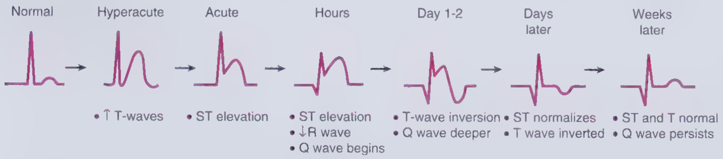
ECG evolution in STEMI:
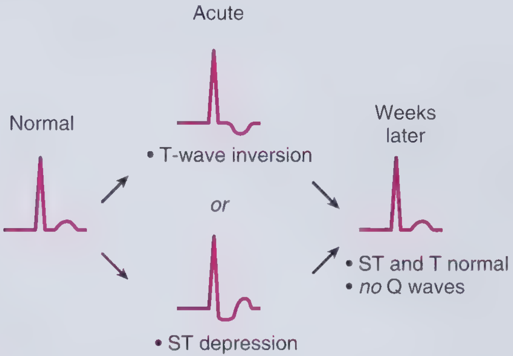
ECG findings in UA/NSTEMI:
Source: Frameworks for Internal Medicine; Harrison's Principles of Internal Medicine 22E
Part 3 - The Newer Paradigm: OMI vs. NOMI
This is a proposed paradigm shift that focuses on the underlying coronary anatomy rather than ECG thresholds.
Definitions
| Term | Full Name | Meaning |
|---|---|---|
| OMI | Occlusion MI | Acute total or near-total occlusion of a major epicardial coronary vessel with insufficient collateral circulation - causing imminent myocyte necrosis. Requires emergent reperfusion. |
| NOMI | Non-Occlusion MI | AMI without angiographic, laboratory, or clinical evidence of acute coronary occlusion. Less emergent anatomy. |
Why the Paradigm Was Proposed
The traditional STEMI/NSTEMI system uses STE on ECG as a surrogate for coronary occlusion. This has two critical failure modes:
-
False negatives (STEMI- OMI): ~25-30% of NSTEMI patients actually have an acute coronary occlusion (OMI) that is missed because they don't meet formal STEMI criteria. These patients suffer delays to catheterization and have approximately double the short- and long-term mortality compared to NSTEMI without occlusion (NOMI). ECG patterns often missed include posterior STEMI, de Winter T-waves, hyperacute T-waves, and subtle STE.
-
False positives: 15-35% of cath lab activations triggered by apparent STEMI criteria find no culprit occlusion.
How OMI and NOMI Map onto the Old System
Old System          New System
─────────────────────────────────────────────────
STEMI               = STEMI(+) OMI  (true OMI with STE - most)
                    + STEMI(+) NOMI (false positive STEs - 15-35%)

NSTEMI              = STEMI(-) OMI  (missed occlusions - 25-30%)
                    + NOMI          (true non-occlusion MIs)
Key Clinical Implication
The real split that determines outcome and treatment urgency is OMI vs. NOMI, not STEMI vs. NSTEMI:
- All OMI patients (whether or not they have STE) benefit from emergent reperfusion
- NOMI patients do not benefit from emergent intervention and can be managed with a more elective early invasive strategy
Advanced ECG interpretation (hyperacute T-waves, de Winter pattern, posterior leads, Wellens pattern) combined with bedside echo and, increasingly, AI tools is the main approach to identifying STEMI(-) OMI.
The DIFOCCULT study (Aslanger et al., 2020) showed ECG reviewers identified ACO in 28.2% of cases originally classified as NSTEMI, with significantly higher in-hospital and long-term mortality in that reclassified group. Meyers et al. (2021) formally compared the two paradigms in a single population, confirming STEMI(-) OMI patients had similarly severe angiographic and peak troponin outcomes as STEMI(+) OMI, but with significantly longer door-to-balloon times.
Quick Summary Table
| STEMI | NSTEMI | OMI | NOMI | |
|---|---|---|---|---|
| Based on | ECG | ECG + troponin | Coronary anatomy | Coronary anatomy |
| Coronary occlusion? | Usually yes | Sometimes (25-30%) | Yes (by definition) | No |
| Emergent PCI needed? | Yes | Not always | Yes (by definition) | No |
| Troponin elevated? | Yes | Yes | Yes | Yes |
The OMI/NOMI framework is not yet universally adopted in guidelines, but it is increasingly influential in emergency cardiology and ECG interpretation training.
This is a shared conversation. Sign in to Orris to start your own chat.