
chronic inflammation
Reading File
Now let me search the medical library and PubMed in parallel for comprehensive coverage of chronic inflammation.
Finding Sources
Searching PubMed
"chronic inflammation"[MeSH Terms] AND mechanisms
Good - I have the main Robbins Pathology sources. Let me read the primary sections in parallel.
Reading File
Reading File
Excellent. Let me now read the detailed morphologic features and cellular mechanisms section.
Reading File
Finding Sources
Let me read more of the chronic inflammation section - the lymphocyte role, other cells, and granulomatous section.
Reading File
I now have comprehensive content. Let me fetch the key diagrams to include in the answer.
Loading Image
Loading Image
I now have all the content needed for a comprehensive answer. Let me also fetch the granuloma image.
Loading Image
I now have everything needed for a thorough, well-illustrated answer on chronic inflammation.
Chronic Inflammation
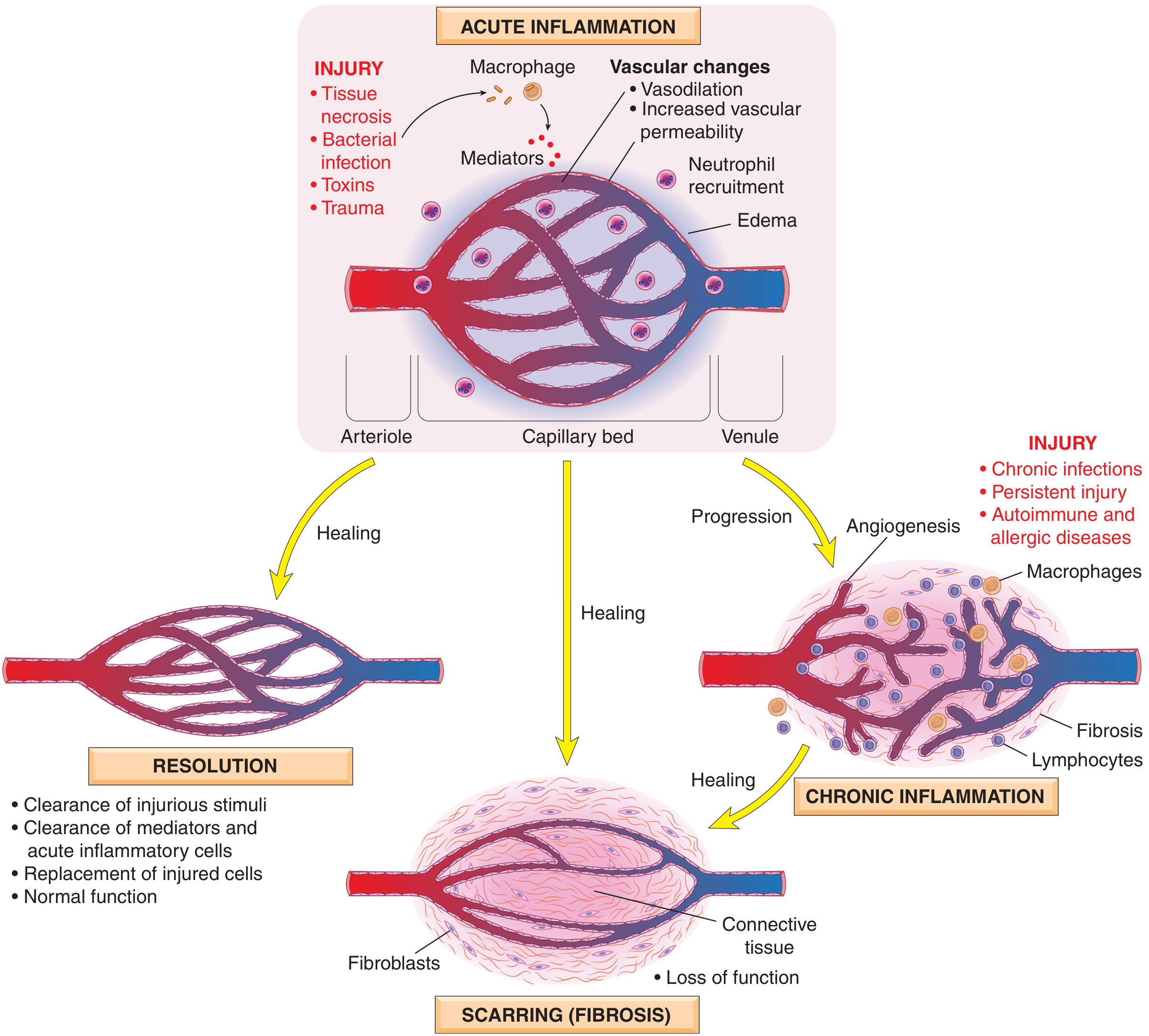
Definition: Chronic inflammation is a response of prolonged duration (weeks to months) in which inflammation, tissue injury, and attempts at repair coexist simultaneously. It may follow unresolved acute inflammation, or arise insidiously without a preceding acute phase - as a smoldering, progressive process. - Robbins & Kumar Basic Pathology, p. 61
Causes
Three main settings trigger chronic inflammation:
-
Persistent infections - Microorganisms that resist eradication: mycobacteria (TB, leprosy), certain viruses, fungi, and parasites. Incompletely resolved acute infection (e.g., bacterial lung abscess) may also evolve into chronicity.
-
Hypersensitivity diseases:
- Autoimmune: Self-perpetuating immune reactions against autoantigens (rheumatoid arthritis, multiple sclerosis, inflammatory bowel disease)
- Allergic: Excessive response to environmental antigens (bronchial asthma)
- These often show mixed acute and chronic patterns due to repeated flares
-
Prolonged exposure to toxic agents:
- Exogenous: inhaled silica particles → silicosis
- Endogenous: excess cholesterol deposition → atherosclerosis
Chronic inflammation is also implicated in diseases not classically thought of as inflammatory, including Alzheimer disease, metabolic syndrome, and type 2 diabetes.
Outcomes of Acute Inflammation
Morphologic Features (Histology)
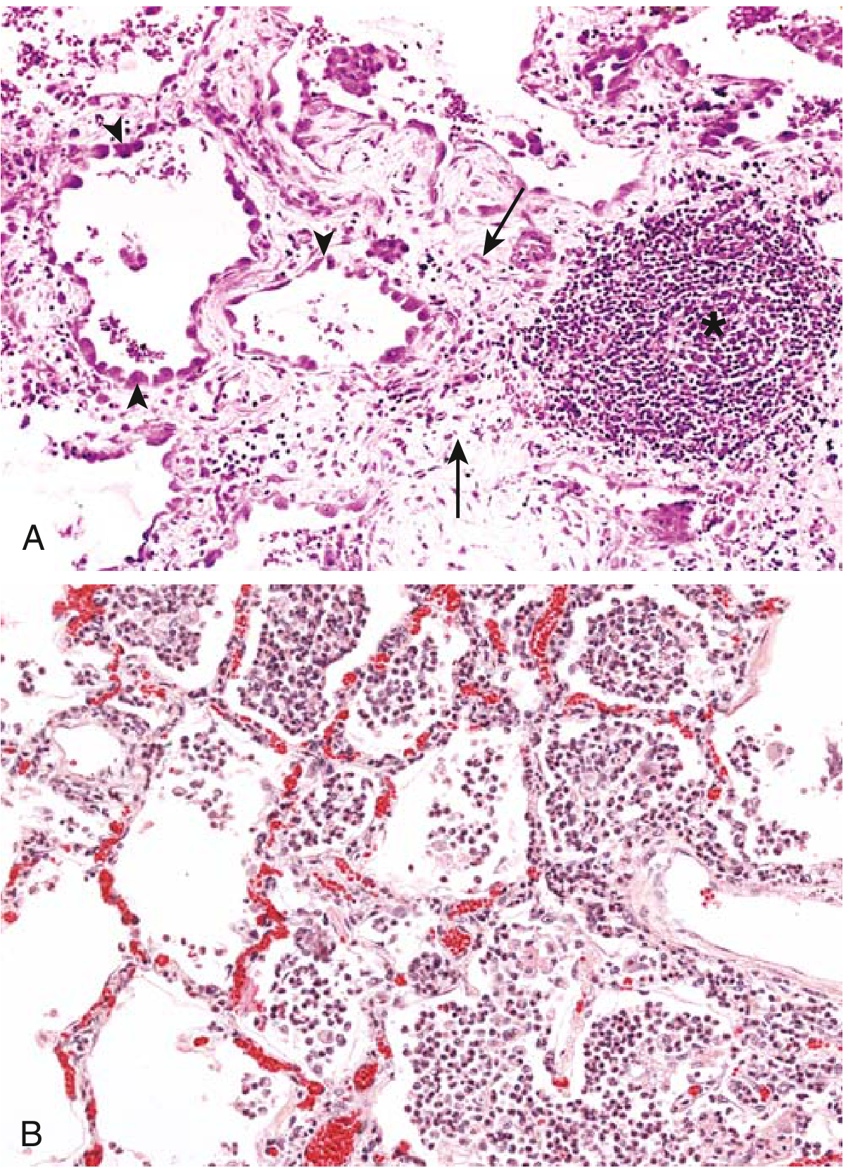
Chronic inflammation is characterized by three hallmark features (seen together):
| Feature | Details |
|---|---|
| Mononuclear cell infiltration | Macrophages, lymphocytes, plasma cells (not neutrophils) |
| Tissue destruction | Caused by the persistent stimulus and inflammatory cells |
| Repair attempts | Angiogenesis + fibrosis → scar formation |
Cells and Mediators
Macrophages (dominant cell)
Macrophages are the dominant cells in most chronic inflammatory reactions. They are derived from blood monocytes (bone marrow origin) that migrate into tissues. Tissue-resident macrophages include Kupffer cells (liver), microglial cells (CNS), alveolar macrophages (lung), and sinus histiocytes (lymph nodes).
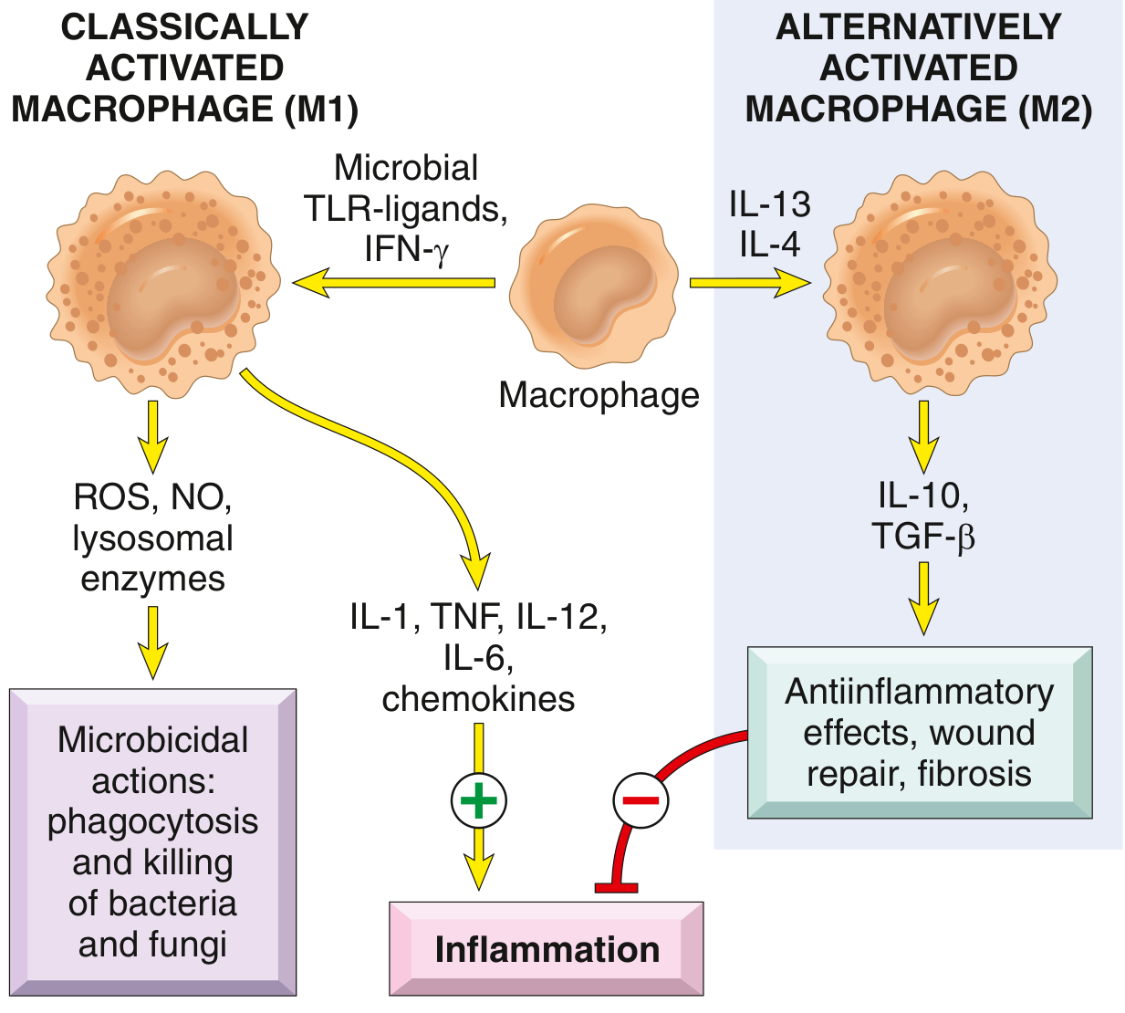
Two functional phenotypes:
| Type | Trigger | Products | Function |
|---|---|---|---|
| M1 (Classical) | Microbial TLR ligands, IFN-γ | ROS, NO, lysosomal enzymes; IL-1, TNF, IL-12, IL-6, chemokines | Microbicidal, pro-inflammatory |
| M2 (Alternative) | IL-4, IL-13 | IL-10, TGF-β | Anti-inflammatory, wound repair, fibrosis |
Key macrophage functions in chronic inflammation:
- Phagocytosis and killing of microbes / dead tissue
- Secretion of cytokines (TNF, IL-1, chemokines) and eicosanoids - propagating the reaction
- Initiation of tissue repair and fibrosis
- Antigen presentation to T lymphocytes, forming a feedback loop
Lymphocytes
- Activated by microbes and antigens; amplify and perpetuate chronic inflammation
- CD4+ T cell subsets drive different inflammatory patterns:
- Th1 → IFN-γ → classical macrophage activation (M1)
- Th2 → IL-4, IL-5, IL-13 → alternative macrophage activation (M2), eosinophil recruitment
- Th17 → IL-17 → chemokine secretion → neutrophil recruitment
- Th1 and Th17 are involved in chronic autoimmune diseases (e.g., rheumatoid arthritis, IBD)
- Th2 drives allergic inflammation and anti-helminth responses
- B lymphocytes and plasma cells secrete antibodies; present in chronic inflammatory foci
Other Cells
- Eosinophils - characteristic of allergic reactions and parasitic infections; IgE and IL-5 mediated
- Mast cells - release histamine and cytokines; present in connective tissue; participate in both acute and chronic phases
- Neutrophils - typically acute, but can persist in some chronic contexts (e.g., bacterial osteomyelitis, certain autoimmune diseases)
Granulomatous Inflammation
A specialized form of chronic inflammation characterized by aggregates of activated macrophages (epithelioid cells), often surrounded by lymphocytes, and sometimes with multinucleated giant cells and central caseous necrosis.
Mechanism: Th1 lymphocytes secrete IFN-γ → activates macrophages → epithelioid transformation. When the agent cannot be eliminated, the granuloma persists.
Causes:
| Category | Examples |
|---|---|
| Infectious | Tuberculosis (caseating granulomas), leprosy, syphilis, fungal infections (histoplasma, coccidioides), schistosomiasis |
| Non-infectious | Sarcoidosis, Crohn disease, berylliosis, foreign body reactions |
Systemic Effects (Acute Phase Response)
Chronic (and acute) inflammation triggers systemic effects via cytokines (mainly IL-1, TNF, IL-6):
| Effect | Mechanism |
|---|---|
| Fever | Cytokines (TNF, IL-1) → PGE2 production in hypothalamus |
| Leukocytosis | Bone marrow release of leukocytes; colony-stimulating factors increase production |
| Acute-phase proteins | IL-6 → liver synthesis of CRP, fibrinogen, serum amyloid A |
| Septic shock (severe) | Very high TNF levels → hypotension, DIC, metabolic abnormalities |
Outcomes of Chronic Inflammation
- Resolution - if the cause is eliminated (rare in true chronic disease)
- Fibrosis / Scarring - the most common outcome; TGF-β is the key fibrogenic mediator
- Tissue loss and organ dysfunction - e.g., cirrhosis, pulmonary fibrosis, joint destruction
Key Diseases with Chronic Inflammation as Core Pathology
| Disease | Dominant Mechanism |
|---|---|
| Rheumatoid arthritis | Autoimmune Th1/Th17 synovitis |
| Bronchial asthma | Th2-driven eosinophilic airway inflammation |
| Atherosclerosis | Lipid-driven vascular macrophage activation |
| Tuberculosis | Granulomatous Th1 response to M. tuberculosis |
| Crohn disease | Th1/Th17 transmural bowel inflammation |
| Cirrhosis | Persistent hepatic injury + stellate cell-driven fibrosis |
| Silicosis | Macrophage activation by indigestible silica particles |
Sources: Robbins & Kumar Basic Pathology (Robbins Pathology), pp. 61-65; Robbins, Cotran & Kumar Pathologic Basis of Disease.
This is a shared conversation. Sign in to Orris to start your own chat.