
Identifying feature of thyroid gland
Reading File
The thyroid gland's identifying histological feature is the presence of follicles - spherical structures lined by follicular epithelial cells and filled with colloid (thyroglobulin). This is the classic hallmark under microscopy.
Searching sources...
Finding Sources
Finding Sources
Finding Sources
Finding Sources
Reading File
Reading File
Loading Image
Loading Image
Reading File
Here is a complete account of the identifying features of the thyroid gland, drawn from authoritative histology and physiology textbooks.
Identifying Feature of the Thyroid Gland
The Thyroid Follicle - The Key Identifying Feature
The thyroid follicle is the structural and functional unit of the thyroid gland - this is its single most important identifying feature under both gross and microscopic examination.
Each follicle is:
- A roughly spherical, cyst-like compartment (diameter 0.2-1.0 mm)
- Walled by a simple cuboidal to low columnar epithelium (follicular epithelium)
- Filled with a gel-like material called colloid (composed predominantly of the glycoprotein thyroglobulin)
Hundreds of thousands of these follicles constitute nearly the entire mass of the human thyroid gland.
Histology (H&E stain):
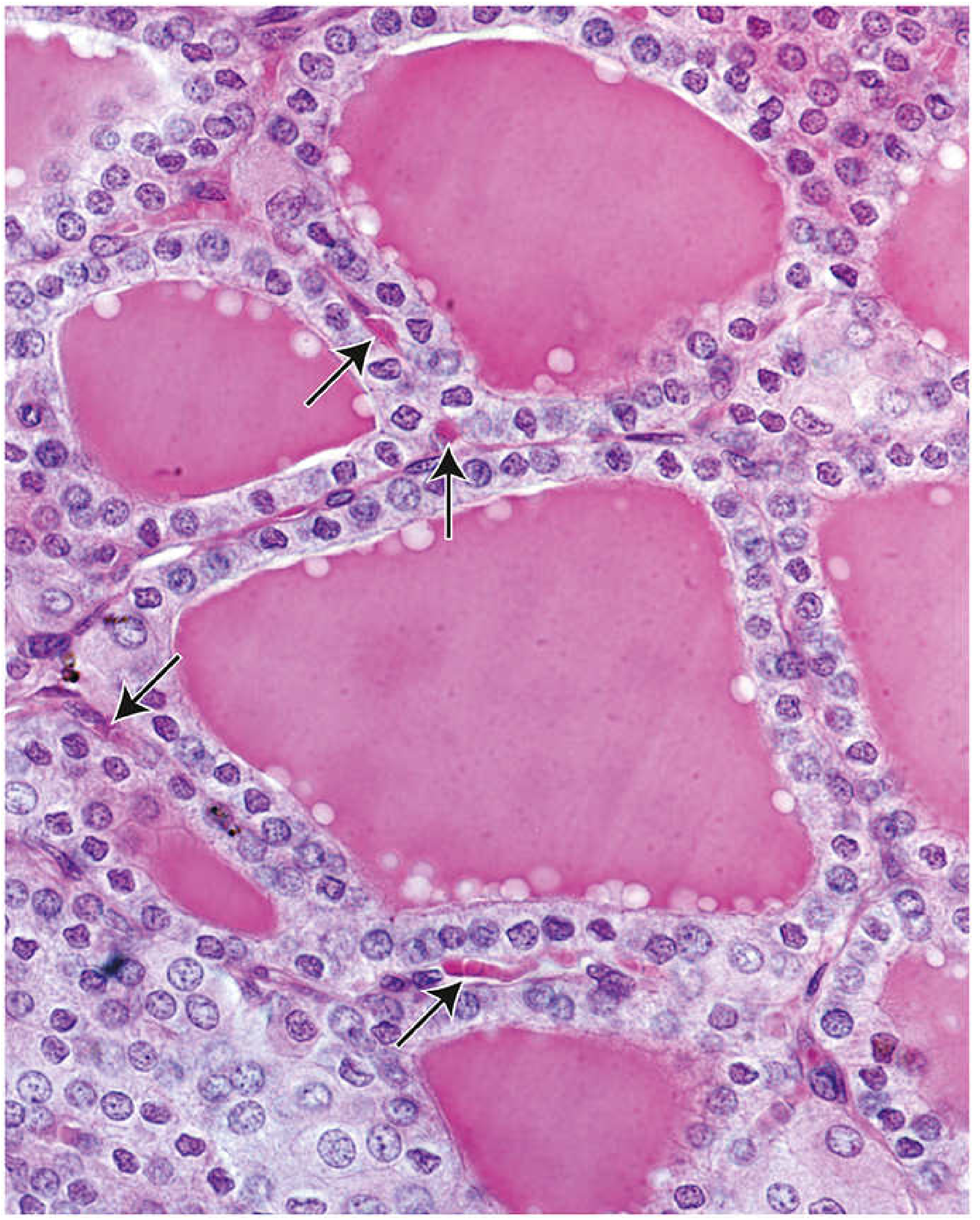
H&E stain of human thyroid. The large pink spaces are colloid-filled follicles, each surrounded by a single layer of follicular epithelial cells. Arrows indicate blood capillaries between follicles. x500. - Histology: A Text and Atlas
Two Cell Types in the Follicular Epithelium
| Cell Type | Also Called | Function | Appearance (H&E) |
|---|---|---|---|
| Follicular cells | Principal cells / Thyrocytes | Produce T3 and T4 | Cuboidal; slightly basophilic cytoplasm; spherical nucleus with prominent nucleoli |
| Parafollicular cells | C cells | Secrete calcitonin | Pale-staining; located at periphery of follicular epithelium, never exposed to follicle lumen |
Functional State Changes the Appearance
The appearance of follicles changes with thyroid activity - this is a classic exam point:
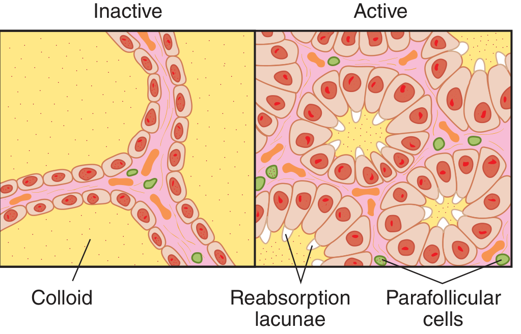
| State | Follicle Size | Epithelial Cell Shape | Colloid | Feature |
|---|---|---|---|---|
| Inactive (resting) | Large | Flat / squamous | Abundant | No reabsorption lacunae |
| Active (stimulated) | Small | Cuboidal to columnar | Reduced | Reabsorption lacunae visible at colloid-cell interface |
Reabsorption lacunae (also called "scalloping") - small punched-out spaces at the periphery of the colloid next to follicular cells - are the hallmark of an actively secreting thyroid gland.
Summary of Key Identifying Features
| Feature | Detail |
|---|---|
| Follicles with colloid | Spherical structures filled with pink, homogeneous colloid on H&E |
| Simple cuboidal epithelium | Lines each follicle; varies with activity |
| Colloid = thyroglobulin | PAS-positive material; precursor of T3/T4 |
| Parafollicular (C) cells | Pale, intercalated between follicular cells; secrete calcitonin |
| Scalloping/Reabsorption lacunae | Seen at colloid periphery in active gland |
| Rich capillary network | Fenestrated capillaries between follicles |
The colloid-filled follicle lined by follicular epithelium is what immediately identifies the thyroid gland in any histological section - no other endocrine gland has this arrangement.
Sources: Histology: A Text and Atlas with Correlated Cell and Molecular Biology; Ganong's Review of Medical Physiology, 26th Edition
This is a shared conversation. Sign in to Orris to start your own chat.