
Ques. A 42-year-old woman comes to the Medicine OPD with a painless swelling in the front of her neck for the past six months. The swelling has gradually increased in size. She has no difficulty in swallowing, no change in voice, and no symptoms of hyperthyroidism or hypothyroidism. On examination, there is a solitary, firm thyroid nodule measuring 3 × 2 cm that moves with swallowing. No cervical lymph nodes are palpable. Q1. What is the most likely diagnosis in this patient? Q2.What is the first laboratory investigation you would order for this patient? Q3.Which investigation is most appropriate to evaluate the characteristics of this thyroid nodule? Q4. If the patient's TSH level is low, what should be the next investigation? Q5.The most appropriate investigation to determine whether this nodule is benign or malignant before surgery is. Q6.Which complication should every patient be informed about before thyroid surgery? Q7.A thyroid nodule is classified as TI-RADS 5 on ultrasound. What is the next best step?
Thyroid Nodule - Clinical Case Analysis
Q1. Most Likely Diagnosis
Q2. First Laboratory Investigation
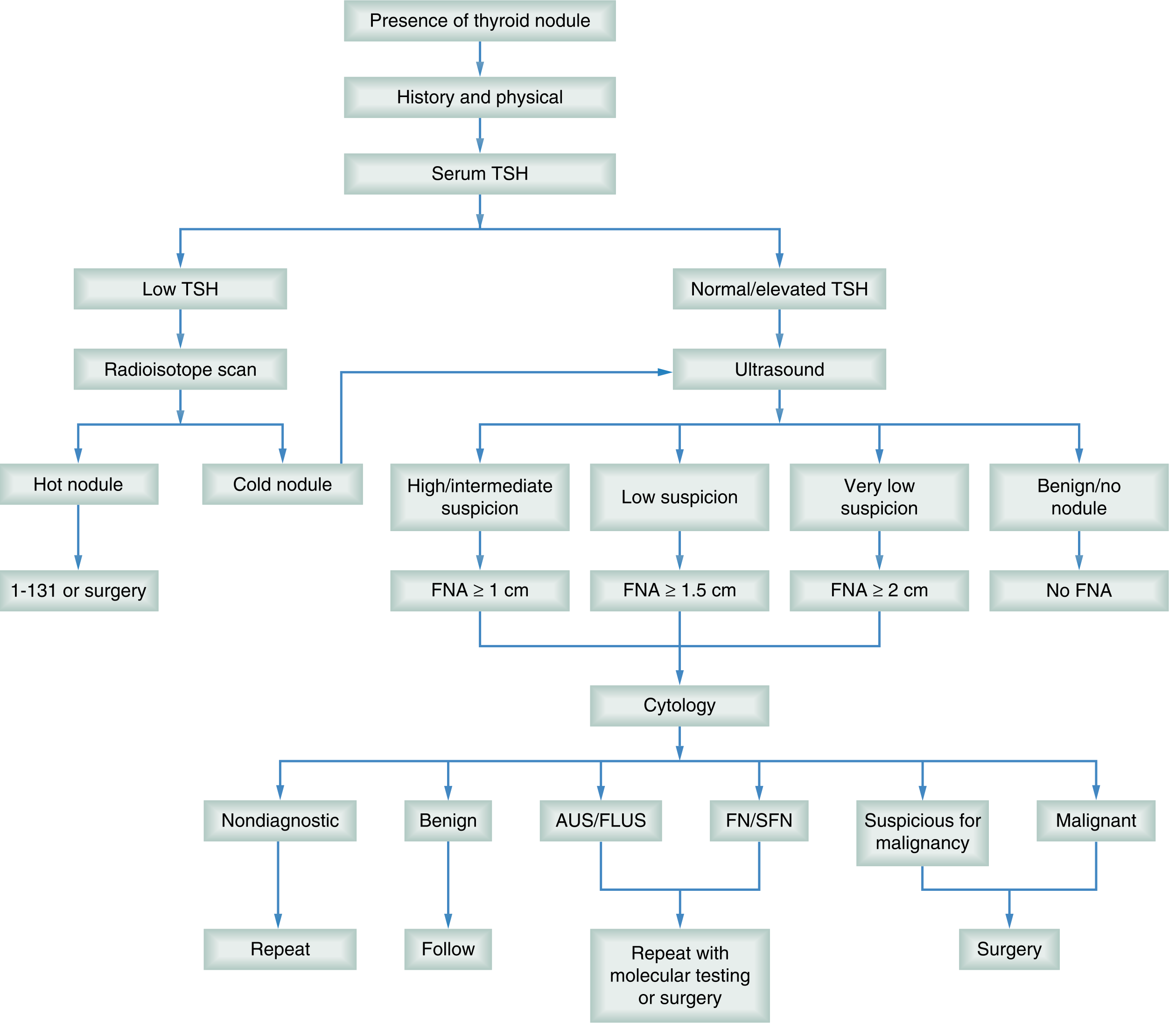
Q3. Most Appropriate Investigation to Evaluate Nodule Characteristics
- Nodule size, location, number
- Cystic vs solid content
- Echogenicity, margins, calcifications
- Doppler vascularity pattern
- Assessment of cervical lymph nodes
- Risk stratification using ACR TI-RADS scoring
- Guidance for FNA biopsy
Q4. If TSH is Low - Next Investigation
- Hot nodule (increased uptake) = autonomous nodule - treatment with I-131 or surgery; FNA generally not needed
- Cold nodule (decreased uptake) = non-functioning nodule - proceed to ultrasound and FNA to rule out malignancy
Q5. Most Appropriate Investigation to Determine Benign vs Malignant (Before Surgery)
- "FNA biopsy is the most accurate and cost-effective invasive procedure in the workup of thyroid nodules. For all thyroid nodules, FNA has a mean sensitivity of over 80% and a mean specificity of over 90%." - Sabiston's
- "Fine-needle aspiration cytology (FNAC) is the diagnostic procedure of choice" - Cummings Otolaryngology
- Results are classified using the Bethesda System for Reporting Thyroid Cytopathology (6 categories), with associated malignancy risk for each category
- "Successful FNAC categorizes nodules as benign, malignant, or suspicious. In 60% to 90% of nodules, FNAC reveals a benign or 'negative' diagnosis. The likelihood of malignancy (false-negative rate) is 1% to 6%." - Cummings Otolaryngology
- Ultrasound-guided FNAC has superior accuracy vs palpation-guided
Q6. Complication Every Patient Must Be Informed About Before Thyroid Surgery
- Transient RLN palsy: 0.7-5.1%
- Permanent RLN palsy: 0.2-1.0%
- Hypocalcaemia/hypoparathyroidism (from parathyroid gland damage, especially with bilateral surgery): persistent in 0.3-6.3%
- Transient hypocalcaemia: more common
- Haemorrhage (rare but life-threatening due to airway compression)
- Wound infection, hypothyroidism (after total thyroidectomy)
Q7. TI-RADS 5 on Ultrasound - Next Best Step
- TR5 nodules ≥1 cm → FNA biopsy is indicated
- The FNA result then guides the decision for surgery
Summary Table
| Question | Answer |
|---|---|
| Q1. Diagnosis | Solitary thyroid nodule (likely benign colloid nodule/follicular adenoma) |
| Q2. First lab investigation | Serum TSH |
| Q3. Best investigation to characterize nodule | USG neck (ultrasonography) |
| Q4. If TSH is low, next investigation | Radionuclide thyroid scan (Tc-99m/I-123 scintigraphy) |
| Q5. Best investigation to determine benign vs malignant | FNAC (Fine Needle Aspiration Cytology) |
| Q6. Complication to inform about before surgery | Recurrent Laryngeal Nerve injury (hoarseness) + Hypoparathyroidism |
| Q7. TI-RADS 5 nodule - next step | FNA biopsy |
- Sabiston Textbook of Surgery, 21st ed., Chapter 73 (Thyroid Nodule workup, TI-RADS, FNA)
- Schwartz's Principles of Surgery, 11th ed., Chapter 38 (Solitary Thyroid Nodule)
- Cummings Otolaryngology Head and Neck Surgery (FNAC, Imaging, RLN)
- Scott-Brown's Otorhinolaryngology Head & Neck Surgery (Surgical complications)