
Bones and joints of thorax in detail
Reading File
Now let me search the medical library for thorax bones and joints content in parallel.
Finding Sources
Finding Sources
Finding Sources
Finding Sources
Gray's Anatomy for Students is the best source. Let me read the thorax section from that book, along with the THIEME Atlas sternum section.
Reading File
Reading File
Good content is coming in. Let me read more from both books - the Gray's Anatomy thorax section from earlier (bones/sternum/ribs parts) and the THIEME Atlas continuing section on ribs and joints.
Reading File
Reading File
Excellent content. Let me read the Gray's Anatomy section on ribs (typical and atypical) and the joints of the thorax from the THIEME Atlas.
Reading File
Reading File
I have excellent, detailed content now. Let me also quickly fetch the key images to include in the answer.
Reading File
Reading File
I now have comprehensive content. Let me read just a bit more on the sternocostal/interchondral joints and fetch the key images.
Reading File
Loading Image
Loading Image
I have excellent, comprehensive content from Gray's Anatomy for Students and the THIEME Atlas. Let me now compose the detailed answer.
Bones and Joints of the Thorax
The thoracic wall is built from four skeletal elements: thoracic vertebrae (12), intervertebral discs, ribs (12 pairs), and the sternum. Together they form the thoracic cage, which protects the thoracic viscera and changes volume during breathing.
1. Thoracic Vertebrae
There are 12 thoracic vertebrae (T1-T12), each uniquely characterized by articulations with ribs.
Typical Thoracic Vertebra (T2-T9)
- Body: Heart-shaped, roughly equal in transverse and anteroposterior dimensions
- Vertebral foramen: Circular (smaller than cervical)
- Spinous process: Long, pointing steeply downward
- Laminae: Broad and overlapping with those of the vertebra below
- Superior articular processes: Flat, articular surfaces facing almost directly posteriorly
- Inferior articular processes: Project from laminae, articular facets facing anteriorly
- Transverse processes: Club-shaped, projecting posterolaterally
Costal Facets on a Typical Vertebra
Each typical thoracic vertebra has three articulation sites per side for ribs:
- Superior costal demifacet (on body) - for part of the head of its own rib
- Inferior costal demifacet (on body) - for part of the head of the rib below
- Transverse costal facet (on transverse process) - for the tubercle of its own rib
Atypical Thoracic Vertebrae
| Vertebra | Special Feature |
|---|---|
| T1 | Superior costal facet is complete (full facet, not a demifacet) for the entire head of rib I; also has inferior demifacet for rib II |
| T10 (often T9) | Only single complete costal facet per side; no inferior demifacets |
| T11 & T12 | Only one complete costal facet per side; no transverse costal facets |
- Gray's Anatomy for Students, p. 179-180
2. Ribs
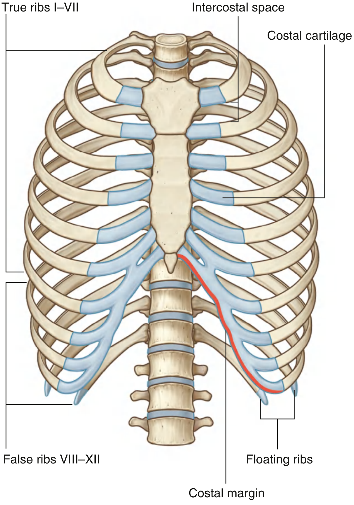
There are 12 pairs of ribs, all articulating posteriorly with the vertebral column and each ending anteriorly in a costal cartilage.
Classification
| Class | Ribs | Anterior attachment |
|---|---|---|
| True ribs | I-VII | Costal cartilages attach directly to sternum |
| False ribs | VIII-XII | Do NOT attach directly to sternum |
| - Vertebrochondral | VIII-X | Costal cartilages join the cartilage of the rib above |
| - Floating ribs | XI-XII | No anterior connection to ribs or sternum |
Structure of a Typical Rib (Ribs III-IX)
Each rib has a posterior end (articulating with vertebrae) and an anterior end (continuing as costal cartilage). The bony segments from posterior to anterior are:
1. Head
- Somewhat expanded, typically presenting two articular facets separated by a crest
- Smaller superior facet: articulates with the inferior costal demifacet of the vertebra above
- Larger inferior facet: articulates with the superior costal demifacet of its own vertebra
- The crest connects via an intra-articular ligament to the adjacent intervertebral disc
2. Neck
- Short, flat region of bone connecting the head to the tubercle
- Bears a sharp superior ridge (crest of the neck) on ribs 2-12
- Directed posterosuperiorly
3. Tubercle
- At the junction of neck and shaft
- Has an articular facet (for the transverse process of its own vertebra) medially
- Has a non-articular roughened area (for ligament attachment) laterally
4. Shaft (Body)
- Curved, with the sharpest bend at the costal angle (just lateral to the tubercle)
- On its inferior inner surface: the costal groove - protects the intercostal vessels and nerves (absent on ribs 1, 11, 12)
- Shafts of ribs 2-12 are twisted about their long axis: external surfaces face slightly downward at the vertebral end and slightly upward at the anterior end
Rib lengths: Rib 1 and 12 are the shortest; rib 7 is the longest. Costal cartilage increases in length from rib 1 to 7, then shortens again.
Atypical Ribs
Rib I
- Broadest and most curved; lies in an almost horizontal plane
- Flat (superior/inferior surfaces rather than lateral/medial)
- Has a scalene tubercle on its superior surface (for scalenus anterior)
- Grooves for the subclavian artery and vein on either side of the scalene tubercle
- Head has a single facet (articulates only with T1)
- No costal groove
Rib II
- Flat like rib I, but twice as long
- Articulates with the vertebral column in a typical fashion (with T1 and T2)
Rib X
- Head has a single facet for articulation with only its own vertebra (T10)
Ribs XI and XII (Floating Ribs)
-
Articulate only with the bodies of their own vertebrae
-
No tubercles, no necks (short and pointed anteriorly)
-
No articulation with transverse processes
-
Gray's Anatomy for Students, p. 180-182; THIEME Atlas of Anatomy, p. 156
3. Sternum
The sternum is a flat, elongated bone lying in the midline of the anterior chest wall. It is slightly convex anteriorly and has multiple costal notches along its lateral borders.
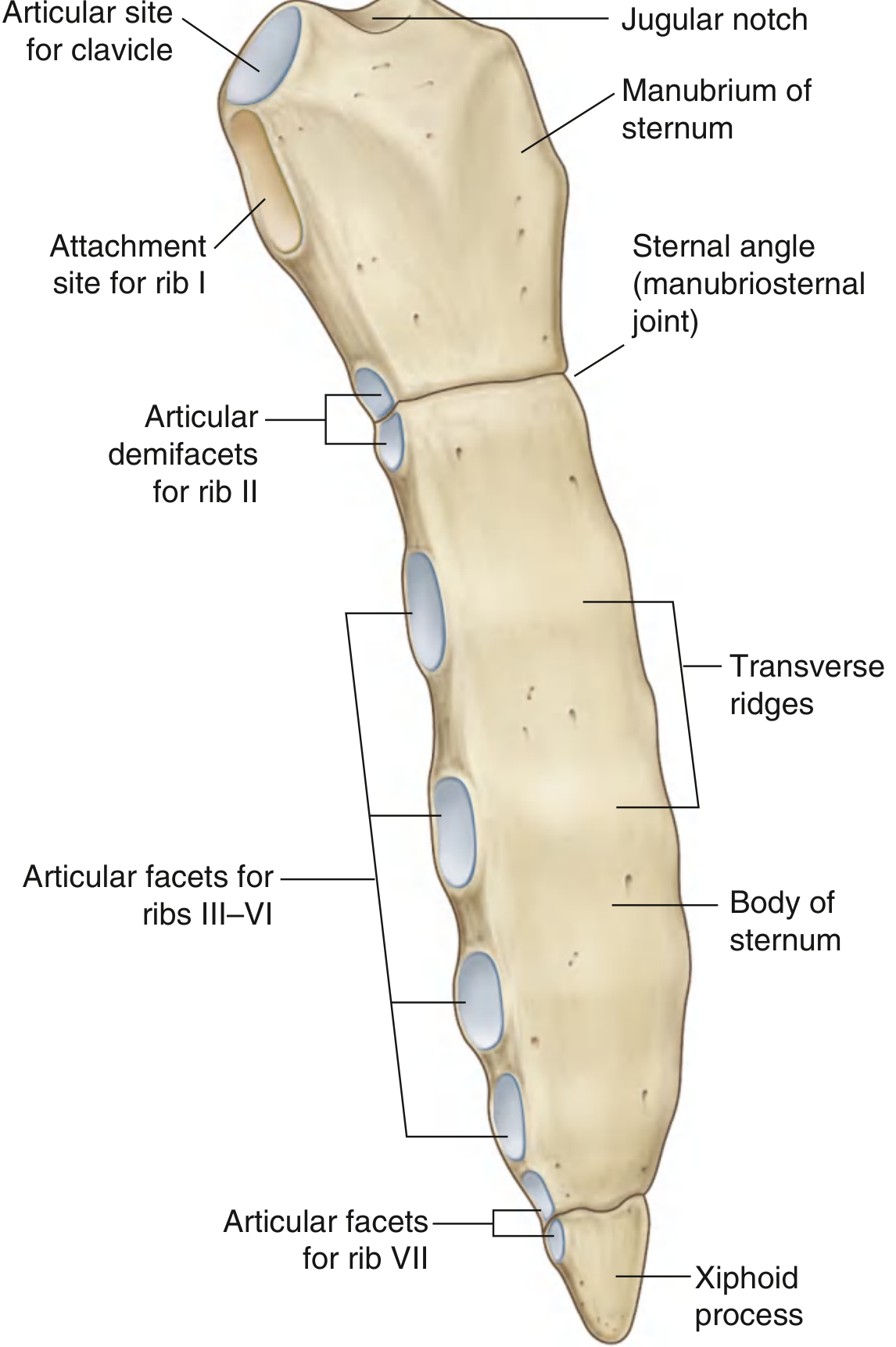
The adult sternum has three parts:
A. Manubrium
- The broadest, most superior part
- Superior border: wide, with a palpable jugular notch (suprasternal notch) in the midline
- On each side of the jugular notch: a large oval clavicular notch for articulation with the clavicle
- Immediately inferior to the clavicular notch on each side: a first costal notch (synchondrosis with rib I)
- Lower lateral border: a demifacet for the upper half of the second costal cartilage
- Clinical: The subcutaneous position of the sternum allows sternal bone marrow aspiration for diagnosis of blood diseases like leukemia
B. Body of the Sternum
- Flat; formed by the fusion of four embryonic sternebrae (visible as transverse ridges on the anterior surface)
- Lateral borders bear costal notches for ribs III to VII
- At its superior end: a demifacet completing the articulation for the second costal cartilage (paired with the manubrium's demifacet)
- At its inferior end: a demifacet for the upper part of the seventh costal cartilage
C. Xiphoid Process
- Smallest and most inferior part
- Highly variable shape: may be wide, thin, pointed, bifid, curved, or perforated
- Begins as cartilage, ossifies in adulthood (but may remain cartilaginous in some adults)
- Upper lateral margin on each side: a demifacet for the seventh costal cartilage
- Has no direct costal cartilage attachments of its own
Sternal Angle (Angle of Louis)
-
Formed at the junction of manubrium and body of sternum - a slight posterior angulation
-
A key clinical landmark: marks the level of the 2nd costal cartilage (the second rib is the most superior palpable rib), the bifurcation of the trachea (T4/T5), and the superior border of the pericardium
-
Gray's Anatomy for Students, p. 182-183; THIEME Atlas of Anatomy, p. 155
4. Joints of the Thorax
A. Costovertebral Joints (Joints of the Rib Head)
Formed between the head of the rib and the costal facets on two adjacent vertebral bodies (plus the intervertebral disc between them).
- Type: Synovial (plane/gliding)
- The joint is divided into two synovial compartments by an intra-articular ligament that attaches the crest of the head of the rib to the intervertebral disc
- Surrounded by a single fibrous joint capsule
Ligaments:
- Radiate (stellate) ligament: Fan-shaped; from the head of the rib to the bodies of both adjacent vertebrae and the intervertebral disc between them. This is the primary stabilizing ligament of the joint.
- Intra-articular ligament: Connects the crest of the rib head to the intervertebral disc; divides the joint into two compartments
Exceptions:
- Rib I, X (and often IX), XI, XII: head articulates with only one vertebra - joint has a single compartment and no intra-articular ligament
Movement: The two compartments allow rotation of the rib neck about its longitudinal axis (upper ribs) or gliding ascent/descent (lower ribs)
B. Costotransverse Joints
Synovial joints between the tubercle of the rib and the transverse process of its own vertebra.
- Type: Synovial (plane/gliding)
- Thin, weak fibrous capsule
- Absent for ribs XI and XII (which have no transverse costal facets on T11/T12)
Ligaments (all extracapsular):
| Ligament | Attachment | Position |
|---|---|---|
| Costotransverse ligament | Neck of rib → transverse process (same level) | Medial to joint |
| Lateral costotransverse ligament | Tip of transverse process → non-articular tubercle | Lateral to joint |
| Superior costotransverse ligament | Superior surface of rib neck → transverse process of vertebra above | Superior |
Movement: Slight gliding movements
C. Sternocostal (Sternocostal) Joints
Between the costal cartilages of ribs I-VII and the sternum.
- Rib I: Not synovial - fibrocartilaginous synchondrosis (primary cartilaginous joint) between manubrium and first costal cartilage
- Ribs II-VII: Synovial joints with thin capsules reinforced by sternocostal ligaments
Special feature of rib II: The joint is divided into two compartments by an intra-articular ligament attaching the second costal cartilage to the manubriosternal junction (sternal angle).
Ligaments:
- Radiate sternocostal ligaments: Radiate from the perichondrium of the costal cartilage to the anterior (and posterior) surface of the sternum, blending with the periosteum to form a dense fibrous membrane called the sternal membrane
D. Interchondral Joints
Between the costal cartilages of adjacent ribs - mainly ribs VII to X (may also include V and VI).
- Type: Usually synovial, with thin fibrous capsules reinforced by interchondral ligaments
- Provide indirect anchorage to the sternum
- Contribute to the smooth inferior costal margin (the oblique ridge running from rib VII to rib X along the lower thorax)
E. Joints Within the Sternum
Manubriosternal joint (Sternal angle / Louis' angle)
- Type: Symphysis (secondary cartilaginous joint)
- Between manubrium and body of sternum
- Slight angular movements during respiration
- Remains cartilaginous in most adults; may ossify in older age
Xiphisternal joint
-
Type: Symphysis
-
Between body and xiphoid process
-
Often ossifies with age
-
Gray's Anatomy for Students, p. 184-186; THIEME Atlas of Anatomy, p. 155
5. Movements at the Thoracic Joints and Respiration
The axes of rib movement run parallel to the necks of the ribs:
- Upper ribs (I-VI): Axes closer to the coronal plane → rib elevation increases the anteroposterior (sagittal) diameter of the thorax ("pump-handle" movement)
- Lower ribs (VII-X): Axes closer to the sagittal plane → rib elevation increases the transverse diameter of the thorax ("bucket-handle" movement)
This coordinated movement at costovertebral and costotransverse joints, combined with diaphragmatic descent, drives the volume changes needed for breathing.
Summary Table: Joints of the Thorax
| Joint | Type | Bones | Key Ligaments |
|---|---|---|---|
| Joint of rib head | Synovial (plane) | Rib head + 2 vertebral bodies | Radiate, intra-articular |
| Costotransverse | Synovial (plane) | Rib tubercle + transverse process | Costotransverse, lateral CT, superior CT |
| Sternocostal (rib I) | Synchondrosis | Rib I cartilage + manubrium | - |
| Sternocostal (ribs II-VII) | Synovial | Costal cartilages + sternum | Radiate sternocostal |
| Interchondral (VII-X) | Synovial | Adjacent costal cartilages | Interchondral |
| Manubriosternal | Symphysis | Manubrium + body | Fibrocartilage disc |
| Xiphisternal | Symphysis | Body + xiphoid | Fibrocartilage disc |
Sources: Gray's Anatomy for Students (9780323934237); THIEME Atlas of Anatomy: General Anatomy and Musculoskeletal System (9781626237186)
This is a shared conversation. Sign in to Orris to start your own chat.