
squamous cell Ca from first aid for Fcps exam prep
Reading File
Squamous cell carcinoma (SCC) is a malignant epithelial tumor arising from squamous cells, occurring in skin, head/neck, lung, esophagus, cervix, and more. Key features include keratin pearls on histology, risk factors like UV exposure/smoking/HPV, and local invasion with potential for metastasis.
Searching sources...
Finding Sources
Finding Sources
The user asks for "First Aid" content - let me check if there's a First Aid book in the library, and also search for pathology-focused SCC content (which is what FCPS exams typically require).
Finding Sources
Finding Sources
No "First Aid" book in the library. I'll use Robbins & Kumar (the primary source for FCPS pathology prep) and also pull from dermatology/surgery sources for comprehensive coverage.
Finding Sources
Finding Sources
Now let me read the key SCC sections from Robbins (both Basic Pathology and Pathologic Basis of Disease) for FCPS-level detail:
Reading File
Reading File
Now let me also read the Robbins, Cotran & Kumar (large Robbins) block8 for SCC - this will cover oral/esophageal/lung SCC:
Reading File
Finding Sources
Now let me read the lung SCC and head/neck SCC sections, plus fetch the histology images:
Reading File
Reading File
Now let me fetch the key histology images for visual reference:
Loading Image
Loading Image
I now have comprehensive data from Robbins. Let me compile the full FCPS-level answer on Squamous Cell Carcinoma.
Squamous Cell Carcinoma (SCC) - FCPS Exam Prep
Based on Robbins & Kumar Basic Pathology and Robbins, Cotran & Kumar Pathologic Basis of Disease
Overview
SCC is a malignant tumor of squamous epithelial cells. It arises at multiple sites - skin, oral cavity/oropharynx, lung, esophagus, cervix, and vulva. The skin is the most common site.
CUTANEOUS SCC
Epidemiology
- Second most common skin malignancy (after BCC)
- Higher incidence in males (except lower leg lesions)
- Arises on sun-exposed sites in older adults with lighter skin types
- Responsible for ~8,000 deaths/year from metastatic disease (roughly equal to metastatic melanoma)
Risk Factors / Etiology
| Factor | Notes |
|---|---|
| UV light (most important) | DNA damage, high mutational load; proportional to lifetime sun exposure |
| Xeroderma pigmentosum | Defective nucleotide excision repair of pyrimidine dimers - exceptionally high risk |
| Immunosuppression | Organ transplant recipients, chemotherapy - reduces immune surveillance |
| HPV (subtypes 5, 8, 16) | Genital skin (HPV-16); rare epidermodyplasia verruciformis (HPV-5/8) |
| Industrial carcinogens | Tars, oils, arsenicals |
| Chronic non-healing ulcers | Marjolin's ulcer - SCC in burn scars, draining osteomyelitis |
| Ionizing radiation | |
| Actinic keratosis | Premalignant precursor lesion |
Pathogenesis / Molecular Genetics
- TP53 mutations - most common; found in actinic keratoses (early event); occur at pyrimidine dimer sites (UV signature)
- RAS activating mutations - increase proliferation
- NOTCH loss-of-function - Notch receptors normally regulate squamous differentiation; loss promotes uncontrolled growth
- UV light also causes transient defect in cutaneous innate immunity
Morphology
Gross:
- In situ: sharply defined, red, scaly plaques (Bowen's disease)
- Invasive: nodular, keratotic/scaly, may ulcerate; raised indurated borders
Microscopy:
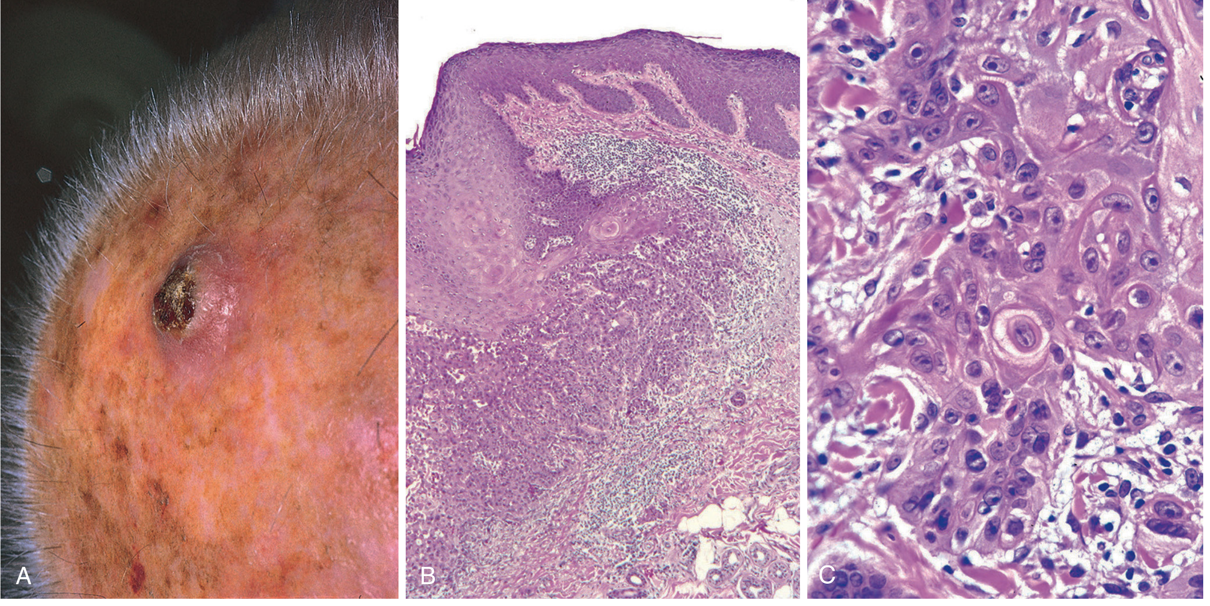
Fig. 25.13 Invasive squamous cell carcinoma. (A) Nodular ulcerated scalp lesion. (B) Atypical squamous epithelium transgressing basement membrane with a keratin pearl at center. (C) High-power showing keratin pearls and individual cell dyskeratosis. (Robbins, Cotran & Kumar)
- In situ: atypical hyperchromatic nuclei at ALL levels of epidermis (full-thickness atypia)
- Invasive (well-differentiated): polygonal cells in orderly lobules, abundant keratinization, keratin pearls (whorled concentric lamellae of keratin - pathognomonic)
- Invasive (poorly differentiated): highly anaplastic cells, foci of necrosis, only abortive single-cell keratinization (dyskeratosis); may need IHC for keratins to confirm
- Variable degrees of differentiation between these extremes
Actinic keratosis (precursor):
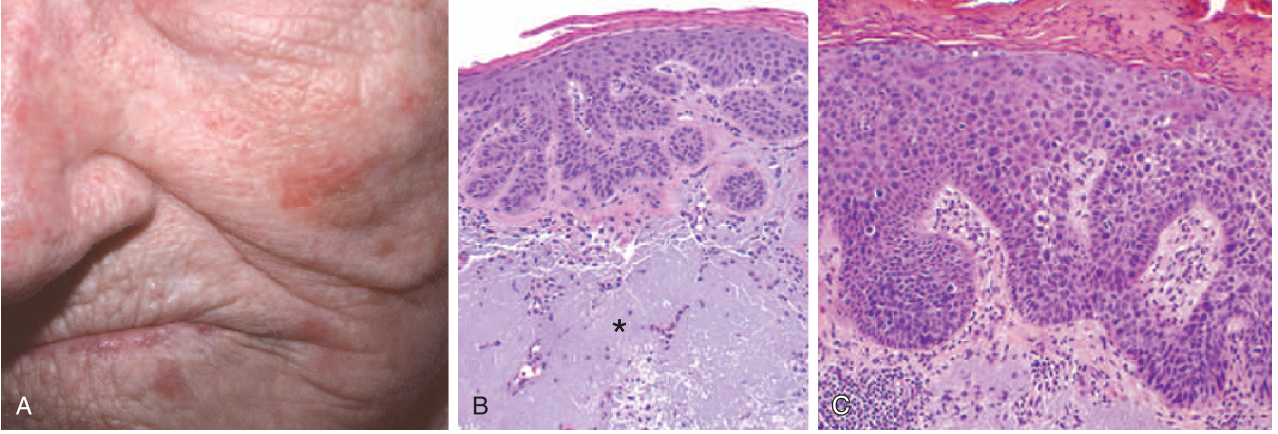
Fig. 22.15 Actinic keratosis and SCC in situ. (A) Red-rough sandpaper lesions on cheek/nose. (B) Basal atypia with hyperkeratosis and dermal solar elastosis (asterisk). (C) Full-thickness epithelial atypia = SCC in situ. (Robbins Basic Pathology)
Clinical Features & Prognosis
- Most discovered small and easily resectable
- ~4-5% have regional lymph node metastases at diagnosis
- Metastasis risk increases with: greater thickness, deeper invasion into subcutis, location near ear/lips
- Actinic keratosis-derived SCC: locally aggressive but slow to metastasize
- Burn scar/ulcer/non-sun-exposed SCC: more aggressive biologically
- Treatment of metastatic disease: high mutational burden → responds well to immune checkpoint inhibitors (first-line for metastatic disease); also sensitive to radiotherapy
HEAD & NECK SCC (HNSCC)
- ~95% of all head and neck cancers are SCC
- 6th most common neoplasm worldwide; >650,000 cases/year globally
Two Distinct Pathogenic Pathways:
1. Carcinogen-associated (tobacco/alcohol):
- Oral cavity (ventral tongue, floor of mouth, lower lip, soft palate, gingiva)
- Mutations: TP53, RAS, NOTCH (same as cutaneous)
- "Field cancerization" concept: multiple independent clones develop throughout mucosa due to chronic carcinogen exposure → risk of synchronous/metachronous second primaries
2. HPV-associated:
- Oropharynx: tonsillar crypts, base of tongue (~90% of tonsillar SCC = HPV+)
- HPV-16 most common subtype (high-risk)
- Spread via orogenital sex
- E6 oncoproteins inhibits p53; E7 inhibits RB
- Overexpress p16 (cyclin-dependent kinase inhibitor) - used as surrogate marker
- Far fewer somatic mutations than tobacco-associated
- Better prognosis than tobacco-associated HNSCC
Additional Regional Risk Factors
- India/SE Asia: betel nut/paan chewing (areca nut + slaked lime + tobacco in betel leaf)
- Lower lip: sunlight and pipe smoking
- In India: chewing tobacco
Morphology
- Early: raised firm pearly plaques or irregular verrucous mucosal thickening, surrounded by leukoplakia/erythroplakia
- Advanced: ulcerated, protruding masses with irregular indurated/rolled borders
- Histology: well-differentiated keratinizing to anaplastic/sarcomatoid
- Degree of keratinization does not correlate with biologic behavior
- Metastasis: cervical lymph nodes (regional); mediastinal lymph nodes, lungs, liver (distant)
LUNG SCC
- Second most common lung carcinoma subtype (after adenocarcinoma, which is now most common)
- Strongly associated with smoking
- Arises centrally near major bronchi from areas of squamous metaplasia → squamous dysplasia → SCC in situ → invasive SCC
- Associated with TP53 and RB mutations
- Often presents with obstructive symptoms, hemoptysis, post-obstructive pneumonia
ESOPHAGEAL SCC
- Common in developing countries and high-risk geographic areas (esophageal cancer belt: Iran, China, parts of Africa)
- Risk factors: alcohol, tobacco, achalasia, Plummer-Vinson syndrome, hot beverages, nitrosamines
- Histology: nests of malignant keratinocytes, glassy cytoplasm, nucleoli, keratin pearls
SUMMARY TABLE - SCC across Sites
| Site | Key RF | HPV? | Key Mutation | Special Feature |
|---|---|---|---|---|
| Skin | UV, immunosuppression | HPV-16 (genital) | TP53, RAS, NOTCH | Keratin pearls; Marjolin's ulcer in scars |
| Oral/Oropharynx | Tobacco, alcohol (carcinogen-assoc); sex (HPV-assoc) | HPV-16 (90% tonsillar) | TP53, RAS, NOTCH | p16 overexpression in HPV+ |
| Larynx/Hypopharynx | Tobacco, alcohol | Variable | TP53 | Field cancerization |
| Lung | Smoking | No | TP53, RB | Central; squamous metaplasia precursor |
| Esophagus | Alcohol, tobacco, achalasia | Rare | TP53 | Geographic clustering |
| Cervix | HPV-16, 18 | HPV (causative) | E6/E7 (p53/RB) | Pap smear for screening |
| Vulva | HPV-16 (younger); lichen sclerosus (older) | HPV-16 | TP53 | Two pathways |
HIGH-YIELD FCPS POINTS
- Keratin pearls (whorled concentric lamellae of keratin) = pathognomonic of well-differentiated SCC
- UV light = most important cause of cutaneous SCC; mutations occur at pyrimidine dimer sites in TP53
- Xeroderma pigmentosum = defective nucleotide excision repair → exceptionally high risk
- Actinic keratosis = premalignant precursor; TP53 mutation is early event
- Marjolin's ulcer = SCC arising in chronic burn scars/ulcers - more aggressive than sun-induced SCC
- HPV-16 = key in oropharyngeal and genital SCC; E6 inhibits p53, E7 inhibits RB; p16 overexpression as IHC marker
- Field cancerization = multiple independent neoplastic clones from chronic mucosal carcinogen exposure
- SCC in situ = full-thickness epithelial atypia (cf. actinic keratosis = only basal layer atypia)
- Metastatic cutaneous SCC responds to immune checkpoint inhibitors (high mutational burden)
- Lung SCC = central, from squamous metaplasia; strongly smoking-associated
- p16 overexpression = surrogate marker for HPV-positive HNSCC; these have better prognosis
Sources: Robbins & Kumar Basic Pathology (Robbins Pathology), p. 891 | Robbins, Cotran & Kumar Pathologic Basis of Disease, p. 1057 | Robbins, Cotran & Kumar Pathologic Basis of Disease, p. 685
This is a shared conversation. Sign in to Orris to start your own chat.