
lesser sac boundries
Reading File
The lesser sac (omental bursa) is a peritoneal cavity behind the stomach with well-defined boundaries on all sides.
Searching sources...
Finding Sources
Finding Sources
Finding Sources
Finding Sources
Reading File
Reading File
Reading File
Loading Image
Here is a complete breakdown of the boundaries of the lesser sac (omental bursa):
Lesser Sac (Omental Bursa) - Boundaries
The lesser sac is a smaller subdivision of the peritoneal cavity lying posterior to the stomach and liver. It communicates with the greater sac through a single opening - the omental (epiploic) foramen / foramen of Winslow.
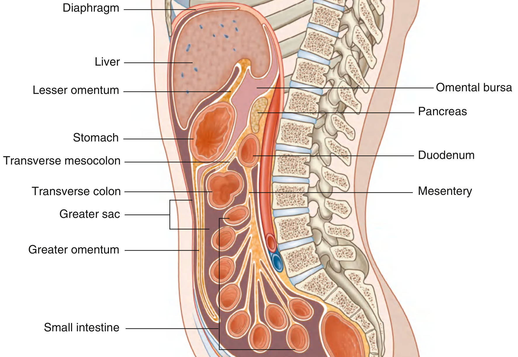
Walls of the Lesser Sac
| Wall | Structure |
|---|---|
| Anterior | Lesser omentum (hepatogastric + hepatoduodenal ligaments), posterior surface of the stomach, and gastrocolic ligament |
| Posterior | Parietal peritoneum covering the pancreas, left adrenal gland, left kidney, and aorta |
| Superior | Caudate lobe of the liver and diaphragm |
| Inferior | Transverse colon and transverse mesocolon |
| Left | Gastrosplenic ligament and splenorenal (lienorenal) ligament |
| Right | Opens into the greater sac via the epiploic foramen |
Boundaries of the Epiploic Foramen (Foramen of Winslow)
This is the only natural entrance to the lesser sac:
| Relation | Structure |
|---|---|
| Anterior | Hepatoduodenal ligament (containing portal vein, hepatic artery proper, and bile duct) |
| Posterior | Inferior vena cava (covered by peritoneum) |
| Superior | Caudate lobe of the liver |
| Inferior | First part of the duodenum (superior part) |
Recesses of the Lesser Sac
The lesser sac has three important recesses:
- Superior recess - extends upward between the esophagus and IVC, as far as the gastric fundus; its upper limit is the left triangular ligament of the liver
- Splenic (left) recess - extends to the left between the gastrosplenic and splenorenal ligaments toward the spleen
- Inferior recess - extends downward between the greater curvature of the stomach and the transverse colon (into the greater omentum)
Clinical Relevance
- Pancreatic pseudocysts commonly form in the lesser sac, as the pancreas forms its posterior wall. The posterior gastric wall can become the anterior wall of a pseudocyst, allowing transgastric drainage.
- Fluid collections in the lesser sac (e.g., from pancreatitis or perforated gastric ulcer) are walled off and may not communicate freely with the rest of the peritoneal cavity.
- The Pringle maneuver (compression of the portal triad to control liver bleeding) exploits the epiploic foramen - a finger is passed posterior to the hepatoduodenal ligament through this foramen.
Sources: Gray's Anatomy for Students, p. 363; Schwartz's Principles of Surgery 11th Ed., p. 1585; Color Atlas of Human Anatomy Vol. 2, p. 373
This is a shared conversation. Sign in to Orris to start your own chat.