
what are the extra nasopharyngeal manifestations of nasopharyngeal angiofibroma? Discuss various surgical options available for complete excision of tumor in detail for 20 marks with diagram and flow charts from Scott Brown, Cummings, stell and marans, Zakir Hussain, dhingra, hazarika and related articles with recent advances according to rguhs university
"juvenile nasopharyngeal angiofibroma" AND surgery
nasopharyngeal angiofibroma extranasopharyngeal extension surgical approaches RGUHS ENT 20 marks
"nasopharyngeal angiofibroma" AND "endoscopic" AND excision
https://pmc.ncbi.nlm.nih.gov/articles/PMC3431533
JUVENILE NASOPHARYNGEAL ANGIOFIBROMA (JNA)
Extranasopharyngeal Manifestations and Surgical Options for Complete Excision
20-Mark Answer - RGUHS Pattern
INTRODUCTION
PART I: EXTRANASOPHARYNGEAL MANIFESTATIONS (8 Marks)
A. Pathways of Spread
ORIGIN: Sphenopalatine Foramen / Pterygoid Process Base
                        |
        ┌───────────────┼───────────────────────┐
        ↓               ↓                       ↓
   MEDIAL           SUPERIOR               LATERAL
   SPREAD           SPREAD                 SPREAD
        ↓               ↓                       ↓
  Nasopharynx    Sphenoid Sinus           Pterygopalatine
  Nasal Cavity   Floor of Sphenoid        Fossa (PPF)
  (via SPF)      (via Vidian Canal)              ↓
                        ↓               Pterygomaxillary
                 Sella Turcica          Fissure
                 (advanced)                     ↓
                                       Infratemporal Fossa
                                               ↓
                                    ┌──────────┴──────────┐
                                    ↓                     ↓
                              Orbit (via              Parapharyngeal
                              IOF/SOF)                Space
                                    ↓
                              Middle Cranial Fossa
                              (via IFS / Maxillary N.)
                              Cavernous Sinus
                              Anterior Cranial Fossa
                              (via Ethmoid - rare)
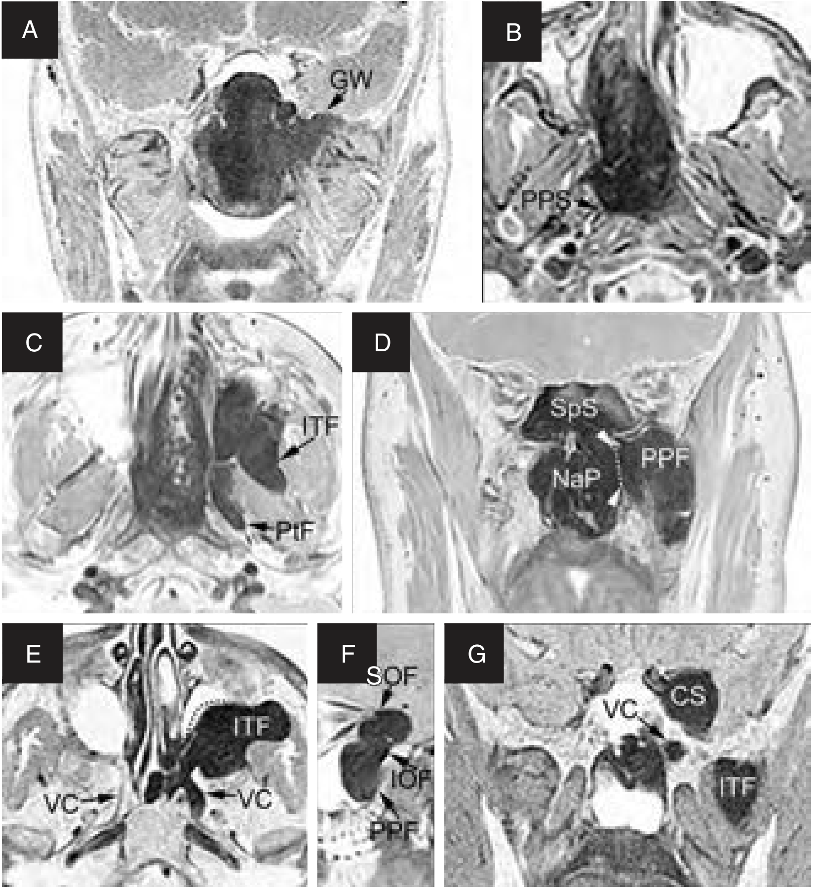
B. Extranasopharyngeal Manifestations by Region
1. PARANASAL SINUSES
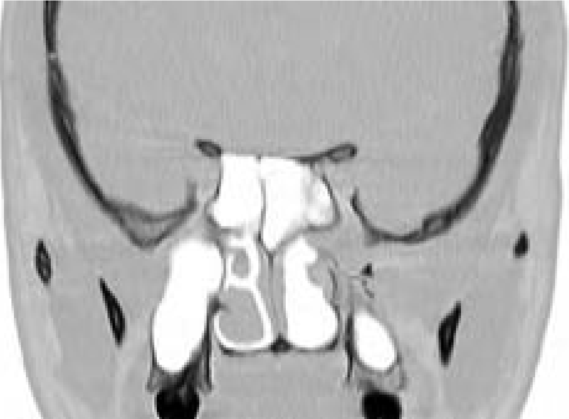
- Maxillary sinus - most common extranasopharyngeal site; anterior bowing of posterior wall = Holman-Miller sign (pathognomonic on X-ray/CT)
- Sphenoid sinus - via floor erosion or vidian canal
- Ethmoid sinus - uncommon; indicates anterior skull base encroachment
- Frontal sinus - rare, via anterior ethmoid spread
2. INFRATEMPORAL FOSSA (ITF)
- Via pterygomaxillary fissure - most important lateral extension
- Tumor may completely fill the ITF in advanced cases
- Clinical features: Cheek swelling/bulge, trismus (pterygoid muscle involvement), palpable mass in temporal fossa
- Bone: permeative intraosseous growth within cancellous bone of greater wing of sphenoid
3. ORBIT
- Via inferior orbital fissure (IOF) - from PPF
- Via superior orbital fissure (SOF) - advanced spread
- Clinical features: Proptosis, diplopia, visual disturbance, chemosis
- Optic canal involvement can cause visual loss
4. INTRACRANIAL EXTENSION
- Via inferior/superior orbital fissure or foramen rotundum (along maxillary nerve) to parasellar/cavernous sinus region
- Via middle cranial fossa floor erosion of greater sphenoid wing
- Via anterior cranial fossa through ethmoid (rare)
- Transdural growth is very rare despite intracranial extension
- Clinical features: Headache, cranial nerve palsies (III, IV, V, VI), visual field defects, raised ICP signs
5. PARAPHARYNGEAL SPACE
- Posterior extension from sphenopalatine region
- Clinical features: Unilateral tonsillar/pharyngeal mass bulge, dysphagia, change in voice (uncommon)
6. NASAL CAVITY (Extranasopharyngeal in same region)
- Anterior extension into nasal cavity, presenting as visible mass at anterior nares
- May present as bilateral involvement of maxillary sinuses and bilateral choanal involvement
7. CHEEK / SOFT TISSUE
- Anterior extension through posterior maxillary wall
- Clinical features: Facial swelling over cheek (Frog-face deformity in large tumors)
8. MIDDLE EAR / EUSTACHIAN TUBE
- Though not direct extension, tubal orifice involvement leads to serous otitis media / conductive hearing loss (seen in early primary nasopharyngeal tumor)
C. Staging Systems (Chandler, Sessions/Radkowski, Andrews/Fisch)
| Stage | Chandler (1984) | Andrews/Fisch | Radkowski |
|---|---|---|---|
| I | Nasopharynx only | Nasopharynx, nasal cavity | Nasopharynx, nasal cavity |
| II | Extends to nasal cavity, sphenoid | PPF, maxillary sinus, ethmoid, sphenoid | PPF/maxillary ± sphenoid |
| IIIa | Maxillary sinus, ethmoid, orbit, ITF, cheek, cavernous sinus | Orbit, ITF - no residual bone erosion | Orbit, ITF |
| IIIb | Same | Orbit, ITF - with residual bone erosion (sella, skull base) | As IIIa + intracranial extradural |
| IV | Intracranial | Intradural ± cavernous sinus | Intracranial with/without ICA encasement |
MRI Diagram: Patterns of Growth
CT showing Holman-Miller Sign
PART II: SURGICAL OPTIONS FOR COMPLETE EXCISION (12 Marks)
A. General Principles of Surgery (applicable to ALL approaches)
"Selection of appropriate approach for adequate exposure, vascular control, and dissection in subperiosteal plane" - PMC 3431533
- Preoperative workup: CT (bone detail), MRI (soft tissue, intracranial), MRA/DSA (vascular supply)
- Preoperative embolization: 24-48 hours before surgery; PVA particles or NBCA/Onyx; reduces intraoperative hemorrhage
- Controlled hypotension anesthesia: target MAP 60-70 mmHg
- Blood grouping and crossmatch: have blood ready
- Subperiosteal dissection plane throughout
- Primary vascular control before tumor delivery
B. Flowchart: Selection of Surgical Approach
JUVENILE NASOPHARYNGEAL ANGIOFIBROMA - CONFIRMED
                          |
                          ↓
          Staging (CT + MRI + MRA/DSA)
                          |
         ┌────────────────┴────────────────────┐
         ↓                                     ↓
   Stage I / II                        Stage III / IV
   (Limited to NP,                     (ITF, Orbit,
   nasal cavity,                       Intracranial
   PPF, sphenoid)                      extension)
         |                                     |
    ┌────┴────┐                     ┌──────────┴──────────────┐
    ↓         ↓                     ↓           ↓             ↓
Endoscopic  Transpalatal     Transmaxillary  Midfacial     Infratemporal
Endonasal   (limited          (Caldwell-Luc  Degloving     Fossa Approach
(Gold        NP disease)      + medial       ± LeFort I    (Fisch Type C)
Standard                      maxillectomy)               + Neurosurgery
Stage I/II)
    ↓                                                         ↓
Combined endoscopic                               Facial Translocation
+ Sublabial / Caldwell-Luc                       (Stage IV with ICA
for medial ITF extension                          involvement)
C. DETAILED SURGICAL APPROACHES
1. ENDOSCOPIC ENDONASAL APPROACH (EEA)
- Patient supine, head elevated 30°; topical decongestant (1:10,000 adrenaline) soaked pledgets
- Wide middle meatal antrostomy + posterior ethmoidectomy for exposure
- Sphenopalatine foramen identified posterior to middle turbinate
- Sphenopalatine artery clipped/coagulated lateral to tumor - critical first step
- Posterior maxillary wall removed (if needed) - Caldwell-Luc port
- Tumor dissected in subperiosteal plane using suction bipolar electrocautery, Freer elevators
- Vidian artery followed and clipped if tumor extends along vidian canal
- Tumor delivered through nasal cavity or mouth
2. TRANSPALATAL APPROACH (Wilson's Approach)
- Patient supine, head extended; Dingman's mouth gag
- U-shaped palatal incision (Wilson) with base at soft palate OR transverse midline incision
- Hard palate mucoperiosteally elevated; palatine vessels preserved if possible
- Posterior hard palate and vomer may be removed for exposure
- Tumor visualized in nasopharynx, dissected under direct vision
- Hemostasis achieved; palate replaced and sutured in layers
3. LATERAL RHINOTOMY + MEDIAL MAXILLECTOMY
- Lynch incision - begins at medial canthus, curves down alongside nose to level of alar base
- Periosteum elevated; nasal bone, frontal process of maxilla, lacrimal bone exposed
- Anterior maxillary wall opened; medial maxillectomy performed
- Posterolateral nasal wall removed to expose pterygopalatine and infratemporal fossa
- Internal maxillary artery/sphenopalatine artery ligated
- Tumor excised under direct vision
- Wound closed; nasal packing
4. MIDFACIAL DEGLOVING APPROACH
- Bilateral intercartilaginous incisions in nasal vestibule + bilateral sublabial incisions connected across the midline
- Facial soft tissue (cheek flap) elevated off maxilla bilaterally - complete facial degloving
- Rhinoplasty incisions and lateral osteotomies NOT required (modification of Casson)
- Excellent bilateral access to nasal cavities, maxillary sinuses, pterygopalatine fossa
- Posterior maxillary wall removed; tumor dissected
- Combined with medial maxillectomy if needed (Fig. 21 - Atlas of Otolaryngology)
- Facial flap replaced; no skin sutures needed (intranasal and sublabial only)
5. TRANSMAXILLARY / CALDWELL-LUC APPROACH
- Sublabial incision in upper buccal sulcus above upper canine
- Anterior maxillary wall opened (canine fossa)
- Posterior maxillary wall removed to access pterygopalatine and infratemporal fossa
- Tumor dissected laterally; internal maxillary artery approached and ligated
- Combined with endoscopic or transpalatal for comprehensive access
6. INFRATEMPORAL FOSSA APPROACH (FISCH TYPE C)
- Pre-auricular (hemicoronal) + temporal incision
- Zygomatic arch osteotomy + temporalis muscle elevation
- Infratemporal fossa exposed widely; mandibular condyle may be displaced
- Middle fossa dura exposed; internal carotid artery identified and controlled in petrous segment
- Tumor dissected from cavernous sinus, parasellar region, ICA
- Combined neurosurgical craniotomy if transdural extension present
7. LEFORT I OSTEOTOMY APPROACH
- Sublabial incision; maxillary osteotomy at LeFort I level
- Maxilla down-fractured and displaced anteriorly - "trapdoor" access
- Excellent exposure of nasopharynx, posterior choanae, pterygopalatine fossa
- Tumor excised; maxilla plated back in anatomical position
8. FACIAL TRANSLOCATION APPROACH
- Weber-Ferguson incision + coronal extension for frontotemporal craniotomy
- Midface osteotomies - nasal, orbital-maxillary, zygomatic arch
- Facial soft tissue rotated laterally as single osteoplastic unit
- Underlying maxilla rotated laterally after osteotomies
- Complete exposure of orbit, anterior/middle skull base, cavernous sinus, ICA
- Tumor excised; osteotomies plated; facial flap replaced
9. TRANSCERVICAL APPROACH
D. Summary Table: Approach vs. Stage
┌─────────────────────────────────────────────────────────────────────┐
│         SURGICAL APPROACH SELECTION TABLE FOR JNA                  │
├─────────────────────────┬──────────────────┬────────────────────────┤
│ APPROACH                │ STAGE            │ ACCESS PROVIDED        │
├─────────────────────────┼──────────────────┼────────────────────────┤
│ Endoscopic Endonasal    │ I, II, IIIa*     │ NP, NC, PPF, SpS,      │
│ (EEA)                   │ (*medial ITF)    │ medial ITF             │
├─────────────────────────┼──────────────────┼────────────────────────┤
│ Transpalatal            │ I                │ NP only                │
├─────────────────────────┼──────────────────┼────────────────────────┤
│ Lateral Rhinotomy       │ II-IIIa          │ NC, Maxillary, PPF     │
├─────────────────────────┼──────────────────┼────────────────────────┤
│ Midfacial Degloving     │ II-IIIa          │ Bilateral NC, Maxillary│
│ ± Medial Maxillectomy   │                  │ PPF, limited ITF       │
├─────────────────────────┼──────────────────┼────────────────────────┤
│ Transmaxillary          │ IIIa             │ Maxillary, lateral PPF │
│ (Caldwell-Luc)          │                  │ ITF                    │
├─────────────────────────┼──────────────────┼────────────────────────┤
│ LeFort I Osteotomy      │ II-IIIb          │ NP, posterior PPF,     │
│                         │                  │ inferior skull base    │
├─────────────────────────┼──────────────────┼────────────────────────┤
│ Infratemporal Fossa     │ IIIb-IV          │ Extensive ITF, skull   │
│ Approach (Fisch Type C) │                  │ base, parasellar, ICA  │
├─────────────────────────┼──────────────────┼────────────────────────┤
│ Facial Translocation    │ IV               │ Complete skull base,   │
│                         │                  │ cavernous sinus, orbit │
└─────────────────────────┴──────────────────┴────────────────────────┘
NC=Nasal Cavity; NP=Nasopharynx; PPF=Pterygopalatine Fossa;
SpS=Sphenoid Sinus; ITF=Infratemporal Fossa
E. PREOPERATIVE EMBOLIZATION - Important Surgical Adjunct
- Polyvinyl alcohol (PVA) particles - transarterial
- NBCA (N-butyl cyanoacrylate) liquid embolic
- Onyx - direct percutaneous puncture technique; best devascularization
- Coils, balloons
- Reduces intraoperative blood loss significantly
- Improves tumor border delineation
- Facilitates complete resection
F. POSTOPERATIVE CONSIDERATIONS
- Recurrence rate: 6-35% depending on stage; higher with intracranial extension
- Follow-up: MRI with contrast (preferred over CT for follow-up - Cummings)
- Spontaneous regression reported post-puberty (androgen-dependent growth)
G. NON-SURGICAL MODALITIES
- Dose: 30-36 Gy (external beam)
- Indications: Unresectable Stage IV, residual disease post-surgery, recurrence with intracranial ICA involvement
- Concerns: Malignant transformation (rare), growth plate disruption in children, radiation-induced malignancy
- Neoadjuvant to reduce tumor volume before surgery
- Used pre-pubertally or in early puberty (Sitenga et al., Int J Dermatol, 2022 - PMID 34748211)
- Evidence: Limited; not standard of care
RECENT ADVANCES (2021-2026)
- Expanded Endoscopic Endonasal Approach (EEA) - now the standard for Stage I-IIIa; even medial infratemporal fossa invasion is no longer a contraindication (Cummings, Hofmann series)
- Coblation-assisted endoscopic excision - reduces bleeding, improves visualization (Chen F et al., 2024, PMID 38858116)
- Preoperative embolization - meta-analysis evidence (Diaz et al., Laryngoscope 2023, PMID 36789781; Segal et al., J Laryngol Otol 2026, PMID 42087760): Embolization beneficial in advanced-stage disease; benefit in early stage remains debated
- Onyx direct percutaneous puncture - superior devascularization vs. transarterial PVA, especially for ICA feeders
- Image-guided surgery (IGS) with neuronavigation for skull base cases
- Molecular targeting - VEGF pathway upregulation documented; FGFR pathway implicated; potential for antiangiogenic therapy (Liu et al., Curr Oncol 2026, PMID 41892175)
- Extranasopharyngeal angiofibroma - recognized as distinct entity arising in sinonasal tract (not nasopharynx); [systematic review - Gazmenga et al., Eur Arch Otorhinolaryngol 2026, PMID 41699246]
SUMMARY DIAGRAM: MANAGEMENT ALGORITHM FOR JNA
SUSPECTED JNA (Adolescent male, unilateral nasal obstruction + epistaxis)
                              ↓
                    Endoscopy + CT/MRI
                              ↓
                   Do NOT biopsy (risk of massive haemorrhage)
                              ↓
                    Staging (Chandler/Fisch/Radkowski)
                              ↓
               ┌──────────────┼──────────────────┐
               ↓              ↓                  ↓
           Stage I         Stage II-IIIa       Stage IIIb-IV
               ↓              ↓                  ↓
          Endoscopic    Preop embolization    Preop embolization
          or            (24-48 hrs)           (DSA guided, Onyx)
          Transpalatal        ↓               Blood ready (4-6 units)
                     Endoscopic ± Caldwell         ↓
                     Luc / MFD               Midfacial Degloving +
                                             ITF approach (Fisch)
                                             or Facial Translocation
                                             ± Neurosurgery
                              ↓
                     Postop MRI (3-6 months)
                              ↓
              Residual / Recurrent disease?
               ↓                    ↓
           Observation           Revision surgery
           (small volume)        or Radiotherapy
REFERENCES
- Cummings Otolaryngology Head and Neck Surgery, 7th ed. - Chapter 50 (Juvenile Angiofibroma) and Chapter 95 - Comprehensive source for staging, surgical techniques, embolization
- Scott-Brown's Otorhinolaryngology Head & Neck Surgery, Vol.1 - Embolization for JNA; histology; surgical decision-making
- Scott-Brown's Vol.2 - Infratemporal fossa approach (Fisch Type C) indications
- KJ Lee's Essential Otolaryngology - Surgical approach overview; radiation indications
- Dhingra - Transpalatal approach; clinical features
- Hazarika ENT - Clinical presentation; surgical principles
- Stell and Maran's Head and Neck Surgery - Advanced surgical approaches
- Andrews JC, Fisch U et al. - Laryngoscope 1989 - Infratemporal fossa approach for extensive JNA
- Diaz A et al. - Laryngoscope 2023 (PMID 36789781) - Meta-analysis: embolization in JNA surgery
- Segal AY et al. - J Laryngol Otol 2026 (PMID 42087760) - Embolization benefits in advanced JNA: meta-analytic comparison
- Gazmenga FP et al. - Eur Arch Otorhinolaryngol 2026 (PMID 41699246) - Extranasopharyngeal angiofibroma systematic review
- Liu X et al. - Curr Oncol 2026 (PMID 41892175) - Molecular mechanisms of JNA; VEGF/FGFR targeting
- PMC 3431533 - Changing trends in management: 150 cases, transmaxillary shift
- Atlas of Otolaryngology - Midfacial degloving + medial maxillectomy; endoscopic technique details
RGUHS Examiner Tip: For a 20-mark answer, the above structure covers: Introduction (1 mark) + Extranasopharyngeal manifestations with pathways (6 marks) + Staging (2 marks) + Surgical approaches in detail (8 marks) + Adjuncts/Recent advances (2 marks) + Diagrams (bonus). Use the flowcharts and tables to score maximum marks.
Chronic rhinosinisitis with nasal polyp in detail for 20 marks with diagram and flow charts from Scott Brown, Cummings, stell and marans, Zakir Hussain, dhingra, hazarika and related articles with recent advances according to rguhs university