
Mouth ulcers diagnosis and treatment full discription
"Stomatitis, Aphthous"[MeSH Terms] AND "therapy"
recurrent aphthous stomatitis minor aphthous ulcer oral mucosa
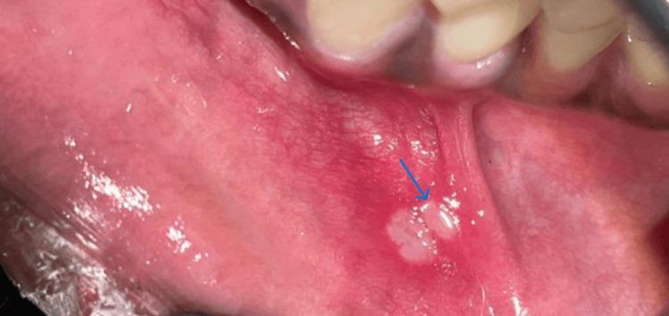
This clinical photograph displays an intraoral view of the lower labial mucosa, highlighting a minor aphthous ulcer (aphthous stomatitis minor). The lesion, indicated by a blue arrow, presents as a small, well-circumscribed, ovoid ulcer approximately 2-5 mm in diameter. It features a characteristic yellowish-white pseudomembranous center (fibrinous slough) surrounded by a distinct, prominent erythematous halo, which signifies localized inflammation. The adjacent mucosal tissue appears moist and healthy, though localized hyperemia is evident in the immediate periphery of the ulcer. This image serves as a classic diagnostic example of minor recurrent aphthous stomatitis, commonly encountered in oral medicine and general clinical practice for differentiating benign oral ulcers from more systemic or infectious conditions.
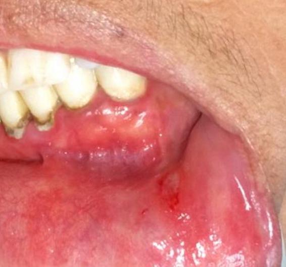
A close-up clinical intraoral photograph showing a pre-operative view of an aphthous ulcer on the internal labial mucosa of the lower left lip. The lesion is ovoid and shallow, approximately 2-3 mm in size, with a central floor covered by a pale, yellowish-white pseudomembranous slough. A well-defined erythematous halo of inflamed tissue surrounds the ulceration, which is a classic clinical sign of recurrent aphthous stomatitis. The surrounding oral mucosa appears moderately inflamed. Adjacent mandibular teeth show evidence of extrinsic staining and gingival inflammation along the cervical margin. The image demonstrates the typical morphology of a minor aphthous ulcer, suitable for educational use in identifying common oral mucosal pathologies in dental and clinical medicine.
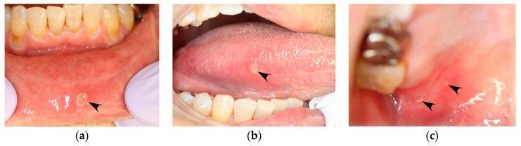
This set of intraoral clinical photographs displays multiple manifestations of Recurrent Aphthous Stomatitis (RAS) in a single patient. Image (a) shows the lower labial mucosa with a solitary, well-defined, shallow, round ulcer characterized by a yellowish-tan necrotic center (pseudomembrane) and a prominent peripheral erythematous halo. Image (b) reveals a similar aphthous ulcer on the right lateral margin of the tongue, demonstrating a pale center and surrounding inflammatory erythema. Image (c) captures the right hard palate mucosa where two smaller, discrete, round ulcers (marked by black arrowheads) are visible against a pinkish-red background, each exhibiting the characteristic necrotic center and erythematous border. These lesions are pathognomonic for minor aphthous ulcers, commonly found in non-keratinized or keratinized oral mucosa. The presentation illustrates the clinical criteria for diagnosing RAS, including lesion morphology, distribution across various anatomical sites in the oral cavity, and the presence of localized inflammation.
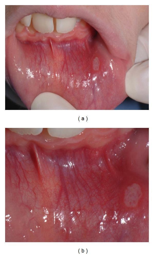
Content Type: Clinical Photograph. Anatomical Region: Oral cavity, specifically the lower labial mucosa. Modality: Macro photography showing a close-up of the oral soft tissue. Key Findings: A single, well-circumscribed, oval-shaped ulcer is visible on the inner surface of the lower lip. The lesion exhibits a central pale-whitish pseudomembranous base with an erythematous halo (peripheral redness), characteristic of Recurrent Aphthous Stomatitis (RAS). The surrounding mucosa shows prominent underlying vascularity and mild generalized erythema. Clinical Significance: This image demonstrates common oral manifestations associated with systemic conditions such as celiac disease (CD). The lesion is a minor aphthous ulcer, typically measuring less than 1 cm in diameter. The positioning near the labial frenum and the distinct morphology are classic diagnostic features for educational indexing in oral pathology and gastroenterology-related dermatology.
Mouth ulcers (oral ulceration): diagnosis and treatment
1. Common causes
| Category | Examples |
|---|---|
| Local trauma | Sharp teeth, denture edges, biting, burns |
| Recurrent aphthous stomatitis (RAS) | The most common cause of recurrent ulcers |
| Infections | Herpes simplex (after the vesicle bursts), chickenpox, zoster, hand-foot-and-mouth disease, herpangina, infectious mononucleosis, HIV, TB, syphilis |
| Skin and mucosal disease | Erosive lichen planus, pemphigus, pemphigoid, erythema multiforme, dermatitis herpetiformis |
| Systemic and GI disease | Coeliac disease, Crohn's disease, ulcerative colitis, Behçet's disease |
| Blood disorders and deficiencies | Low iron/ferritin, folate or B12; leukaemia; neutropenia |
| Drugs and treatments | NSAIDs, beta-blockers, nicorandil (potassium-channel openers), chemotherapy, radiotherapy |
| Malignancy | Squamous cell carcinoma, lymphoma |
2. Recurrent aphthous stomatitis (canker sores)
- Stress
- Local trauma
- Haematinic deficiency (iron, folate, B12)
- Food sensitivity (chocolate, nuts, tomatoes, spices)
- Sodium lauryl sulfate toothpaste
- Menstrual cycle
- Xerostomia
- H. pylori (eradication may reduce recurrences)
- Drugs (NSAIDs, beta-blockers, COX-2 inhibitors, sertraline)
- Systemic disease (Crohn's, coeliac disease, Behçet's, HIV, PFAPA, Sweet syndrome)
Three clinical types
| Feature | Minor | Major | Herpetiform |
|---|---|---|---|
| Frequency | Most common (~80%) | Less common | Least common |
| Size | Under 10 mm (usually under 5 mm) | Over 10 mm, deep, raised borders | 1 to 3 mm pinpoint |
| Number | Up to about 10 | Usually 1 to 3 | Up to 100, may coalesce |
| Site | Non-keratinized mucosa (lips, cheeks, ventral tongue), usually anterior | Any site, including keratinized mucosa, usually posterior (soft palate, fauces) | Non-keratinized mucosa, sometimes keratinized |
| Healing | 1 to 3 weeks, no scar | Weeks to 3 months, may scar | About 2 weeks, no scar |
3. Diagnosis
History
- Duration and pattern. Is it a single persistent ulcer or recurrent episodes? Do ulcers overlap in time?
- Site. A repeated ulcer in the same spot suggests a local cause. Ulcerated lichen planus can also do this.
- Red flags. Duration over 2 weeks, an indurated or rolled edge, a painless ulcer, leukoplakia or erythroplakia nearby, a neck node, or a smoking and alcohol history.
- Systemic review.
- GI symptoms: diarrhoea, weight loss, bloating.
- Genital ulcers or eye problems (Behçet's).
- Skin rash, joint pain, fever.
- HIV risk.
- Drug history. Ask about NSAIDs, beta-blockers, nicorandil, methotrexate and chemotherapy.
- Family history.
Examination
| Herpes simplex | Aphthae | |
|---|---|---|
| Site | Keratinized mucosa (hard palate, gingiva, lip border) | Non-keratinized mucosa |
| Vesicle phase | Yes | No |
| Cause | HSV-1/2 | Immune dysregulation |
Investigations (directed by history)
- Full blood count, ESR, ferritin, folate and B12 levels.
- Coeliac serology (tissue transglutaminase IgA) and consideration of HIV testing.
- Viral swab, PCR or Tzanck smear if herpes is suspected.
- Colonoscopy if inflammatory bowel disease is suspected.
- Biopsy for any ulcer lasting over 2 weeks, any suspicious feature, or a major aphthous ulcer that does not respond to treatment. Biopsy is also needed to confirm vesiculobullous disease, with direct immunofluorescence.
- Pathergy test and Behçet's criteria if there are genital or eye features.
4. Treatment
A. Treat the underlying cause
- Replace deficient iron, folate or B12.
- Treat coeliac disease with a gluten-free diet, and treat IBD.
- Stop causative drugs. Withdrawing them stops recurrences and separates drug-induced ulcers from true idiopathic RAS. - Cummings, Ch. 87
- Smooth sharp teeth and fix dentures.
- Treat HSV with antivirals (aciclovir or valaciclovir) if confirmed.
B. Supportive and self-care measures
- Avoid spicy, salty, acidic or crusty food.
- Use a soft toothbrush.
- Consider switching to an SLS-free toothpaste.
- Use a multivitamin with iron.
- Reduce trauma and stress.
C. Mild or infrequent minor ulcers
- Chlorhexidine 0.2% rinse, plus analgesics and topical anaesthetics (such as 2% viscous lidocaine or benzydamine). - Cummings; Tintinalli's Emergency Medicine
- Protective agents: sucralfate or bismuth. - Sleisenger and Fordtran's
- Bioadhesive pastes containing steroid and anaesthetic, such as Orobase. - Tintinalli's
D. Moderate disease: topical corticosteroids (first line)
- Fluocinonide, fluocinolone or clobetasol gel or ointment, or triamcinolone in Orabase. They work only if started at the prodrome or first sign of an ulcer. - Goldman-Cecil
- A tetracycline or doxycycline mouth rinse at onset shortens the course. - Goldman-Cecil
E. Severe, major or frequent disease
- Major aphthae. A short course of prednisone (for example 40 mg daily for 3 days). If there is no good response, do an incisional biopsy to exclude neoplasia. - Goldman-Cecil
- Second-line and steroid-sparing options. Colchicine 0.6 mg three times daily, tetracycline 250 mg four times daily, cimetidine 400 to 800 mg daily, azathioprine 50 mg daily, and thalidomide for refractory cases. - Sleisenger and Fordtran's
- Other options in specialist care: dapsone, pentoxifylline and apremilast (for example in Behçet's).
- HIV-associated major aphthae. Refractory cases may need thalidomide, under specialist supervision.
F. Other conditions
- Herpetic stomatitis. Antivirals, fluids, analgesia and mouthwash.
- Lichen planus, pemphigus and pemphigoid. Topical or systemic steroids, with immunosuppression guided by a specialist.
- Suspected malignancy. Urgent referral to an oral and maxillofacial or head and neck team.
5. When to refer
- Ulcer lasting over 2 weeks without a clear cause
- Hard, indurated or fixed ulcer, or neck lymph nodes
- Suspected systemic disease (Behçet's, IBD, coeliac, HIV, haematological)
- Failure of standard treatment
- Large or scarring major aphthae