
Upper motor neuron lesion
Reading File
Finding Sources
Finding Sources
Reading File
Reading File
Upper Motor Neuron (UMN) Lesion
What is the upper motor neuron?
UMNs are neurons whose cell bodies lie mainly in the motor cortex. Their axons form the corticospinal and corticobulbar tracts, which descend through the subcortical white matter and internal capsule to the brainstem and spinal cord, where they synapse on lower motor neurons (LMNs) - the brainstem cranial nerve nuclei and spinal cord anterior horn cells that actually innervate muscle. Indirect (extrapyramidal) pathways contributing to UMN function include the rubrospinal, reticulospinal, vestibulospinal, and tectospinal tracts (Frameworks for Internal Medicine, p. 597-598).
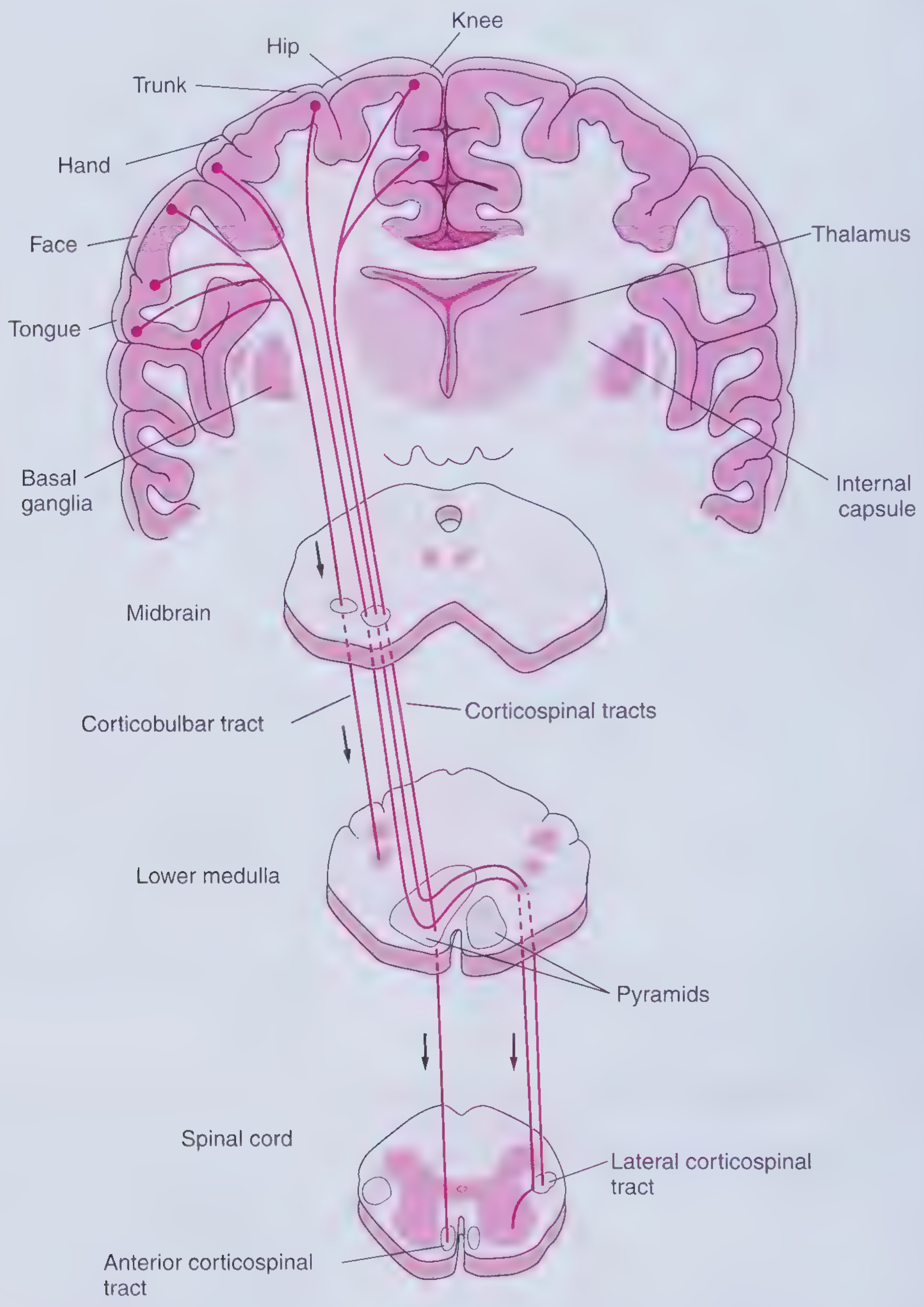
Where can UMN lesions occur?
Anywhere along this pathway "above" the anterior horn cell/cranial nerve nucleus:
- Brain: cerebral cortex, subcortical white matter, internal capsule, brainstem
- Spinal cord: at or above the level of the anterior horn cells
Clinical features of a UMN lesion
| Feature | UMN lesion |
|---|---|
| Tone | Increased (spasticity) - "clasp-knife" quality |
| Reflexes | Hyperreflexia |
| Pathological reflexes | Babinski sign (extensor plantar response), Hoffmann sign, jaw jerk |
| Bulk | Atrophy is absent or minimal (disuse atrophy only, no denervation atrophy) |
| Fasciculations | Absent |
| Pattern of weakness | Tends to affect groups of muscles rather than individual muscles - classically weaker extensors than flexors in the arm, weaker flexors than extensors in the leg (pyramidal pattern) |
| Distribution | Often hemiparesis/hemiplegia (brain lesion) or paraparesis with a sensory "level" (spinal cord lesion) |
This contrasts with LMN lesions (affecting anterior horn cells, nerve roots, or peripheral nerves), which cause hypotonia/flaccidity, hyporeflexia or areflexia, marked atrophy, and fasciculations (Rheumatology, 2-Vol Set, p. and Neuroanatomy through Clinical Cases).
Localizing clues
- Ipsilateral cranial nerve signs + contralateral hemiparesis → brainstem lesion (corticobulbar tract for that nerve has already crossed or synapsed above the lesion, while the corticospinal tract has not yet crossed).
- Hemiparesis without cranial nerve involvement → lesion above the brainstem (cortex, subcortical white matter, or internal capsule).
- Bilateral limb involvement with a sensory level, bladder/bowel dysfunction → spinal cord lesion.
- Facial UMN lesions spare the forehead (bilateral cortical innervation of the upper face), whereas LMN facial nerve lesions (e.g., Bell's palsy) involve the whole face including the forehead (Neuroanatomy through Clinical Cases, Ch. 12.3).
Common causes
- Cortical/subcortical/internal capsule: ischemic or hemorrhagic stroke, tumor, demyelination
- Brainstem: stroke (producing classic crossed syndromes), demyelination (multiple sclerosis), tumor
- Spinal cord: cervical spondylotic myelopathy/cord compression, multiple sclerosis, transverse myelitis, trauma, tumor
- Motor neuron disease (e.g., ALS, primary lateral sclerosis) can produce mixed or pure UMN signs, distinguishing it from pure LMN disorders like progressive muscular atrophy or multifocal motor neuropathy (Harrison's Principles of Internal Medicine, 22nd ed.)
Recent evidence check
This is core, stable neuroanatomy/neurology content rather than an area with shifting evidence base, so no recent systematic review/meta-analysis search was needed to validate it.
If you want, I can go deeper into a specific angle - e.g., UMN vs LMN facial palsy, cervical myelopathy localization, or ALS's mixed UMN/LMN presentation.
Lower motor neuron lesion
Finding Sources
Finding Sources
Lower Motor Neuron (LMN) Lesion
What is the lower motor neuron?
LMNs are the motor neurons whose cell bodies lie in the brainstem cranial nerve nuclei and the anterior (ventral) horn of the spinal cord. They receive input from upper motor neurons (corticospinal/corticobulbar tracts) and directly innervate skeletal muscle via peripheral nerves/cranial nerves and the neuromuscular junction. A lesion anywhere from the anterior horn cell/cranial nerve nucleus outward - nerve root, plexus, peripheral nerve, or the neuron itself - produces an LMN pattern of weakness.
Clinical features of an LMN lesion
| Feature | LMN lesion |
|---|---|
| Tone | Decreased (hypotonia/flaccidity) |
| Reflexes | Reduced or absent (hyporeflexia/areflexia) |
| Bulk | Marked atrophy (denervation atrophy) |
| Fasciculations | Present (spontaneous motor unit discharges visible under skin) |
| Fibrillations/spontaneous activity | Present on EMG, along with insertional activity |
| Pattern of weakness | Tends to be focal, following a specific root, nerve, or muscle group rather than a global hemibody pattern |
| Sensory involvement | Often accompanies weakness if a mixed sensorimotor nerve or root is affected (unlike pure UMN lesions) |
This is the mirror image of the UMN pattern discussed previously (spasticity, hyperreflexia, Babinski sign, minimal atrophy, no fasciculations) - Neuroanatomy through Clinical Cases, 3rd ed.; Bradley and Daroff's Neurology in Clinical Practice.
Anatomic localization
LMN signs arise from pathology at any of these levels:
- Anterior horn cell: poliomyelitis, other acute flaccid paralyses, amyotrophic lateral sclerosis (ALS, when LMN-predominant), spinal muscular atrophy, progressive muscular atrophy
- Nerve root: disc herniation, radiculopathy
- Plexus: brachial/lumbosacral plexopathy
- Peripheral nerve: diabetic neuropathy, multifocal motor neuropathy with conduction block, entrapment neuropathies, Guillain-Barre syndrome
- Neuromuscular junction/muscle: technically produce a similar flaccid, hyporeflexic picture, though myasthenia gravis classically causes fatigable weakness without atrophy or fasciculations, and myopathies typically spare reflexes until late and don't cause fasciculations
Distinguishing LMN weakness clinically
- Distribution: LMN lesions localize to a specific root, plexus, or peripheral nerve territory (e.g., a foot drop from peroneal nerve palsy), whereas UMN lesions produce more diffuse hemiparesis or paraparesis.
- Sensory loss: acute LMN weakness from nerve root or peripheral nerve disease is often accompanied by pain and sensory loss in a matching distribution, unlike pure motor cortex/corticospinal tract lesions.
- EMG/NCS: insertional activity and spontaneous denervation potentials (fibrillations, positive sharp waves) are classic electrodiagnostic markers of LMN pathology - Bradley and Daroff's Neurology in Clinical Practice, Ch. 36 (Clinical Electromyography).
Combined UMN + LMN signs
Some diseases affect both levels simultaneously - most notably ALS, which shows hyperreflexia, spasticity, and Babinski sign (UMN) together with fasciculations, atrophy, and weakness (LMN) in overlapping myotomes. Finding both sign patterns in the same body region, without a sensory level or single anatomic explanation, is a key diagnostic clue pointing toward motor neuron disease rather than a single structural lesion (ROSEN's Emergency Medicine, Box 93.9; case discussion in Ch. 9 Q&A).
Quick comparison: UMN vs LMN
| UMN | LMN | |
|---|---|---|
| Tone | ↑ (spastic) | ↓ (flaccid) |
| Reflexes | ↑ | ↓/absent |
| Babinski | Present | Absent |
| Atrophy | Minimal | Marked |
| Fasciculations | Absent | Present |
| Distribution | Diffuse (hemibody/level) | Focal (root/nerve/segment) |
This is core neuroanatomy content rather than a rapidly evolving evidence area, so no additional systematic-review search was needed to validate it. Let me know if you'd like to go deeper into a specific LMN disorder (e.g., ALS, Guillain-Barre, radiculopathy localization) or into EMG/NCS findings in more detail.
myasthenia gravis
Finding Sources
Finding Sources
Searching PubMed
"myasthenia gravis"[MeSH Terms] AND treatment
Searching Images
myasthenia gravis ptosis clinical photo
Reading File
Myasthenia Gravis (MG)
Pathophysiology
MG is an autoimmune disorder of the neuromuscular junction (NMJ). Autoantibodies (most commonly against the postsynaptic acetylcholine receptor, AChR) bind and disrupt normal neuromuscular transmission, reducing the number of functional AChRs available at the endplate. Less common antibody targets include MuSK (muscle-specific kinase) and LRP4. The result is a reduced safety margin for neuromuscular transmission, producing weakness that worsens with repeated use and improves with rest (Bradley and Daroff's Neurology in Clinical Practice, p. 1958-1960).
Epidemiology
- US prevalence ~20/100,000 (roughly 60,000 patients)
- Bimodal age/sex distribution: women affected ~3x more than men before age 40; incidence higher in men after age 50; roughly equal during puberty
- Prevalence has risen over the past 50 years, partly from better detection and increased longevity - most US patients are now over 50 (Bradley and Daroff's Neurology in Clinical Practice, p. 1959)
Clinical presentation
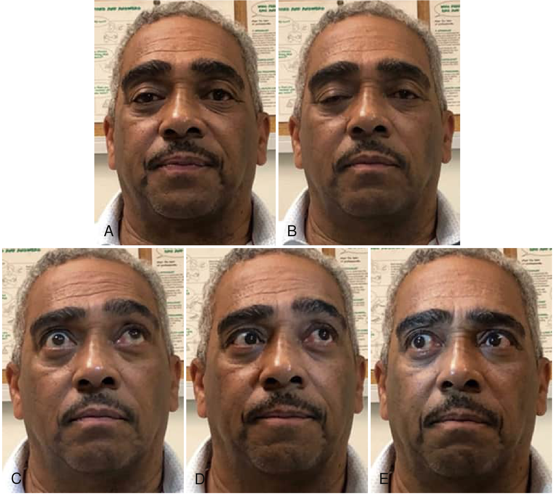
- Hallmark: fluctuating, fatigable weakness - worse with sustained or repeated activity, worse later in the day, better with rest
- Ocular involvement is the initial symptom in about two-thirds of patients (ptosis, diplopia from extraocular muscle weakness), and nearly all patients develop ocular signs within 2 years
- Bulbar involvement: dysarthria, dysphagia, chewing fatigue (initial symptom in ~1/6 of patients)
- Limb weakness: initial symptom in ~10%, typically proximal > distal
- Weakness can occasionally start focally (neck extensors, elbow/finger extensors, hip flexors, ankle dorsiflexors)
- Myasthenic crisis: severe generalized/bulbar weakness causing respiratory failure requiring ventilatory support - a medical emergency
Diagnosis
- Antibody testing: anti-AChR antibodies (most patients with generalized MG); anti-MuSK or anti-LRP4 in a subset of seronegative patients
- Electrodiagnostics: repetitive nerve stimulation shows a decremental response; single-fiber EMG shows increased jitter (most sensitive test, especially in ocular MG)
- Edrophonium (Tensilon) test: rapid, transient improvement in weakness - largely of historical interest now given availability of antibody/EMG testing
- Ice pack test: improvement of ptosis with cooling, a simple bedside test
- Imaging: CT chest to evaluate for thymoma, present in ~10-15% of AChR-antibody-positive patients
Treatment
- Symptomatic: acetylcholinesterase inhibitors (pyridostigmine) to increase acetylcholine availability at the NMJ
- Immunosuppression: corticosteroids, azathioprine, mycophenolate mofetil, and other steroid-sparing agents for long-term control
- Rapid/severe disease or crisis: IVIG or plasma exchange (plasmapheresis)
- Thymectomy: recommended for thymoma; also beneficial in non-thymomatous AChR-antibody-positive generalized MG in appropriately selected patients
- Newer targeted biologics: complement inhibitors (eculizumab, ravulizumab) and FcRn antagonists (efgartigimod) approved for AChR-antibody-positive generalized MG, used when conventional immunosuppression is inadequate
Recent evidence update (2024-2025 systematic reviews)
Newer network meta-analyses of biologic therapies suggest differences in efficacy and tolerability among the novel targeted agents (complement inhibitors vs. FcRn antagonists vs. other biologics), which may refine second-line treatment choice beyond what older textbook chapters describe:
- Guan et al. 2025, systematic review/network meta-analysis of novel biologics in MG (PMID: 40346603)
- Zhong et al. 2024, systematic review/network meta-analysis on initiation response and therapeutic efficacy of biological targeted therapies in MG (PMID: 39529623)
These support that biologics have real comparative efficacy differences worth discussing with a neurologist when standard immunosuppression fails or is poorly tolerated - this is an evolving area, so specifics of drug choice should be confirmed against current guidelines rather than relying solely on textbook chapters.
Let me know if you want to go deeper into MG crisis management, MuSK-antibody MG (which behaves somewhat differently), or the specific dosing/monitoring of any of these agents.
This is a shared conversation. Sign in to Orris to start your own chat.