
Application of Electrical Stimulation (Galvanic and Faradic Currents) in Specific Clinical Conditions
iontophoresis electrical stimulation treatment hyperhidrosis
TENS transcutaneous electrical nerve stimulation electrode placement pain
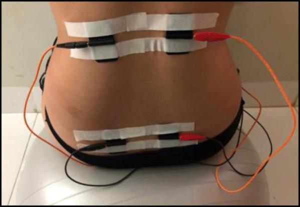
Clinical photograph demonstrating the electrode placement for Transcutaneous Electrical Nerve Stimulation (TENS) on the posterior trunk of a patient. Four black silicone electrodes are arranged in two horizontal pairs: a superior pair located in the thoracolumbar region (approximately T10–L1) and an inferior pair located in the sacral region (approximately S2–S4). The electrodes are secured to the skin using horizontal strips of white medical masking tape. Electrical leads are attached to each electrode, featuring a combination of black and orange wires that connect to a portable electrostimulation unit. This setup is typical for obstetric or physiotherapeutic pain management protocols, where the placement targets dermatomes associated with pelvic and lower back pain. The patient is shown in a seated position, likely on a therapeutic ball, consistent with labor pain management or pelvic floor rehabilitation settings.
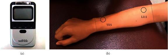
Educational panel illustrating Transcutaneous Electrical Nerve Stimulation (TENS) for lateral epicondylitis treatment. Figure (a) displays a portable TENS device featuring a black upper section with a digital LCD screen and control buttons, and a silver lower section branded 'wellLife'. Figure (b) is a clinical photograph of a patient's right dorsal forearm, demonstrating the specific electrode placement sites according to WHO guidelines for acupuncture-based therapy. Two key anatomical points are circled and labeled: TE5 (Waiguan), located on the posterior aspect of the forearm approximately two inches above the wrist crease between the radius and ulna, and LI11 (Quchi), situated at the lateral end of the cubital crease when the elbow is flexed. Dotted vertical lines demarcate the wrist and elbow regions to provide anatomical orientation. The imagery illustrates the clinical application of electrical stimulation at traditional acupoints for pain management and functional recovery in upper extremity musculoskeletal conditions.
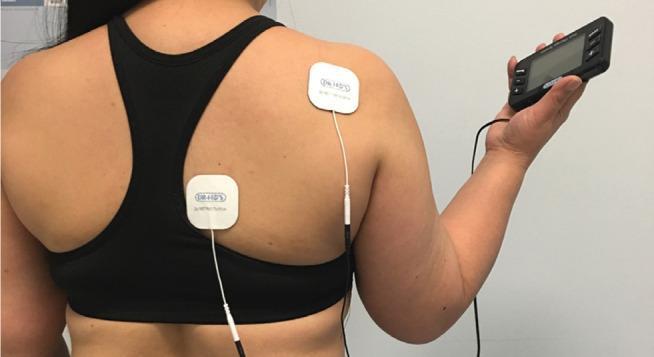
This clinical photograph demonstrates the application and setup of a Transcutaneous Electrical Nerve Stimulation (TENS) unit for pain management. The image shows a patient's posterior upper torso with two white, square-shaped, self-adhesive electrode pads positioned on the right trapezius and rhomboid regions. The patient is holding a handheld, black digital controller featuring an LCD screen and tactile interface buttons for adjusting intensity and mode. Lead wires connect the portable pulse generator to the surface electrodes. This procedural image illustrates the non-invasive placement of electrotherapy components used in physical medicine and rehabilitation to modulate pain signals. The positioning reflects common clinical use for musculoskeletal pain or muscle relaxation in the thoracic and shoulder regions. The visual content serves as an educational example of patient-administered electroanalgesia and the interface between medical devices and cutaneous application.
Application of Electrical Stimulation (Galvanic and Faradic Currents) in Specific Clinical Conditions
1. Fundamental Properties of Each Current Type
Galvanic Current (Direct Current, DC)
- Flows continuously in one direction (unidirectional)
- Requires application for several milliseconds to elicit a muscle contraction
- Used when muscles are denervated (and therefore no longer respond to brief faradic pulses)
- Produces electrochemical effects at both electrodes (anode/cathode), which form the basis for iontophoresis and electrolysis
- Can create tissue ionic changes, alter pH locally, and drive charged drug molecules into tissue
Faradic Current (Alternating/Interrupted Current)
- Brief pulse (<1 millisecond) of rapidly alternating current
- Effectively stimulates normally innervated muscle via intact motor nerves
- Cannot produce contraction in denervated muscle (requires prolonged galvanic stimulus instead)
- Forms the basis of TENS, NMES, and electrodiagnostic studies
"The electrical pulse required is brief, less than a millisecond, and is most effectively induced by rapidly alternating (faradic) current. If there has been muscle denervation, an electrical pulse of several milliseconds induced by a constant electrical (galvanic) stimulus is required to produce the same response." - Adams and Victor's Principles of Neurology, 12th Ed.
2. Electrodiagnosis: Denervation Testing
| Finding | Normal (Innervated) Muscle | Denervated Muscle |
|---|---|---|
| Faradic response | Strong contraction | No contraction |
| Galvanic response | Brisk, brief contraction | Slow, prolonged "worm-like" contraction (reaction of degeneration) |
| Minimum current needed | Low | High |
- Adams and Victor's Principles of Neurology, 12th Edition
3. TENS (Transcutaneous Electrical Nerve Stimulation)
Mechanism
- Gate control theory: TENS bombards interneurons in the substantia gelatinosa, presynaptically inhibiting T-cells that transmit nociception to higher brain centers, effectively "closing the gate"
- Endorphin release: Particularly with low-frequency (acupuncture-like) TENS, stimulates release of endogenous opioid peptides
- Vasodilation: Direct local vasodilation may reduce relative ischemia contributing to pain
- The treatment may gradually break the pain-spasm-pain cycle
Clinical Indications
- Musculoskeletal pain syndromes (chronic joint pain, myalgias)
- Post-operative pain
- Labor pain (electrode placement over T10-L1 and S2-S4 dermatomes is used in obstetric settings)
- Fibromyalgia (sometimes helpful)
- Lateral epicondylitis and other soft tissue conditions
Technique
- Trim hair and cleanse skin with 70% isopropyl alcohol
- Apply conductive gel and place electrodes over the affected area
- Begin with the lowest comfortable setting; gradually increase intensity
- Treatment duration: 30-60 minutes, once or twice daily
- After treatment: turn unit off, return settings to zero
Contraindications
- Cardiac pacemaker (especially older models - risk of malfunction)
- Over the carotid sinus or anterior neck
- Pregnancy (over the abdomen/pelvis)
- Active skin infection or open wounds at electrode site
- Areas of impaired skin sensation (patient must give accurate feedback on intensity)
Complications
- Skin irritation or contact dermatitis at electrode sites
- Pacemaker interference
- Inadvertent motor nerve stimulation (twitch response)
Current Evidence
- Pfenninger and Fowler's Procedures for Primary Care, 3rd Ed.
- Goldman-Cecil Medicine
- Firestein & Kelley's Textbook of Rheumatology
4. NMES (Neuromuscular Electrical Stimulation)
Clinical Applications
- Overcome muscle activation deficits (e.g., quadriceps inhibition after knee injury or surgery)
- Prevent muscle atrophy in non-ambulatory patients
- Strengthen muscles alongside exercise programs when voluntary activation is limited by pain
- Prevent pressure ulcers in neurological patients with paralysis (maintain circulation and muscle bulk)
- Respiratory rehabilitation in severe COPD (home-based NMES has been studied in RCTs)
In Denervated Muscle
- Firestein & Kelley's Textbook of Rheumatology
5. Iontophoresis (Galvanic Current for Drug Delivery)
Mechanism
- Based on the repulsion of similarly charged ions: positively charged drugs are driven from the anode (positive electrode); negatively charged drugs from the cathode (negative electrode)
- Also alters cell membrane permeability via acoustic streaming-like ionic effects
Electrical Dosing
- Dose = current intensity (mA) × time (minutes) = mA·min
- Most treatments: 10-20 minutes, repeated 3-8 times
- Current range: 1-4 mA depending on drug and electrode size
Common Drugs and Their Indications
| Ion / Drug | Polarity | Therapeutic Use |
|---|---|---|
| Dexamethasone (1-4 mA × 15-20 min) | Negative | Tendinitis, tenosynovitis, bursitis, arthritis |
| Methylprednisolone (1-4 mA × 15-20 min) | Negative | Postherpetic neuralgia |
| Lidocaine 4% (4 mA × 20-30 min) | Positive | Topical anesthesia (pre-procedure) |
| Acetic acid (3-4 mA × 10-20 min) | Negative | Calcified tendinitis, calcium deposits |
| Sodium chloride (4 mA × 20-45 min) | Negative | Keloids, scar tissue softening |
| Copper sulfate (4 mA × 20-30 min) | Positive | Fungal infections (tinea pedis) |
| Hyaluronidase (1-2 mA × 20-40 min) | Positive | Edema, lymphedema, scleroderma |
| Iodine (2 mA × 1 min → 4 mA × 5 min) | Negative | Fibrosis, scar tissue, trigger finger |
| Salicylate (4 mA × 45 min) | Negative | Analgesia, myalgia, plantar warts |
| Zinc (4 mA × 15 min) | Positive | Wound healing, chronic ulcers |
Specific Clinical Applications
Hyperhidrosis
- Battery-powered home units (e.g., Drionic) are available for ongoing maintenance
- More effective for palms and soles than axillae
- Goldman-Cecil Medicine; Dermatology 2-Volume Set 5th Ed; Fitzpatrick's Dermatology
Musculoskeletal Conditions
- Bursitis, tendinitis, fasciitis, sprains, carpal tunnel syndrome, de Quervain's disease, trigger points
- Works best for superficial, localized inflammatory lesions (penetration limited to ~1.5 cm)
- Pfenninger and Fowler's Procedures for Primary Care
Analgesia and Skin Anesthesia
- Lidocaine iontophoresis provides needle-free topical anesthesia before procedures (venipuncture, skin procedures)
- Roberts and Hedges' Clinical Procedures in Emergency Medicine
Contraindications for Iontophoresis
- Allergy to the therapeutic agent
- Cardiac pacemakers
- Abnormal skin sensation (impairs feedback on current tolerance)
- Superficial abrasions, cuts, bruises
- Areas of recent bleeding
- Overlying metallic implants (screws, staples, wires - risk of enhanced current delivery)
- Recent scars or skin grafts
- Over the heart or carotid sinus
6. Galvanic Electrolysis / Electroepilation
- Inserting a fine needle electrode (anode/negative pole) into the hair follicle
- The patient holds a moistened pad as the cathode
- DC application generates sodium hydroxide via electrochemical reaction at the needle tip
- NaOH acts as a caustic agent, destroying the hair root (30-60 seconds per follicle)
- Dermatology 2-Volume Set 5th Ed (Elsevier)
7. Electrical Stimulation in Wound Healing
Mechanisms
- Increased blood flow: Low-frequency ES promotes sensory nerve activation → vasodilation → improved nutrient delivery
- Cellular recruitment: Promotes migration of neutrophils, macrophages, lymphocytes, and fibroblasts during different wound healing phases
- Collagen remodeling: Encourages fibroblast migration via the PI3K/PTEN pathway; enhances collagen fiber structure and ECM arrangement
- Antibacterial effect: Decreases bacterial load in wound tissue
- Gene expression: Alters expression of chemokines, cytokines, and collagen genes via intracellular polyamine and KCNJ15/Kir4.2 pathways
Delivery Methods
- Electrodes placed directly on or near the wound
- Current may be direct (unidirectional galvanic) or pulsatile (monophasic or biphasic)
- Parameters (frequency, polarity, amplitude, placement) tailored to wound characteristics
- Typical protocol: ~5 hours/week
Evidence
- Sabiston Textbook of Surgery, 8th Ed
8. Bone Healing (Electromagnetic and DC Stimulation)
- Direct current stimulation promotes osteogenesis through piezoelectric effects in bone and alteration of local ionic environment
- Indicated in: non-union fractures, failed previous surgery, avascular necrosis
- Both basic science research and high-quality clinical evidence suggest a clinical benefit for bone healing, though optimal dosage and patient selection remain unclear
- Campbell's Operative Orthopaedics 15th Ed 2026; Rockwood and Green's Fractures in Adults 10th Ed
9. Summary Table: Galvanic vs. Faradic Applications
| Current Type | Key Characteristic | Primary Clinical Applications |
|---|---|---|
| Galvanic (DC) | Continuous, unidirectional | Iontophoresis, hyperhidrosis, wound healing, electrolysis/epilation, bone healing, denervated muscle testing |
| Faradic (AC/interrupted) | Brief pulses <1 ms | TENS (pain), NMES (muscle strengthening), nerve conduction studies, innervated muscle stimulation |
| Both / Combined | Varies | Electroepilation (blend), wound healing ES, rehabilitation |
Key Clinical Precautions Across All Modalities
- Never use over implanted cardiac pacemakers or defibrillators
- Avoid over the carotid sinus, anterior neck, or directly over the heart
- Do not apply to areas of compromised skin sensation (burn risk with galvanic; inability to report excess current with iontophoresis)
- Avoid during pregnancy over the abdomen/pelvis
- Skin integrity must be assessed before and during treatment
- Avoid over metallic implants (plates, screws, staples) for iontophoresis
- Adams and Victor's Principles of Neurology, 12th Ed
- Pfenninger and Fowler's Procedures for Primary Care, 3rd Ed
- Dermatology 2-Volume Set 5th Ed (Elsevier)
- Fitzpatrick's Dermatology, 9th Ed
- Goldman-Cecil Medicine, International Ed
- Firestein & Kelley's Textbook of Rheumatology, 2-Volume Set
- Sabiston Textbook of Surgery, Biological Basis of Modern Surgical Practice
- Campbell's Operative Orthopaedics 15th Ed 2026
- Rockwood and Green's Fractures in Adults 10th Ed
- Roberts and Hedges' Clinical Procedures in Emergency Medicine