
ashey and walsham forceps use
Walsham forceps nasal bone fracture reduction
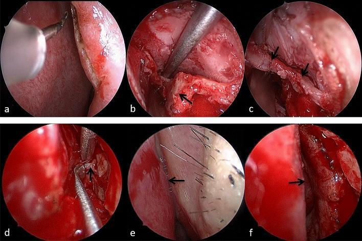
A series of six intranasal endoscopic photographs (a–f) documenting the Endoscopic Endonasal Incisional Reduction (EIIR) of a nasal fracture. (a) Displays the initial 1 cm arc incision at the rim of the piriform aperture using an electric stylet. (b) Shows the exposed displaced fracture of the frontal process of the maxilla (FPM). (c) Demonstrates the reduction of the fracture fragments, with arrows indicating the restored fracture lines. (d) Highlights the intraoperative removal of trapped soft tissue (black arrow) from between bone fragments using surgical forceps. (e) Provides a preoperative endoscopic view of a narrowed internal nasal valve (arrow) caused by the collapsed FPM obstructing the airway. (f) Provides a postoperative view following successful reduction, demonstrating a significantly widened internal nasal valve (arrow) and restored patency. This sequence illustrates the surgical management of traumatic nasal obstruction and aesthetic deformity by addressing displaced maxillary and nasal bone fragments.
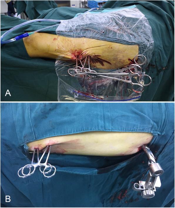
Two-panel clinical photograph illustrating a minimally invasive surgical technique for femoral shaft fracture reduction. Both panels show a patient's limb (thigh) in a sterile field with blue drapes and clear plastic adhesive drapes. In Panel A, two long, curved 26-cm haemostatic forceps are inserted into a percutaneous incision to act as levers for bone alignment. The forceps handles are interlocked to create a stable, self-maintaining structure, further secured by a Kocher forceps clamped across their shafts to prevent sliding. Blood and surgical tubing are visible at the operative site. Panel B demonstrates the same interlocking forceps technique in a separate patient case, showing the stable configuration of the two haemostatic forceps without the addition of a Kocher clamp. A second incision is visible distally, containing a larger orthopaedic instrument, likely related to intramedullary nail fixation. This visual sequence demonstrates the 'lever principle' using common surgical instruments to maintain fracture reduction percutaneously.
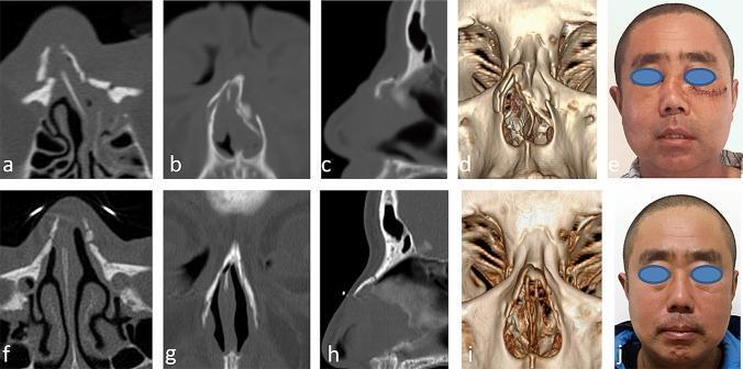
A multi-panel medical comparison set illustrating a nasal fracture case before and after surgical intervention. The top row (a-e) displays preoperative diagnostic and clinical views: (a) axial, (b) coronal, and (c) sagittal CT scans showing comminuted nasal bone fractures with lateral displacement and septal deviation; (d) a 3D CT reconstruction highlighting the skeletal deformity; and (e) a clinical photograph showing visible nasal bridge depression and deviation. The bottom row (f-j) presents postoperative outcomes: (f-h) corresponding CT planes demonstrating anatomical realignment and stable fracture reduction; (i) a 3D reconstruction showing restored nasal pyramid symmetry; and (j) a clinical photograph at 6-month follow-up showing a well-corrected nasal bridge with improved aesthetic contour. The images collectively demonstrate the efficacy of endonasal incision internal reduction (EIIR) for complex nasal fractures involving the nasal bones and frontal process of the maxilla. This content is relevant for otolaryngology and maxillofacial surgery training.
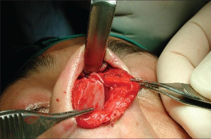
This clinical photograph captures an intraoperative view of an open rhinoplasty procedure, demonstrating a severe fracture-dislocation of the nasal septum in a 20-year-old male patient. The image shows the internal nasal structures exposed following the elevation of the nasal skin envelope. The nasal septum exhibits a clear transverse fracture, with a significant segment of the septal cartilage displaced from its anatomical midline position. Surgical exposure is maintained using an Aufricht retractor superiorly to lift the soft tissue, while two pairs of surgical forceps are used laterally for tissue stabilization and manipulation of the fractured septal segments. The visual highlights the complexity of post-traumatic secondary nasal deformity, specifically the disruption of the internal nasal valve and the loss of cartilaginous support. This clinical case illustrates the necessity for surgical reduction and reconstructive techniques, such as the use of spreader grafts, to restore functional airway patency and aesthetic nasal structure.
Ashe's (Asch's) Forceps and Walsham's Forceps
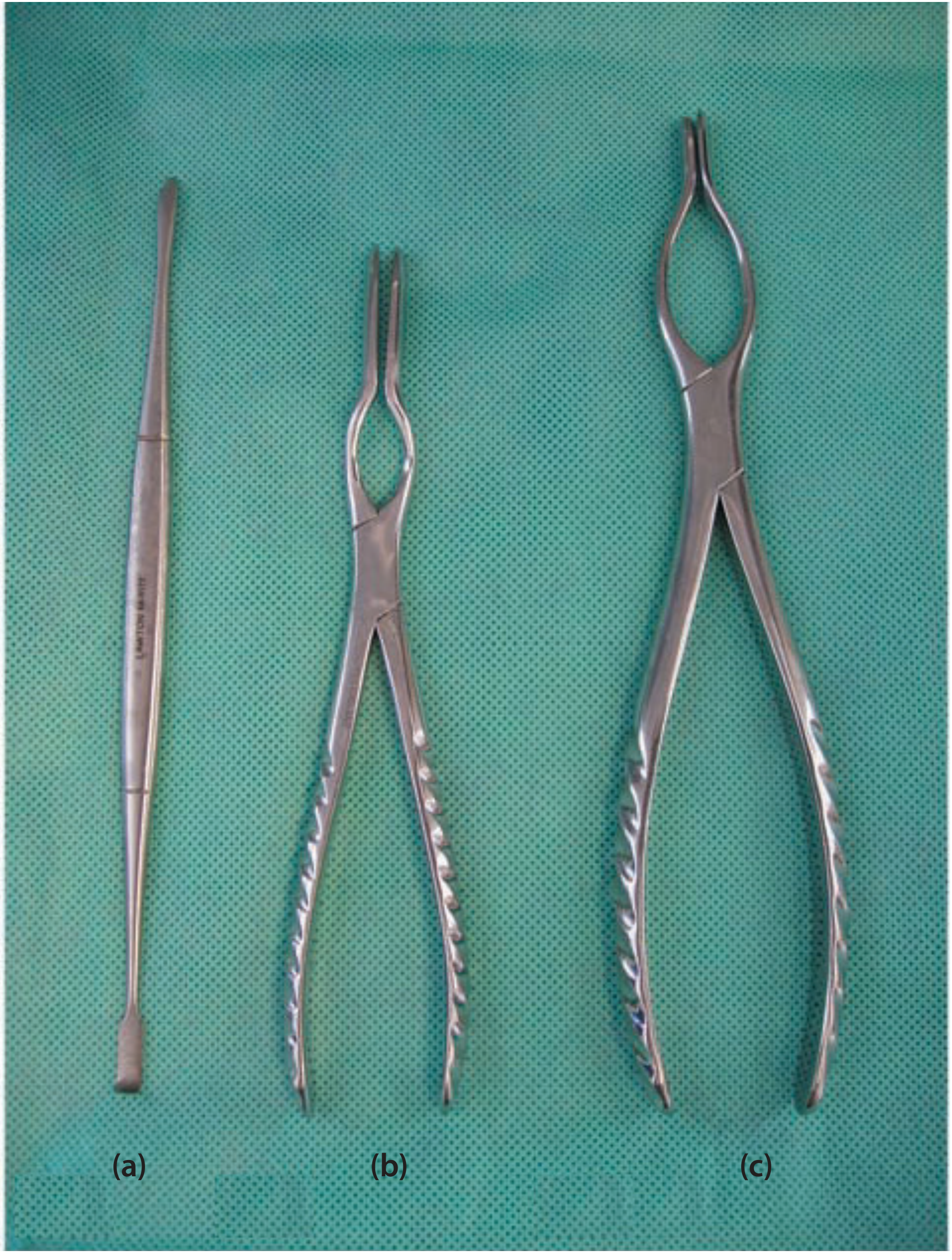
Ashe's (Asch's) Forceps
- Both arms of the instrument are inserted on either side of the septum, positioned superiorly just underneath the dorsum.
- An upward and outward force perpendicular to the plane of the dorsum is applied - this lifts the septum out of overlap and the arms push the ends back into realignment.
- Septal reduction is often more challenging than nasal bone reduction because of the complexity of the surrounding forces and the frequency of pre-existing deformities.
- Note the critical surgical principle: "where goes the septum, so does the nose" - satisfactory reduction of nasal bones is often not possible without first improving the position of the septum, so septum should be reduced before nasal bones.
- If Ashe's forceps alone cannot achieve septal reduction, a Killian or hemitransfixion incision with mucosal flap elevation (i.e., formal septoplasty) may be needed.
Walsham's Forceps
- One arm is inserted intranasally (into the nasal cavity); the other arm is placed against the external skin surface.
- The displaced nasal bone is grasped firmly (but without crushing the intervening tissue).
- The segment is disimpacted first, then repositioned into its anatomic location.
- They allow manipulation with greater versatility than elevators but provide less fine tactile feedback.
- Excessive torqueing of fragments should be avoided to prevent additional fracture.
- They commonly provoke mucosal bleeding during reduction due to their traumatic nature.
- Used when initial reduction with a blunt elevator (Boies, Joker, Howarth) has failed, or when overlapping bone fragments require grasping rather than simple elevation.
Summary Table
| Feature | Ashe's (Asch's) Forceps | Walsham's Forceps |
|---|---|---|
| Target structure | Nasal septum | Nasal bones |
| Blade placement | Both arms on either side of septum (bilateral intranasal) | One arm intranasal, one external |
| Mechanism | Upward + outward force to lift septum out of overlap | Grasp, disimpact, then reposition bone fragment |
| When used | Septal fracture-dislocation | Displaced/overlapping nasal bone fractures |
| Limitation | May not suffice for complex septal fractures | Less tactile feedback than elevators; causes mucosal bleeding |
| Order of use | First (septum before bones) | After septal reduction |
Clinical Context
- First increase the deformity slightly (disimpact the fragments)
- Then move back toward/past the midline
- Scott-Brown's Otorhinolaryngology Head & Neck Surgery, Vol 1, p. 1235
- Cummings Otolaryngology Head and Neck Surgery, p. 592