
Larynx anatomy
Reading File
This is a broad anatomy topic - I'll search for dedicated larynx anatomy sections across the medical library.
Finding Sources
Finding Sources
Excellent - found dedicated anatomy sections. Let me read the two most authoritative sources in parallel: Scott-Brown's dedicated "ANATOMY OF THE LARYNX" and Gray's Anatomy for Students.
Reading File
Reading File
Good start. Now let me read more of Gray's Anatomy (which has the most structured coverage) and the Scott-Brown's section for more detail.
Reading File
Reading File
Good - I have strong cartilage content. Now let me read more about muscles, membranes, and neurovascular supply from both sources.
Reading File
Reading File
Excellent detail. Let me now get the muscles and neurovascular sections.
Reading File
Reading File
I have comprehensive content. Let me now fetch some key anatomical images to include in the response.
Loading Image
Loading Image
Loading Image
I now have comprehensive textbook content and high-quality anatomical diagrams. Let me compile the full answer.
Larynx Anatomy
Overview
The larynx is a hollow musculoligamentous structure with a cartilaginous framework that caps the lower respiratory tract. It lies in front of the C3-C6 vertebrae (slightly higher in women), extending from the laryngeal inlet down to the inferior border of the cricoid cartilage. It serves two primary functions:
- Valve/sphincter to protect the lower airway during swallowing
- Sound production (phonation)
Motor and sensory innervation is via the vagus nerve [CN X].
(Gray's Anatomy for Students)
Divisions of the Larynx
The larynx is divided into three compartments by the false and true folds:
| Region | Boundaries |
|---|---|
| Supraglottis | Epiglottis + aryepiglottic folds down to the apex of the laryngeal ventricle |
| Glottis | From apex of ventricle, includes the true vocal folds, anterior and posterior commissures |
| Subglottis | From ~1 cm below the free edge of vocal folds to the inferior border of the cricoid |
Note: "Vocal cord" is anatomically inaccurate - "vocal fold" is preferred, as these structures vibrate in a multiplanar (not simply cord-like) fashion. (Scott-Brown's Otorhinolaryngology)
Laryngeal Cartilages
The framework includes 3 unpaired and 3 paired cartilages:
Unpaired Cartilages
1. Cricoid Cartilage
- Most inferior laryngeal cartilage; the only one that completely encircles the airway
- Shaped like a signet ring: broad posterior lamina + narrow anterior arch
- Posterior lamina has two oval depressions (for posterior cricoarytenoid muscles) separated by a vertical ridge (for esophageal attachment)
- Has articular facets for arytenoid cartilages (superolateral lamina) and inferior thyroid horns (lateral lamina)
2. Thyroid Cartilage (largest)
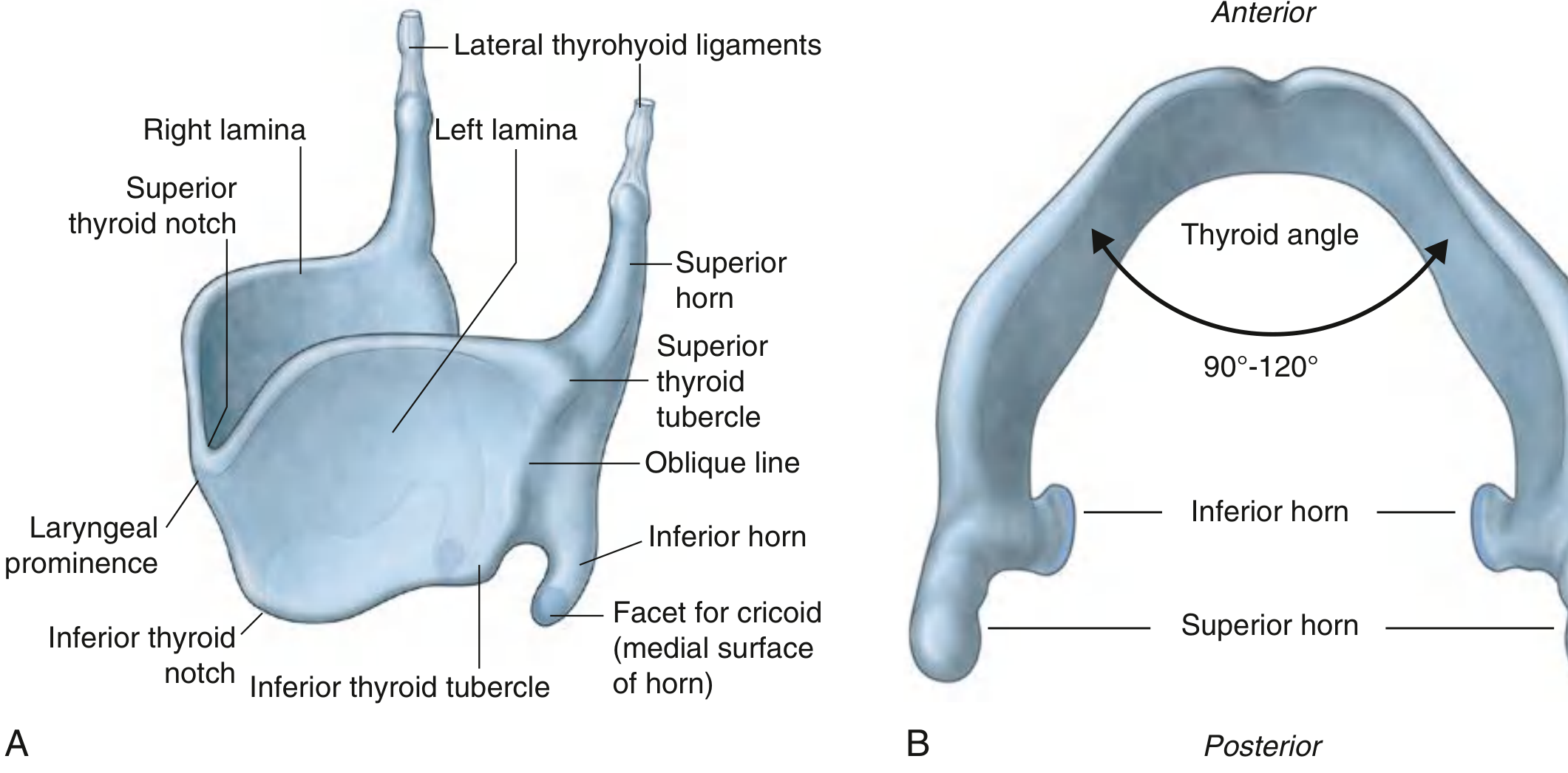
- Formed by right and left laminae that fuse anteriorly as the laryngeal prominence (Adam's apple)
- Thyroid angle: ~90° in men, ~120° in women - this is why the prominence is more visible in men
- Each lamina has superior and inferior horns (cornua):
- Superior horn connects via the lateral thyrohyoid ligament to the greater horn of hyoid
- Inferior horn articulates with the cricoid at the cricothyroid joint (synovial)
- Oblique line on the lateral surface: attachment for sternothyroid, thyrohyoid, and inferior constrictor muscles
3. Epiglottis
- Leaf-shaped; attached by its stem via the thyroepiglottic ligament to the posterior thyroid angle, midway between laryngeal prominence and inferior thyroid notch
- Projects posterosuperiorly; upper margin lies behind the pharyngeal tongue
- Also attached to hyoid by hyoepiglottic ligament - the space between these two ligaments is the pre-epiglottic space
- Its posterior surface has an epiglottic tubercle and numerous mucosal pits for mucous glands
- The mucous membrane reflects onto the tongue as the glossoepiglottic fold (midline) and lateral glossoepiglottic folds, forming the valleculae
Paired Cartilages
4. Arytenoid cartilages - pyramid-shaped, sit on the sloping shoulders of the cricoid lamina; have a vocal process (attachment of vocal ligament) and a muscular process (attachment of most intrinsic muscles)
5. Corniculate cartilages - small, at the apex of arytenoids; form the posterior limit of the aryepiglottic folds
6. Cuneiform cartilages - within the aryepiglottic folds, providing support
Membranes and Ligaments
Extrinsic
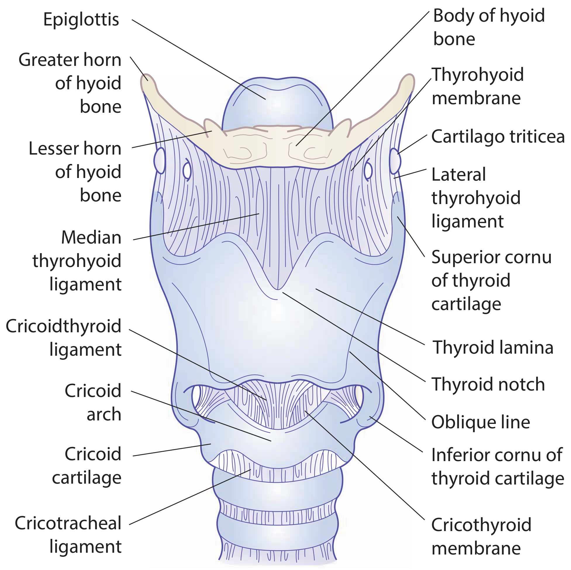
- Thyrohyoid membrane: fibroelastic sheet from upper thyroid cartilage to hyoid bone; reinforced centrally as median thyrohyoid ligament and posteriorly as lateral thyrohyoid ligaments (which often contain a sesamoid nodule, the cartilago triticea). Pierced by the internal branch of the superior laryngeal nerve and superior laryngeal vessels
- Cricotracheal ligament: joins inferior cricoid border to first tracheal ring
Intrinsic (Fibroelastic Membrane)
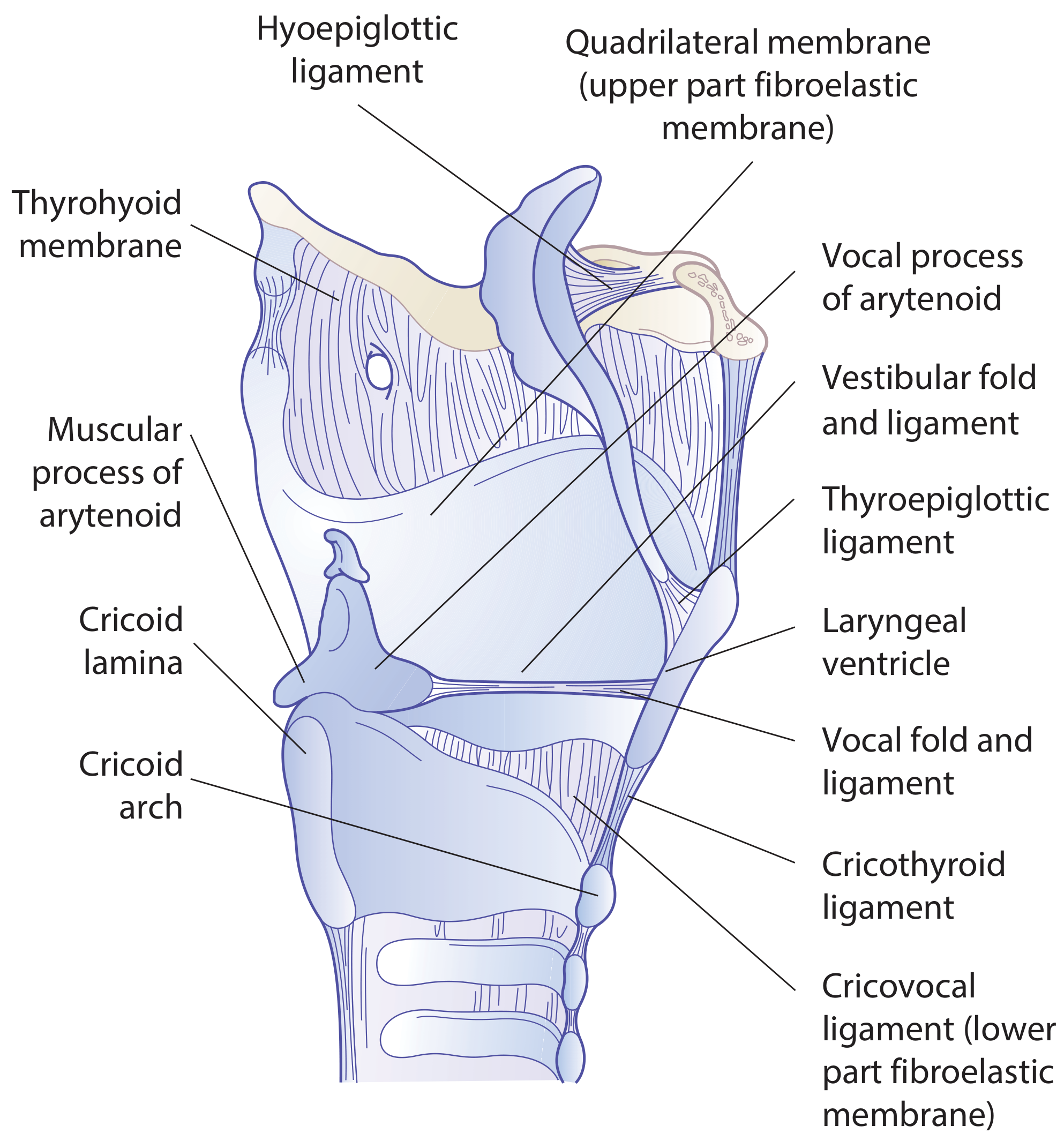
Divided by the laryngeal ventricle into upper and lower parts:
| Part | Name | Free Margin |
|---|---|---|
| Upper | Quadrangular membrane | Lower free margin = vestibular ligament (under false vocal cord) |
| Lower | Conus elasticus (cricothyroid ligament) | Upper free margin = vocal ligament (under true vocal cord) |
- The median cricothyroid ligament is the thickened anterior midline portion of the conus elasticus - the site used for emergency cricothyrotomy
- The laryngeal ventricle (sinus) is the lateral recess between the vestibular and vocal folds
Joints
| Joint | Type | Movement |
|---|---|---|
| Cricothyroid | Synovial | Thyroid tilts forward/downward on cricoid → lengthens and tenses vocal folds |
| Cricoarytenoid | Synovial | Arytenoids rock and rotate → abducts/adducts vocal folds |
Intrinsic Muscles
All intrinsic muscles are innervated by the recurrent laryngeal nerve (RLN) - except the cricothyroid, which is innervated by the external branch of the superior laryngeal nerve (SLN).
| Muscle | Action | Nerve |
|---|---|---|
| Posterior cricoarytenoid (PCA) | Abducts vocal folds (opens rima glottidis) - the ONLY abductor | RLN |
| Lateral cricoarytenoid (LCA) | Adducts vocal folds | RLN |
| Transverse arytenoid | Adducts arytenoids (closes posterior glottis); only unpaired muscle | RLN |
| Oblique arytenoid | Sphincter of laryngeal inlet | RLN |
| Thyroarytenoid | Shortens/relaxes vocal folds; sphincter of vestibule | RLN |
| Vocalis (medial part of thyroarytenoid) | Fine tension adjustment of vocal folds | RLN |
| Cricothyroid | Tilts thyroid forward, elongating and tensing vocal folds (raises pitch) | External SLN |
The cricothyroid is the only intrinsic muscle that lies outside the cartilaginous framework of the larynx. (Scott-Brown's)
Neurovascular Supply
Innervation (both from CN X - Vagus)
- Superior laryngeal nerve (SLN):
- Internal branch: sensory to larynx above the vocal folds; pierces thyrohyoid membrane
- External branch: motor to cricothyroid muscle
- Recurrent laryngeal nerve (RLN): motor to all other intrinsic muscles; sensory below vocal folds. On the right, loops around the subclavian artery; on the left, loops around the aortic arch
Blood Supply
- Superior laryngeal artery (from superior thyroid artery, branch of external carotid) - accompanies the internal branch of the SLN through the thyrohyoid membrane
- Inferior laryngeal artery (from inferior thyroid artery, branch of thyrocervical trunk)
Lymphatic Drainage
- Supraglottis: rich lymphatic drainage to upper and middle deep cervical nodes - this is why supraglottic tumors spread early
- Glottis: virtually no lymphatics - explains why glottic cancers metastasize late
- Subglottis: drains to paratracheal and lower deep cervical nodes
Clinical Notes
- Infant larynx: funnel-shaped with the narrowest point at the subglottic-tracheal junction (not the glottis as in adults); positioned higher in the neck; softer cartilages prone to collapse
- After puberty, the AP diameter of the male larynx nearly doubles (~36 mm men vs ~26 mm women)
- The pre-epiglottic space and the paraglottic spaces are filled with loose connective tissue - important routes for tumor spread
- RLN injury (e.g., from thyroid surgery, mediastinal tumors) causes ipsilateral vocal fold paralysis; bilateral injury causes respiratory distress
Sources: Scott-Brown's Otorhinolaryngology Head & Neck Surgery; Gray's Anatomy for Students (Gray's Anatomy for Students, p. 1192-1201)
This is a shared conversation. Sign in to Orris to start your own chat.