
Digital subtraction angiography in detailed simplified manner from standard ENT textbook
Reading File
Digital Subtraction Angiography (DSA) is a fluoroscopic technique used to visualize blood vessels by electronically subtracting bony and soft-tissue background, leaving only the contrast-filled vessels visible. It is used in ENT primarily for vascular tumors (e.g., juvenile nasopharyngeal angiofibroma, glomus tumors), pre-operative embolization, and vascular anomalies.
Searching sources...
Finding Sources
Finding Sources
Now let me search specifically for ENT-context DSA content and also retrieve the best radiology/ENT-relevant sections:
Finding Sources
Reading File
Reading File
Excellent - found the key ENT content in Scott-Brown's and Cummings. Let me now read those sections in detail:
Reading File
Reading File
Finding Sources
Now let me get more detail on the Cummings chapter on DSA/endovascular procedures in H&N surgery, and also the Grainger & Allison radiology text for the technical details:
Reading File
Reading File
Now let me read more from Cummings on the technical setup of DSA in H&N, and search for images:
Reading File
Searching Images
digital subtraction angiography head neck DSA technique
Searching Images
juvenile nasopharyngeal angiofibroma DSA angiogram embolization
Now let me get the key section in Cummings about the DSA technique setup and the glomus jugulare embolization procedure, plus fetch the images:
Reading File
I now have enough content from multiple standard ENT and radiology textbooks to give a thorough, well-structured answer. Let me fetch the key DSA images referenced in the textbooks:
Loading Image
Loading Image
I have comprehensive data now. Let me compose the full, detailed, simplified explanation of DSA from the ENT perspective.
Digital Subtraction Angiography (DSA) - ENT Perspective
Based on Cummings Otolaryngology Head and Neck Surgery, Scott-Brown's Otorhinolaryngology Head & Neck Surgery, and Grainger & Allison's Diagnostic Radiology
1. What is DSA? - The Core Concept
DSA is a fluoroscopic (real-time X-ray) imaging technique that electronically removes (subtracts) the background of bones and soft tissues, leaving only the contrast-filled blood vessels visible as bright, clear images against a black background.
The simple analogy: Imagine taking two photographs of the same scene - one before a person arrives, and one after. By subtracting the "before" image from the "after" image, you see only the person, with the background completely gone. DSA does exactly this with blood vessel images.
2. How Does It Work? - Step by Step
Step 1 - Mask Image (Reference Image)
- Before contrast is injected, a plain fluoroscopic image is captured.
- This image shows bones, soft tissues, and all background structures.
- It is digitized (converted into a grid of numbers) and stored in a computer.
Step 2 - Contrast Injection
- A contrast agent (iodine-based) is injected into a blood vessel via a catheter.
- A second fluoroscopic image is captured when the vessels fill with contrast.
Step 3 - Digital Subtraction
- The computer subtracts the first (mask) image from the second (contrast) image pixel by pixel.
- All background structures that appear in both images cancel out to zero (black).
- Only the contrast-filled vessels remain - shown as bright white.
As Murray & Nadel's Textbook of Respiratory Medicine explains: "The background body structures are subtracted, leaving an image of the contrast-filled vessel... Because the body background does not obscure the final image, angiography can be accomplished with smaller amounts of contrast than otherwise necessary."
3. Equipment Used
According to Cummings Otolaryngology Head and Neck Surgery:
| Component | Details |
|---|---|
| C-arm fluoroscopy unit | Variable projections; biplanar units allow two simultaneous views |
| Image intensifier + TV camera | High-resolution; captures and digitizes fluoroscopic frames |
| Flat-detector angiography machine | Modern standard; enables 3D rotational DSA and CT-like parenchymal images |
| Diagnostic catheters | 4-Fr or 5-Fr polyethylene catheters (Seldinger technique) |
| Microcatheters | 2-3 Fr, highly flexible; for superselective catheterization of tiny tumor-feeding vessels |
| Micro-guidewires | 0.008-inch caliber; used with microcatheters |
Road mapping - a critical DSA feature in ENT: real-time high-resolution subtracted fluoroscopy during injection of embolic agents. It allows the operator to visualize, in real time, exactly where embolic material is going - preventing reflux into normal vessels or dangerous collateral channels.
4. Vascular Access - The Seldinger Technique
- A needle punctures the common femoral artery (transfemoral route, most common) or occasionally the radial or brachial artery.
- A flexible guidewire is passed through the needle.
- The needle is removed; a sheath and catheter are advanced over the guidewire.
- The catheter is steered under fluoroscopic guidance into the target vessel (e.g., external carotid artery, internal carotid artery).
- Contrast is injected manually or via a pump injector.
For a complete diagnostic cerebral/H&N study, up to 6 vessels may be selectively catheterized:
- Both internal carotid arteries (ICA)
- Both external carotid arteries (ECA)
- Both vertebral arteries (if needed)
5. Imaging Parameters
- Frame rate: 2-3 images/second for routine; 4-7/second for fast-flow lesions (AVMs, high-flow tumors)
- 3D DSA: Automatic pump injection of 15-20 mL contrast at 3-5 mL/sec; the C-arm rotates 180-200° during injection, creating a 3D rotational angiogram
- Biplanar units: Allow simultaneous AP and lateral (or two oblique) projections in a single injection, reducing contrast dose and procedure time
6. ENT-Specific Indications
DSA is used in ENT/Head & Neck Surgery for the following conditions:
A. Juvenile Nasopharyngeal Angiofibroma (JNA)
- JNA is a highly vascular tumor arising near the sphenopalatine foramen in adolescent males.
- Intranasal biopsy is contraindicated due to risk of life-threatening hemorrhage.
- Selective DSA elegantly maps the feeding vessels and their exact supply territory.
- Pre-operative embolization of feeder vessels is performed at the same sitting.
- Post-embolization surgical resection is then the treatment of choice.
- (Scott-Brown's Otorhinolaryngology, Vol. 2)
B. Glomus Tumors (Paragangliomas)
- Glomus jugulare - arises in the jugular foramen; fed predominantly by the ascending pharyngeal artery and posterior division of the middle meningeal artery; less commonly by the stylomastoid branch of the occipital and posterior auricular arteries.
- Glomus tympanicum - small middle ear tumor; usually no embolization needed.
- Glomus vagale / Carotid body tumor - fed by multiple ECA branches.
DSA maps each arterial feeder compartment-by-compartment. Superselective catheterization of each feeder allows embolization with microspheres (e.g., 400-μm Embosphere microspheres), producing marked reduction in tumor blush before surgery. (Cummings Otolaryngology)
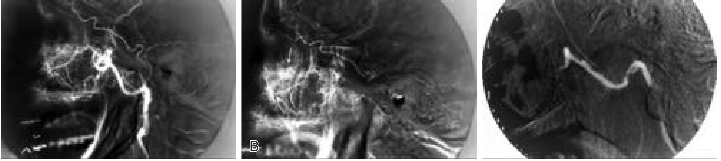
C. Intractable Epistaxis
- When conservative measures and packing fail, DSA identifies the offending vessel.
- The culprit vessel (usually the internal maxillary artery or a sphenopalatine branch) is visualized.
- Embolization with polyvinyl alcohol (PVA) particles (e.g., 150-μm) is performed under DSA road map guidance.
- (Cummings Otolaryngology)
D. Meningiomas and Hypervascular Skull Base Tumors
- DSA maps dural blood supply and dangerous ECA-ICA anastomoses.
- Pre-operative embolization reduces intraoperative blood loss.
- If ICA sacrifice is contemplated (tumor encasing the ICA), DSA is used for the Balloon Occlusion Test (BOT) - temporarily inflating a balloon in the ICA to test if the hemisphere can tolerate permanent occlusion.
E. Vascular Malformations
- Arteriovenous malformations (AVMs)
- Arteriovenous fistulas (AVFs) - including carotid-cavernous fistulas
- Venous malformations
- DSA provides definitive mapping of the nidus, feeders, and draining veins.
F. Traumatic Vascular Injuries
- Pseudoaneurysms, carotid laceration, vertebral artery injury
- DSA confirms diagnosis and guides endovascular repair (coils, stents)
7. Embolic Agents Used Under DSA Guidance
| Agent | Type | ENT Use |
|---|---|---|
| PVA particles (150, 250, 400 μm) | Particulate | Epistaxis, JNA, glomus tumors |
| Embosphere microspheres (calibrated) | Particulate | Glomus tumors, meningiomas |
| Coils (platinum/steel) | Mechanical | Pseudoaneurysms, fistulas, vessel sacrifice |
| NBCA (N-butyl cyanoacrylate, "glue") | Liquid | AVMs, fast-flow fistulas |
| Onyx (ethylene vinyl alcohol) | Liquid | AVMs, dural AVFs |
| Absolute ethanol | Liquid sclerosant | Venous/lymphatic malformations, recurrent tumors |
| Detachable balloons | Mechanical | Carotid-cavernous fistulas, vessel occlusion |
Key principle from Cummings: Particulate agents (PVA, microspheres) are preferred for preoperative tumor embolization because they are temporary enough to allow tissue handling without permanent ischemia to adjacent cranial nerves. True liquid agents like cyanoacrylates and ethanol carry too high a risk for cranial nerve injury or passage through dangerous anastomoses.
8. Dangerous Anastomoses in H&N DSA
This is an ENT-critical concept. The ECA and ICA communicate via several anastomotic channels. If embolic material flows through these, it can cause stroke or cranial nerve injury:
| ECA Branch | Anastomoses With |
|---|---|
| Middle meningeal artery | Ophthalmic artery (ICA territory) |
| Ascending pharyngeal artery | Vertebral artery branches |
| Internal maxillary artery | Ophthalmic artery |
| Occipital artery | Vertebral artery |
Superselective microcatheter placement beyond these anastomotic points is essential before injecting any embolic agent. High-resolution DSA road mapping allows real-time monitoring for any unwanted reflux.
9. Advantages of DSA Over Other Methods
| Feature | DSA | CTA | MRA |
|---|---|---|---|
| Spatial resolution | Highest | High | Moderate |
| Temporal resolution (flow dynamics) | Excellent | Limited | Limited |
| Allows intervention in same sitting | Yes | No | No |
| Superselective vessel mapping | Yes (microcatheter) | No | No |
| Radiation | Yes (significant) | Yes (moderate) | No |
| Contrast needed | Less (due to subtraction) | More | None (MRA) or gadolinium |
| Real-time road mapping | Yes | No | No |
As Grainger & Allison's Diagnostic Radiology notes: "Due to the quality of CTA and MRA, there are fewer indications for purely diagnostic DSA. DSA may be used to resolve discrepancies between or indeterminate findings of other noninvasive methods and serves as an integral part of endovascular interventional procedures."
10. Contraindications and Risks
Contraindications:
- Documented anaphylaxis to iodine contrast (most important; relative if pre-medicated)
- Severe renal failure (relative; haemofiltration can follow the procedure)
- Severe uncorrected coagulopathy (relative in elective settings)
Risks specific to H&N DSA:
- Cerebral thromboemboli / stroke - from catheter manipulation in the aortic arch and cervical vessels
- Vessel spasm, dissection, or thrombosis - from catheter/guidewire trauma
- Cranial nerve injury - from inadvertent embolization of vasa nervorum (especially cranial nerves IX, X, XI in glomus embolization)
- Reflux of embolic agent into ECA-ICA anastomoses causing blindness or stroke
- Groin hematoma at puncture site
- Contrast nephropathy - minimized by hydration
11. Summary - When to Order DSA in ENT
| Scenario | Purpose |
|---|---|
| JNA confirmed on MRI/CT | Map feeders + pre-op embolization |
| Glomus jugulare/vagale | Map feeders + pre-op embolization |
| Recurrent intractable epistaxis | Identify vessel + therapeutic embolization |
| Skull base meningioma | Map supply + embolize before surgery |
| Suspected vascular malformation | Diagnose type + plan treatment |
| Traumatic vascular injury in neck | Confirm + endovascular repair |
| Carotid body tumor | Map + embolize bilaterally if bilateral |
Key Takeaway for ENT: DSA in head and neck surgery is not merely a diagnostic tool - it is most powerful as an interventional platform that combines precise vascular mapping with therapeutic embolization, significantly reducing surgical blood loss and morbidity for vascular tumors of the skull base and neck. The principle of DSA (mask image subtraction) makes it uniquely suited for this role by providing real-time, high-contrast visualization of small feeding vessels and dangerous anastomoses that no other imaging modality can match.
Sources: Cummings Otolaryngology Head and Neck Surgery (Chapter 136, Endovascular Management); Scott-Brown's Otorhinolaryngology Head & Neck Surgery, Vol. 2 (Chapter 22, Epistaxis/JNA); Grainger & Allison's Diagnostic Radiology (Chapter 53, Neuroangiography).
This is a shared conversation. Sign in to Orris to start your own chat.