
Explain cryptogenic organising pneumonia
organizing pneumonia histology CT imaging
"cryptogenic organizing pneumonia"
Cryptogenic Organising Pneumonia (COP)
Epidemiology
- Mean age at presentation: ~50-60 years
- No sex predominance
- Estimated US incidence: 6-7 cases per 100,000/year
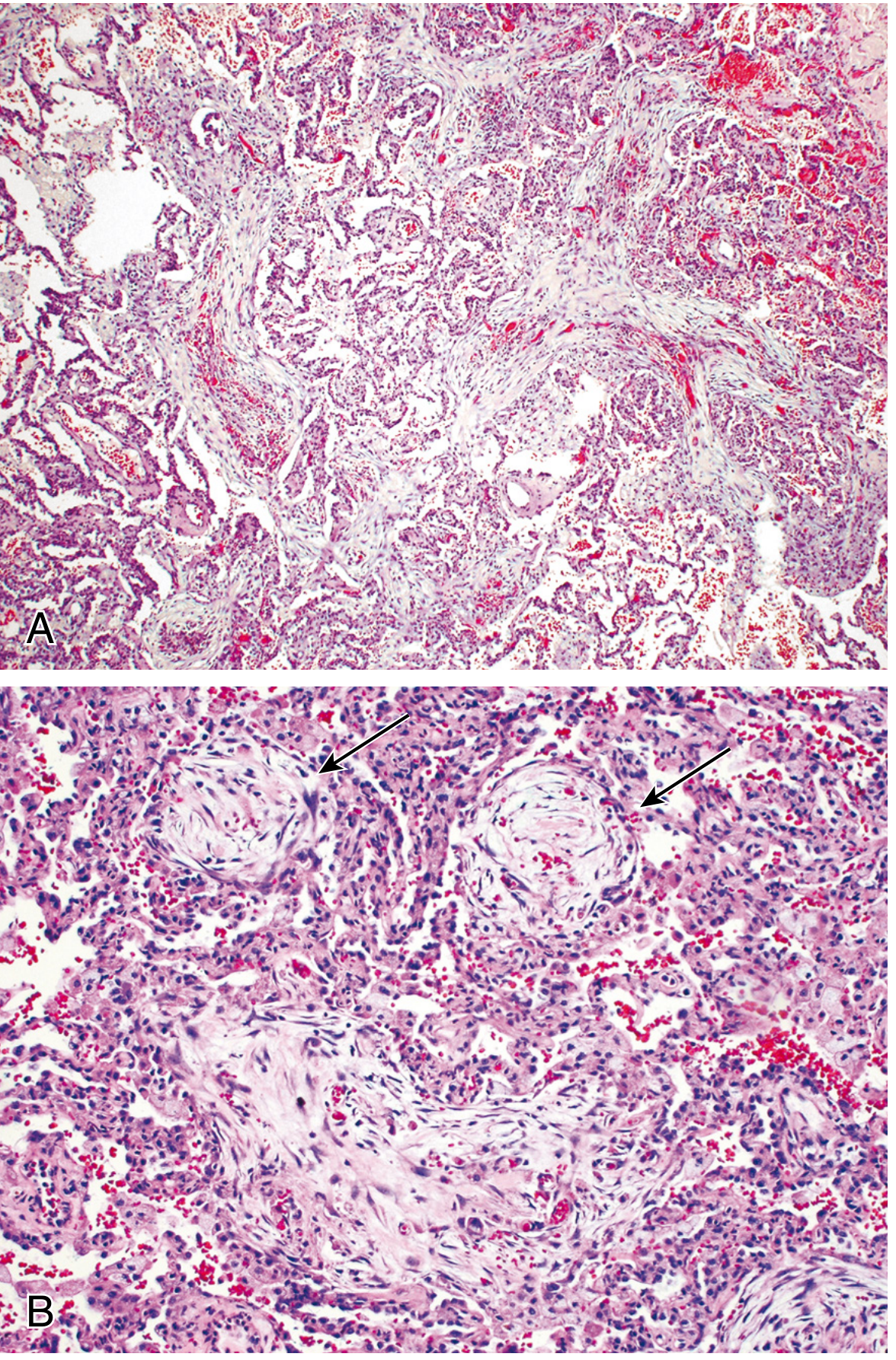
Pathophysiology / Histopathology
- The connective tissue plugs are all the same age (temporally uniform)
- The underlying alveolar architecture is preserved
- There is no honeycombing or established interstitial fibrosis (this distinguishes it from UIP/IPF)
Clinical Presentation
- Subacute, flu-like illness over days to weeks: cough, dyspnea, fever, fatigue, sometimes weight loss
- Inspiratory crackles/rales on exam
- Restrictive pattern on pulmonary function testing with hypoxemia
- Commonly misdiagnosed as community-acquired pneumonia that fails to respond to antibiotics
- Rarely, presents fulminantly with a clinical picture resembling ARDS
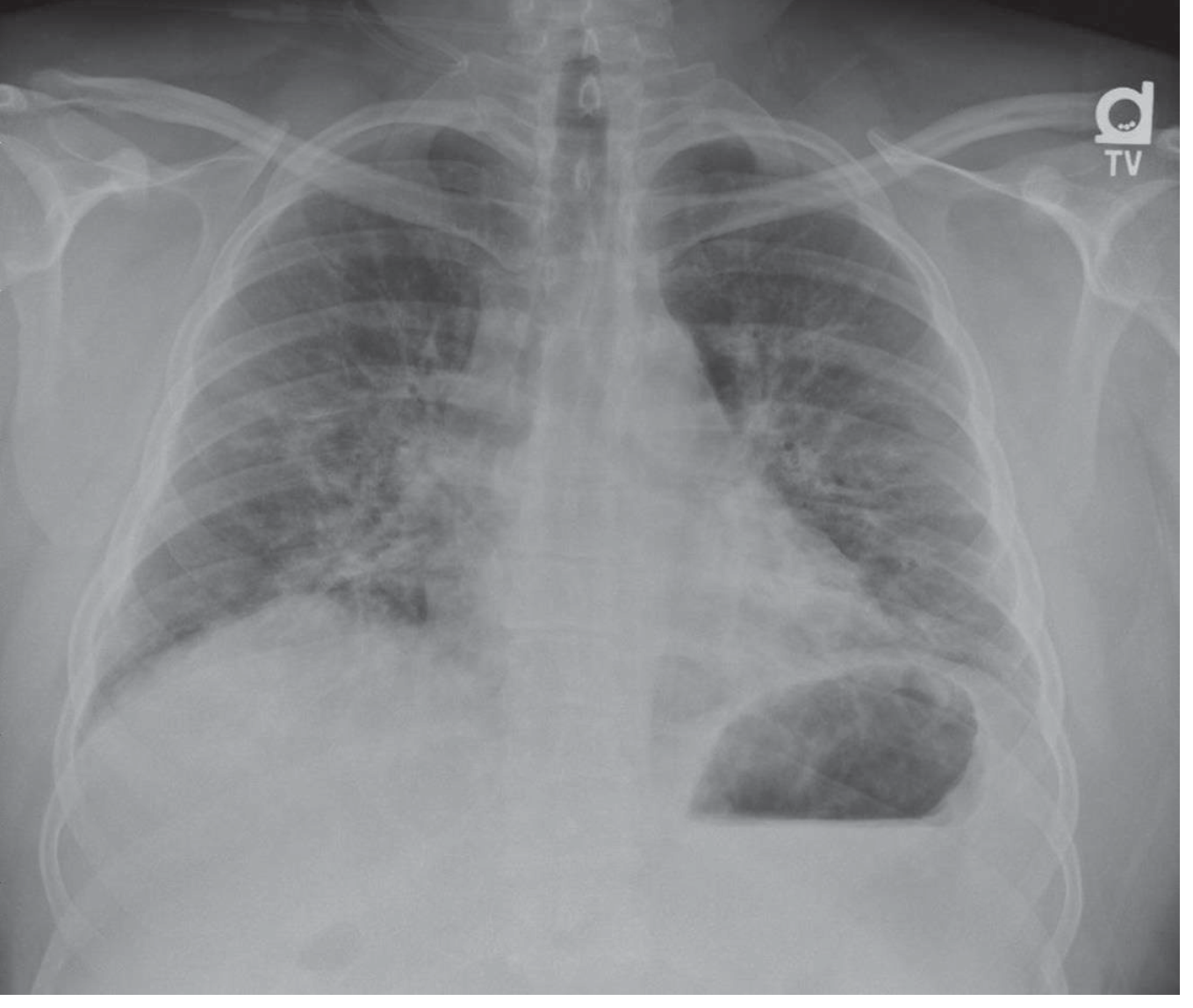
Imaging
Diagnosis
Treatment
- Corticosteroids are first-line and often produce a dramatic clinical and radiographic response
- Therapy is usually continued for at least 6 months, since relapse rates are high when steroids are tapered or stopped
- For relapsing or refractory disease, or to reduce steroid burden, alternative agents such as macrolides (mild disease), mycophenolate, cyclophosphamide, or rituximab (biologic) are increasingly used
- Secondary forms may need longer-term therapy directed at the underlying cause
Prognosis
Vs necrotizing pneumonia
necrotizing pneumonia CT cavitation lung abscess
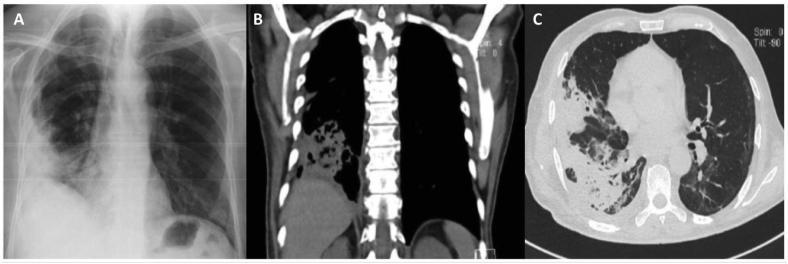
A composite of three clinical diagnostic images illustrating necrotizing pneumonia in the right lung. (A) Posteroanterior chest X-ray shows a large, heterogeneous opacity in the right basal region with ill-defined borders, indicating consolidation. (B) Coronal CT scan in mediastinal window demonstrates a hyperdense area in the right lower lobe consistent with lung consolidation, featuring internal low-attenuation areas suggestive of tissue breakdown or early cavitation. (C) Axial CT scan in lung window reveals a large, peripheral pulmonary opacity in the right lung with intimate pleural contact. The opacity exhibits internal air-filled lucencies and a distinct central cavitation, characteristic of necrotizing pneumonia and pulmonary abscess formation. This series is an educational example of progressive infectious lung disease, highlighting the transition from radiographic opacification to CT-confirmed cavitation and parenchymal necrosis. Key concepts include imaging signs of consolidation, cavitation, and pleural involvement in lower respiratory tract infections.
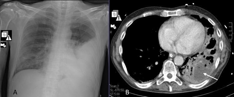
The composite image displays two diagnostic modalities for the chest. Image A is a frontal chest X-ray (radiography) demonstrating extensive opacification and consolidation in the left lower lung field, consistent with lobar pneumonia. The heart border is obscured, and the left costophrenic angle is obliterated. Image B is an axial computed tomography (CT) scan of the chest at the level of the heart, showing the evolution of the pathology. It reveals a large area of consolidation in the left posterior lung containing multiple low-density, hypodense regions. A white arrow points to a well-defined pulmonary abscess and surrounding cavitation within the necrotizing lung tissue. The right lung remains relatively clear on both modalities. These images illustrate the clinical progression from initial community-acquired pneumonia to necrotizing pneumonia and abscess formation, a significant complication often associated with specific bacterial pathogens or co-infections.
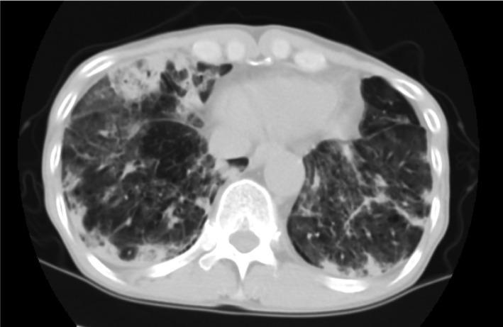
This diagnostic image is an axial chest computed tomography (CT) scan in a lung window, demonstrating severe bilateral pulmonary pathology. The scan shows extensive, multifocal areas of consolidation and patchy ground-glass opacities (GGO) distributed primarily in the lower lobes. In the right lung, particularly within the lower lobe, there are distinct cavitary lesions characterized by thick-walled, low-attenuation air-filled spaces, some of which exhibit internal air-fluid levels suggesting necrotizing pneumonia or abscess formation. The left lung also displays significant parenchymal involvement with dense opacification that obscures underlying vascular structures. The radiographic patterns are consistent with a severe inflammatory or infectious process, such as necrotizing community-acquired pneumonia or secondary bacterial infection (e.g., Pseudomonas aeruginosa). This visual material serves as an educational example of rapidly progressive pulmonary cavitation and the complex interplay between consolidation and ground-glass patterns in critical respiratory illness.
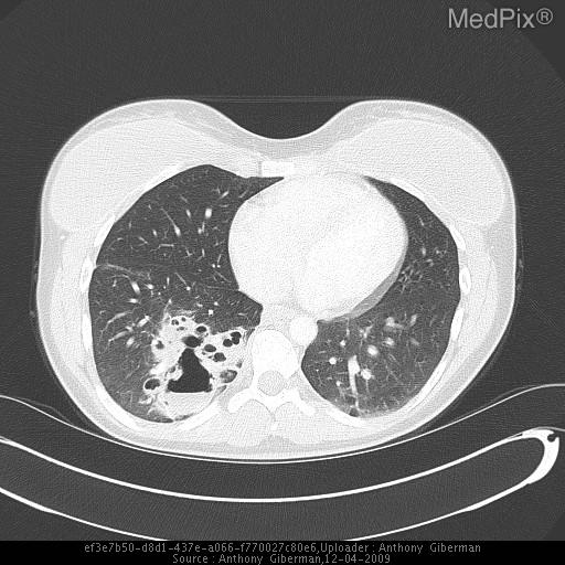
**Imaging Modality:** Axial Computed Tomography (CT) of the chest, lung window.
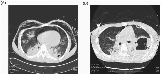
This side-by-side comparison displays axial chest computed tomography (CT) scans of a patient with necrotizing pneumonia, illustrating disease progression from day 7 (A) to day 26 (B) of hospitalization. Image A reveals bilateral, patchy alveolo-interstitial infiltrates with multiple small, low-density cavitations in the left lung consistent with early abscess formation. Additionally, there is evidence of subcutaneous emphysema and a small pneumothorax. Image B shows significant evolution of the pathology; the multiple small cavitations in the left lung have coalesced into a single, large, thick-walled abscess cavity. This mature abscess demonstrates a prominent air-fluid level, a classic radiological sign of a lung abscess containing both gas and inflammatory liquid. The surrounding lung parenchyma remains significantly compromised. These images serve as a teaching tool for recognizing the radiological stages of MRSA-related pulmonary destruction, from initial cavitation and septic complications to the formation of organized abscesses with air-fluid levels.
"necrotizing pneumonia" children OR adults
Cryptogenic Organizing Pneumonia vs. Necrotizing Pneumonia
Side-by-side comparison
| Feature | Cryptogenic Organizing Pneumonia (COP) | Necrotizing Pneumonia |
|---|---|---|
| Nature | Idiopathic interstitial lung disease - non-infectious | Infectious complication of pneumonia/lung abscess; sometimes called "lung gangrene" |
| Cause | Unknown by definition (organizing pneumonia pattern with no identified trigger) | Bacterial (S. aureus incl. MRSA, Klebsiella pneumoniae, Pseudomonas, anaerobes like Bacteroides/Fusobacterium), occasionally fungal (Mucor, Aspergillus) |
| Core pathology | Polypoid plugs of loose granulation tissue (Masson bodies) fill alveolar ducts/alveoli/bronchioles; underlying lung architecture is preserved, no true tissue destruction | True liquefactive necrosis of lung parenchyma with destruction of architecture; multiple small (<2 cm) cavities/microabscesses that may coalesce into a larger abscess |
| Onset | Subacute, over weeks - flu-like cough, dyspnea, fever, fatigue | Acute, often fulminant - high fever, purulent/foul sputum, hemoptysis, sepsis |
| Risk factors | Can be idiopathic, or trigger includes CTD (polymyositis, RA), drugs, radiation, HSCT/GVHD | Aspiration (alcoholism, poor dentition, altered consciousness, achalasia), post-influenza S. aureus, immunodeficiency, bronchial obstruction |
| Imaging | Patchy, migratory, subpleural/peribronchial consolidation with ground-glass; "reversed halo/atoll" sign; no cavitation | Consolidation progressing to cavitation, air-fluid levels, may show pneumatoceles, empyema, pneumothorax |
| Cultures/labs | Sterile - no organism identified; BAL/biopsy needed to exclude infection | Positive sputum/blood cultures or pleural fluid; often polymicrobial/anaerobic |
| Diagnosis | Clinical + HRCT + biopsy showing Masson bodies, after excluding infection | Clinical + imaging showing cavitation + microbiologic confirmation |
| Treatment | Corticosteroids (often >6 months given high relapse rate); steroid-sparing agents (mycophenolate, rituximab) for refractory disease. Antibiotics are NOT effective. | Prolonged antibiotics (anaerobic + broad-spectrum coverage), drainage of abscess/empyema, occasionally surgical resection |
| Complications | Relapse on steroid taper; rarely progresses to fibrosis | Empyema, bronchopleural fistula, sepsis, massive hemoptysis, respiratory failure |
| Mortality/prognosis | Generally good with treatment; most recover | Historically ~40% mortality untreated (preantibiotic era); still serious even now, though improved with modern antimicrobials and drainage |
Key conceptual distinction
Why it matters clinically
- Necrotizing pneumonia will show cavitation/abscess on CT and positive cultures - it needs antibiotics ± drainage, and worsens without them.
- COP will show migratory ground-glass/consolidation without cavitation, negative cultures, and dramatically improves with steroids, not antibiotics - giving antibiotics alone will fail because there is no infection to treat.
Create a diagnostic decision tree or flowchart comparing COP vs necrotizing pneumonia
python3 -c "import graphviz; print('graphviz ok')" 2>&1; which dot 2>&1; python3 -c "import matplotlib; print('mpl ok')" 2>&1
mkdir -p /tmp/workspace/cop-vs-necpna/output && ls -la /tmp/workspace/
~/cop-vs-necpna/build_flowchart.py
import graphviz

dot = graphviz.Digraph('COP_vs_NecPna', format='png')
dot.attr(rankdir='TB', splines='ortho', bgcolor='white', fontname='Helvetica')
dot.attr('node', fontname='Helvetica', fontsize='11')
dot.attr('edge', fontname='Helvetica', fontsize='10')

# ---------- Colors ----------
COP_FILL = '#dbe9f6'
COP_BORDER = '#1f5fa8'
NEC_FILL = '#fbdcdc'
NEC_BORDER = '#b5271f'
DECISION_FILL = '#fff2cc'
DECISION_BORDER = '#b58e00'
START_FILL = '#e8e8e8'
START_BORDER = '#444444'
TEST_FILL = '#e6f2e6'
TEST_BORDER = '#3a7d3a'

# ---------- Start ----------
dot.node('start', 'Patient with cough, dyspnea \u00b1 fever\nand patchy consolidation on CXR/CT\n("pneumonia" not fitting typical CAP course)',
          shape='box', style='rounded,filled', fillcolor=START_FILL, color=START_BORDER, width='3.5')

# ---------- Decision 1: Onset / tempo ----------
dot.node('d1', 'Onset & toxicity?', shape='diamond', style='filled', fillcolor=DECISION_FILL, color=DECISION_BORDER)
dot.edge('start', 'd1')

dot.node('acute', 'ACUTE (days), toxic-appearing\nfever, purulent/foul sputum \u00b1 hemoptysis\nRisk factors: aspiration, alcoholism, poor\ndentition, post-influenza, immunodeficiency',
          shape='box', style='filled', fillcolor=NEC_FILL, color=NEC_BORDER)
dot.node('subacute', 'SUBACUTE (weeks), flu-like\ncough, dyspnea, fatigue, low-grade fever\nOften "failed" empiric antibiotics\n\u00b1 CTD / drug / radiation / HSCT history',
          shape='box', style='filled', fillcolor=COP_FILL, color=COP_BORDER)
dot.edge('d1', 'acute', label='  acute/septic')
dot.edge('d1', 'subacute', label='  indolent')

# ---------- Decision 2 (necrotizing branch): imaging ----------
dot.node('d2', 'CT chest:\ncavitation / air-fluid level /\nmicroabscesses?', shape='diamond', style='filled', fillcolor=DECISION_FILL, color=DECISION_BORDER)
dot.edge('acute', 'd2')

dot.node('nec_yes', 'YES\nMultiple <2cm cavities or\nabscess with air-fluid level,\n\u00b1 empyema / pneumothorax',
          shape='box', style='filled', fillcolor=NEC_FILL, color=NEC_BORDER)
dot.edge('d2', 'nec_yes', label='  yes')

dot.node('recheck1', 'Reconsider: uncomplicated CAP,\nseptic emboli, malignancy,\nfungal/mycobacterial disease',
          shape='box', style='filled', fillcolor='#f0f0f0', color='#666666')
dot.edge('d2', 'recheck1', label='  no')

# ---------- Test node (necrotizing branch): micro ----------
dot.node('t1', 'Sputum / blood / pleural\nfluid culture + Gram stain\n(\u00b1 anaerobic culture)', shape='box', style='filled', fillcolor=TEST_FILL, color=TEST_BORDER)
dot.edge('nec_yes', 't1')

dot.node('nec_dx', 'DIAGNOSIS:\nNECROTIZING PNEUMONIA\n(bacterial: S. aureus/MRSA, Klebsiella,\nanaerobes, Pseudomonas; rarely fungal)',
          shape='box', style='rounded,filled', fillcolor=NEC_FILL, color=NEC_BORDER, penwidth='2')
dot.edge('t1', 'nec_dx', label='  organism identified\n  or high clinical suspicion')

dot.node('nec_tx', 'TREATMENT\nProlonged IV\u2192PO antibiotics\n(anaerobic + broad-spectrum coverage)\nDrain abscess/empyema if needed\nSurgery if refractory',
          shape='box', style='filled', fillcolor=NEC_FILL, color=NEC_BORDER)
dot.edge('nec_dx', 'nec_tx')

# ---------- Decision 2b (COP branch): imaging ----------
dot.node('d3', 'CT chest:\nmigratory subpleural/peribronchial\nconsolidation + GGO \u00b1 reversed\nhalo (atoll) sign, NO cavitation?',
          shape='diamond', style='filled', fillcolor=DECISION_FILL, color=DECISION_BORDER)
dot.edge('subacute', 'd3')

dot.node('cop_yes', 'YES - pattern suggestive\nof organizing pneumonia', shape='box', style='filled', fillcolor=COP_FILL, color=COP_BORDER)
dot.edge('d3', 'cop_yes', label='  yes')

dot.node('recheck2', 'Reconsider: NSIP, hypersensitivity\npneumonitis, chronic eosinophilic\npneumonia, atypical infection',
          shape='box', style='filled', fillcolor='#f0f0f0', color='#666666')
dot.edge('d3', 'recheck2', label='  no')

# ---------- Test node (COP branch): exclude infection + biopsy ----------
dot.node('t2', 'Bronchoscopy + BAL culture\n(exclude infection) AND\nTransbronchial/surgical biopsy\nor cryobiopsy', shape='box', style='filled', fillcolor=TEST_FILL, color=TEST_BORDER)
dot.edge('cop_yes', 't2')

dot.node('d4', 'Biopsy: granulation-tissue plugs\n(Masson bodies) in alveolar ducts/\nalveoli, architecture PRESERVED,\ncultures NEGATIVE?', shape='diamond', style='filled', fillcolor=DECISION_FILL, color=DECISION_BORDER)
dot.edge('t2', 'd4')

dot.node('d5', 'Secondary cause found?\n(CTD serologies, drug/\nradiation history, malignancy,\nGVHD post-HSCT)', shape='diamond', style='filled', fillcolor=DECISION_FILL, color=DECISION_BORDER)
dot.edge('d4', 'd5', label='  yes')

dot.node('sec_op', 'DIAGNOSIS:\nSECONDARY ORGANIZING PNEUMONIA\n(treat underlying cause + steroids)', shape='box', style='rounded,filled', fillcolor='#fde9c8', color='#b58e00')
dot.edge('d5', 'sec_op', label='  yes')

dot.node('cop_dx', 'DIAGNOSIS:\nCRYPTOGENIC ORGANIZING PNEUMONIA (COP)\nNo identifiable cause after workup',
          shape='box', style='rounded,filled', fillcolor=COP_FILL, color=COP_BORDER, penwidth='2')
dot.edge('d5', 'cop_dx', label='  no')

dot.node('cop_tx', 'TREATMENT\nOral corticosteroids \u2265 6 months\n(taper slowly - high relapse rate)\nSteroid-sparing agents if refractory/\nrelapsing: macrolide, mycophenolate,\nrituximab, cyclophosphamide',
          shape='box', style='filled', fillcolor=COP_FILL, color=COP_BORDER)
dot.edge('cop_dx', 'cop_tx')

dot.edge('d4', 'recheck1', label='  no (destruction/\n  organisms found)')

# ---------- Legend ----------
with dot.subgraph(name='cluster_legend') as c:
    c.attr(label='Legend', fontsize='11', style='dashed', color='#999999')
    c.node('leg1', 'Necrotizing pneumonia pathway', shape='box', style='filled', fillcolor=NEC_FILL, color=NEC_BORDER)
    c.node('leg2', 'COP pathway', shape='box', style='filled', fillcolor=COP_FILL, color=COP_BORDER)
    c.node('leg3', 'Decision point', shape='diamond', style='filled', fillcolor=DECISION_FILL, color=DECISION_BORDER)
    c.node('leg4', 'Confirmatory test', shape='box', style='filled', fillcolor=TEST_FILL, color=TEST_BORDER)

dot.render('/tmp/workspace/cop-vs-necpna/output/COP_vs_Necrotizing_Pneumonia', format='png', cleanup=True)
print("done")
cd /tmp/workspace/cop-vs-necpna && python3 build_flowchart.py && ls -la output/
COP vs Necrotizing Pneumonia
Image · PNG

- Entry point - any patient with cough/dyspnea/fever and patchy consolidation that doesn't fit a routine CAP course.
- First branch point - tempo of illness: acute and toxic-appearing (days, purulent sputum, aspiration/alcoholism/post-flu risk factors) pushes you toward necrotizing pneumonia; subacute and flu-like (weeks, failed empiric antibiotics, ± CTD/drug/radiation history) pushes toward the organizing pneumonia pathway.
- Imaging split: cavitation, air-fluid levels, or microabscesses on CT confirm the necrotizing pathway; migratory ground-glass/consolidation with a reversed halo (atoll) sign and no cavitation supports organizing pneumonia.
- Confirmatory testing differs completely: cultures (sputum/blood/pleural fluid) for necrotizing pneumonia vs. bronchoscopy/BAL (to exclude infection) plus biopsy showing Masson bodies for organizing pneumonia.
- Final split for the organizing pneumonia branch: if a secondary trigger is found (CTD, drug, radiation, GVHD), it's labeled secondary organizing pneumonia rather than true COP - COP is a diagnosis of exclusion.
- Treatment endpoints are deliberately opposite: antibiotics ± drainage/surgery for necrotizing pneumonia vs. prolonged corticosteroids ± steroid-sparing agents for COP - giving antibiotics for COP or steroids alone for an undrained necrotizing process would both fail.
If cop is non infectious why it called pneumonia
Why "pneumonia" if it's not infectious?
Two different meanings of "pneumonia"
- Clinical/lay usage today: "pneumonia" = infectious inflammation of the lung, almost always what people mean when they say it.
- Original pathologic usage: "pneumonia" simply means inflammation/consolidation of the lung parenchyma with the alveolar air spaces filled by something (exudate, cells, tissue) - regardless of cause. Under this older definition, "organizing pneumonia" describes a specific histologic pattern: the alveolar spaces and distal airways get filled and "organized" by loose fibroblastic/granulation tissue plugs (Masson bodies), producing lung consolidation that looks like pneumonia radiographically and even clinically (fever, cough, consolidation on imaging) - but the filling material is scar-forming granulation tissue, not pus or infectious exudate.
The naming history makes this clearer
- Epler introduced the term bronchiolitis obliterans organizing pneumonia (BOOP) in 1985 to describe this histologic pattern - fibroblastic plugs organizing within alveolar ducts/alveoli, often with associated small-airway (bronchiolar) involvement.
- BOOP caused confusion because clinicians kept mistaking it for the very different disease "bronchiolitis obliterans" (constrictive, irreversible airway scarring - a distinct condition).
- The American Thoracic Society/European Respiratory Society consensus classification (1998, Katzenstein and Myers; later ATS/ERS statements) recommended replacing BOOP with cryptogenic organizing pneumonia (COP), since "organizing pneumonia" better captures the essential pathology - organizing granulation tissue filling airspaces - without implying the fibrotic airway disease, and "cryptogenic" specifies that no cause is found.
- Murray & Nadel's Textbook of Respiratory Medicine notes explicitly: "The term organizing pneumonia defines this organizing fibroblastic tissue in alveoli" - the word choice is about the tissue reaction pattern, not about an infectious etiology.
Practical takeaway
- COP behaves clinically like pneumonia (fever, cough, consolidative opacities, sometimes even looks infective on a chest X-ray) which is exactly why it gets misdiagnosed as bacterial pneumonia and treated with antibiotics that don't work.
- But pathologically it is a non-infectious immune/fibroproliferative reaction - granulation tissue organizing within alveoli - that happens to produce a "pneumonia-like" clinical and radiographic picture.
- This is analogous to how "eosinophilic pneumonia," "lipoid pneumonia," or "chemical pneumonitis/pneumonia" are also non-infectious conditions that borrow the word "pneumonia" because they cause lung consolidation, not because bacteria or viruses are involved.
How many days of antibiotics in necrotizing pneumonia
"necrotizing pneumonia" antibiotic duration treatment
Antibiotic Duration for Necrotizing Pneumonia
General range
- 3 to 12 weeks total antimicrobial therapy is the range cited for necrotizing pneumonia and lung abscess (Schwartz's Principles of Surgery, 11th ed.)
- Many sources anchor around ~6 weeks of systemic antibiotics as a typical course for necrotizing pneumonia from bacterial, mycobacterial, fungal, or parasitic pathogens, targeted to the organism identified on blood/BAL/needle aspirate cultures (Goldman-Cecil Medicine)
- Compare this to uncomplicated pneumonitis, where 5-10 days of total treatment is usually adequate - necrotizing pneumonia, abscess, and empyema explicitly require prolonged parenteral therapy followed by oral step-down, not the short course (Murray & Nadel's Textbook of Respiratory Medicine)
How duration is actually decided (not a fixed number)
- Start with IV (parenteral) therapy while the patient is acutely ill/bacteremic
- Switch to oral therapy once clinically stable and tolerating oral intake
- Continue oral therapy for weeks, with repeat imaging (CXR or CT) to confirm the cavity/consolidation is resolving or resolved before stopping
- If empyema or abscess is present, drainage (chest tube, VATS, or percutaneous) is often needed alongside antibiotics - antibiotics alone with an undrained collection will prolong or fail therapy
Pediatric consideration
Practical bottom line
| Scenario | Typical duration |
|---|---|
| Uncomplicated CAP | 5-7 days |
| Necrotizing pneumonia / lung abscess | 3-6 weeks minimum, up to 12 weeks in refractory cases |
| Endpoint marker | Clinical improvement + radiographic resolution/near-resolution of cavity, not a calendar date |
Excuberent honeycombing