
Anion gap 10 mark answer
Reading File
Searching sources...
Finding Sources
Finding Sources
Now let me read the most comprehensive sections in parallel:
Reading File
Reading File
Reading File
Reading File
Reading File
Reading File
Now let me get the images and the urine anion gap section:
Loading Image
Loading Image
Finding Sources
Reading File
Reading File
I now have comprehensive material to write the 10-mark answer. Let me compile everything into a structured, exam-quality response.
Anion Gap
Definition
The anion gap (AG) is a calculated value representing the difference between the major measured serum cation (Na⁺) and the major measured serum anions (Cl⁻ and HCO₃⁻):
AG = [Na⁺] - ([Cl⁻] + [HCO₃⁻])
Using normal values: AG = 140 - (104 + 24) = 12 mEq/L
The normal range is 8-12 mEq/L (some sources cite 7-14 mEq/L).
Physiological Basis
Electrical neutrality must be maintained in plasma - the total cation charge must equal the total anion charge. Only three electrolytes are measured routinely (Na⁺, Cl⁻, HCO₃⁻). The apparent "gap" reflects the net charge of unmeasured anions minus unmeasured cations:
AG = Unmeasured anions - Unmeasured cations
- Unmeasured cations: K⁺, Ca²⁺, Mg²⁺
- Unmeasured anions: albumin (largest component, ~11 mEq/L), phosphate, sulfate, organic acid anions (lactate, urate)
The normal AG exists primarily because of the net anionic charge of albumin. - National Kidney Foundation Primer on Kidney Diseases, 8e
Diagrammatic Representation
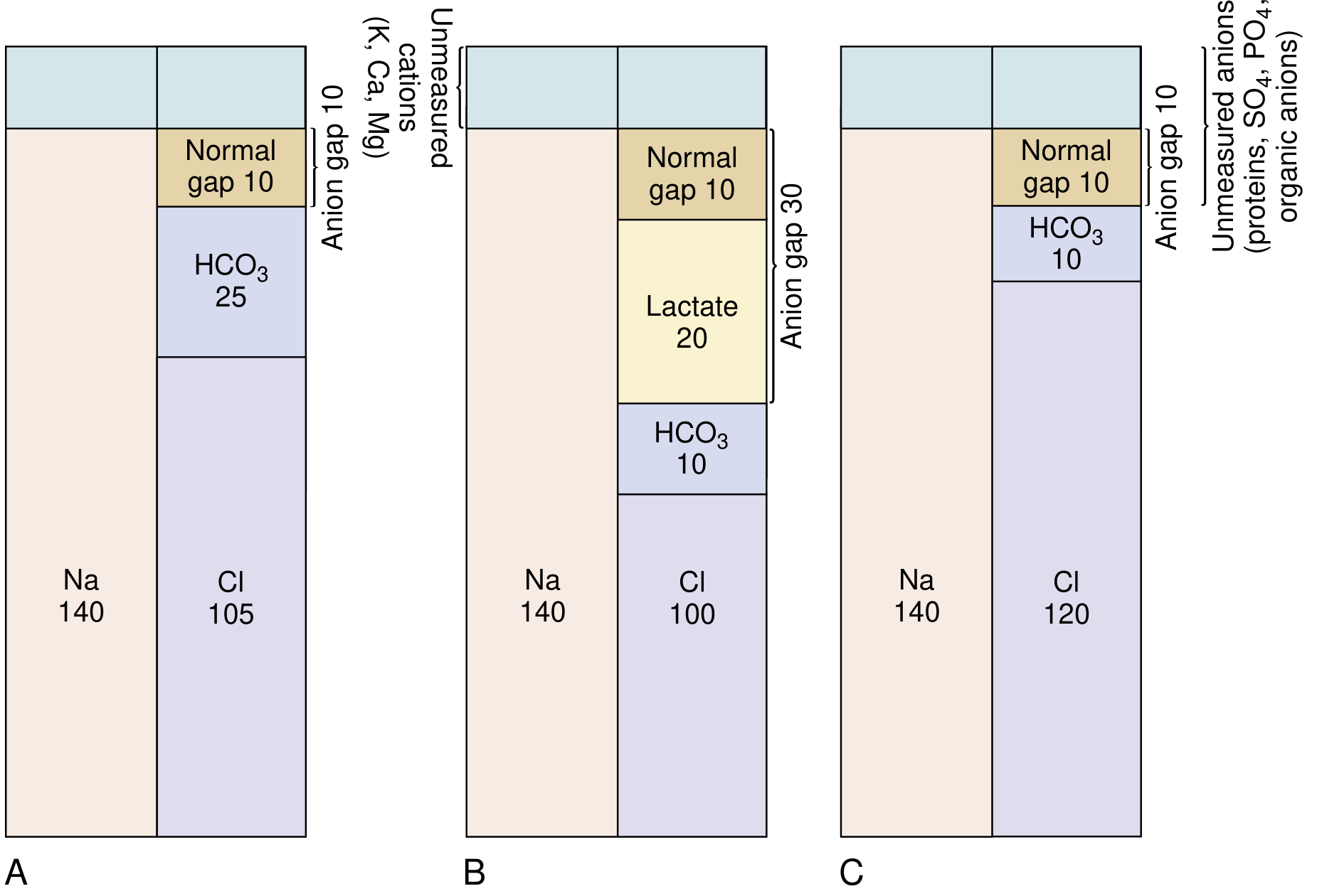
Fig: (A) Normal AG = 10. (B) High AG acidosis - HCO₃⁻ falls, unmeasured organic anion (lactate) rises, AG = 30. (C) Hyperchloremic (normal AG) acidosis - HCO₃⁻ falls, Cl⁻ rises, AG unchanged.
Corrected Anion Gap (for Hypoalbuminemia)
Albumin accounts for the bulk of the normal AG. Hypoalbuminemia reduces the AG and can mask a true high-AG acidosis. Correction formula:
Corrected AG = AG + 2.5 × (4 - serum albumin in g/dL)
The AG falls approximately 2.5 mEq/L for each 1 g/dL drop in albumin below normal. This correction is critical in critically ill, malnourished, or catabolic patients. - NKF Primer; Goldman-Cecil Medicine
Clinical Uses of the Anion Gap
1. Classifying Metabolic Acidosis
| Type | AG | Mechanism |
|---|---|---|
| High AG metabolic acidosis | Elevated (>12) | Accumulation of non-chloride acid anions |
| Normal AG (hyperchloremic) acidosis | Normal (8-12) | HCO₃⁻ loss replaced by Cl⁻ rise |
2. High Anion Gap Metabolic Acidosis - Causes
The mnemonic GOLDMARK is widely used:
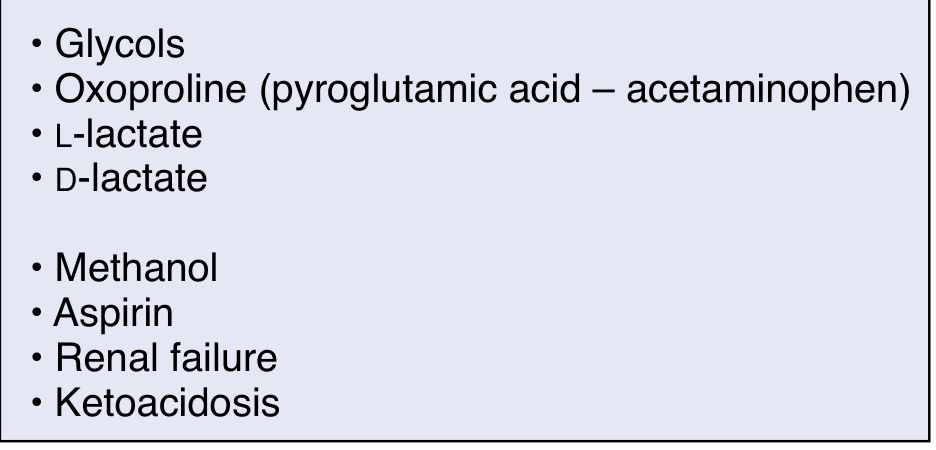
- G - Glycols (ethylene glycol, propylene glycol)
- O - Oxoproline (pyroglutamic acid - from chronic acetaminophen use)
- L - L-lactate (lactic acidosis)
- D - D-lactate
- M - Methanol
- A - Aspirin (salicylate poisoning)
- R - Renal failure
- K - Ketoacidosis (DKA, alcoholic, starvation)
In high AG acidosis, non-volatile acids dissociate into H⁺ + conjugate anion; the H⁺ consumes HCO₃⁻ while the anion accumulates, raising the AG. - Morgan & Mikhail's Clinical Anesthesiology, 7e
3. Normal Anion Gap (Hyperchloremic) Acidosis - Causes
When HCO₃⁻ falls but Cl⁻ rises by the same amount, no organic anion accumulates and the AG stays normal. Causes include:
- GI losses: diarrhea, small bowel fistula, ileostomy (HCO₃⁻-rich fluid lost)
- Renal tubular acidosis (RTA): Types 1, 2, and 4
- Acetazolamide (carbonic anhydrase inhibitor)
- Excessive normal saline (iatrogenic hyperchloremia)
- Ureterosigmoidostomy
The Delta-Delta Ratio (Δ/Δ)
When a high AG acidosis is identified, the delta-delta ratio helps detect mixed acid-base disorders:
Δ/Δ = (Measured AG - Normal AG) / (Normal HCO₃⁻ - Measured HCO₃⁻) = (AG - 12) / (24 - [HCO₃⁻])
| Ratio | Interpretation |
|---|---|
| 1-2 | Pure high AG metabolic acidosis |
| >2 | Coexisting metabolic alkalosis (or chronic respiratory acidosis) |
| <1 | Coexisting normal AG (hyperchloremic) acidosis |
If the rise in AG is larger than the fall in HCO₃⁻, an additional process is raising HCO₃⁻ (e.g., metabolic alkalosis). If the fall in HCO₃⁻ exceeds the rise in AG, a simultaneous hyperchloremic acidosis is present. - Goldman-Cecil Medicine
Urine Anion Gap
In normal AG (hyperchloremic) acidosis, the urine anion gap helps distinguish renal from extrarenal causes:
Urine AG = (Urine Na⁺ + Urine K⁺) - Urine Cl⁻
The principal unmeasured urinary cation is NH₄⁺, which increases (with Cl⁻) during systemic acidosis.
| Urine AG | Interpretation |
|---|---|
| Negative (Cl⁻ > Na⁺+K⁺) | Increased NH₄⁺ excretion - extrarenal cause (diarrhea) - appropriate renal response |
| Positive (Cl⁻ < Na⁺+K⁺) | Impaired NH₄⁺ secretion - renal cause (RTA, renal failure) |
The urine AG is a surrogate marker for urinary NH₄⁺ excretion. Its utility is limited when non-chloride anions are present in urine (keto-anions, hippurate), in which case the urine osmole gap is more reliable. - NKF Primer on Kidney Diseases, 8e; Morgan & Mikhail
Causes of LOW Anion Gap
A reduced AG (<6 mEq/L) is clinically important and may indicate:
- Hypoalbuminemia (most common)
- Multiple myeloma with cationic immunoglobulins (IgG type - unmeasured cations)
- Lithium or bromide toxicity (spurious Cl⁻ elevation)
- Hypercalcemia, hypermagnesemia (increased unmeasured cations)
A mild AG elevation (up to 20 mEq/L) may not be diagnostically helpful; values >30 mEq/L almost always indicate a true high AG acidosis. - Morgan & Mikhail's Clinical Anesthesiology, 7e
Osmolar Gap and the Anion Gap
In certain high AG acidoses (methanol and ethylene glycol poisoning), an osmolar gap is also present:
Osmolar gap = Measured osmolality - Estimated osmolality
where: Estimated osmolality = 2[Na⁺] + glucose/18 + BUN/2.8
These low-molecular-weight alcohols add significant moles of solute to plasma, widening the measured-calculated difference. Other high AG causes (ketoacids, lactic acid, salicylate) do not produce a significant osmolar gap due to their higher molecular weights. - Costanzo Physiology, 7e
Summary Table
| Parameter | Normal AG Acidosis | High AG Acidosis |
|---|---|---|
| AG | 8-12 mEq/L (normal) | >12 mEq/L (elevated) |
| Plasma Cl⁻ | Elevated (hyperchloremic) | Normal or low |
| Urine AG | Negative (diarrhea) / Positive (RTA) | Not applicable |
| Common causes | Diarrhea, RTA, saline | DKA, lactic acidosis, toxins, renal failure |
Sources: National Kidney Foundation Primer on Kidney Diseases, 8e | Morgan & Mikhail's Clinical Anesthesiology, 7e | Goldman-Cecil Medicine | Costanzo Physiology, 7e
This is a shared conversation. Sign in to Orris to start your own chat.