
Hymen and its type in forensic medicine
types of hymen forensic medicine diagram
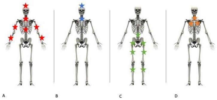
This Comparison Chart consists of four anatomical skeletal diagrams (A, B, C, and D) illustrating the distribution of skeletal lesions based on different mechanisms of trauma, commonly used in forensic medicine and traumatology. Diagram A shows a fall onto the head with defense attempt, indicated by star markers on the skull, cervical spine, shoulders, upper arms, and distal forearms (wrists). Diagram B represents a fall onto the head without defense attempt, showing localized markers on the skull and cervical spine only. Diagram C depicts a fall onto the feet (axial loading), with markers concentrated on the pelvic girdle, proximal femurs, and distal femurs/knees. Diagram D illustrates a specific clinical case where lesions are isolated to the cervical spine, sternum, and bilateral rib cage, which clinically excludes a standard fall mechanism. These illustrations serve to educate on the correlation between injury patterns and primary impact zones in blunt force trauma and fall scenarios.
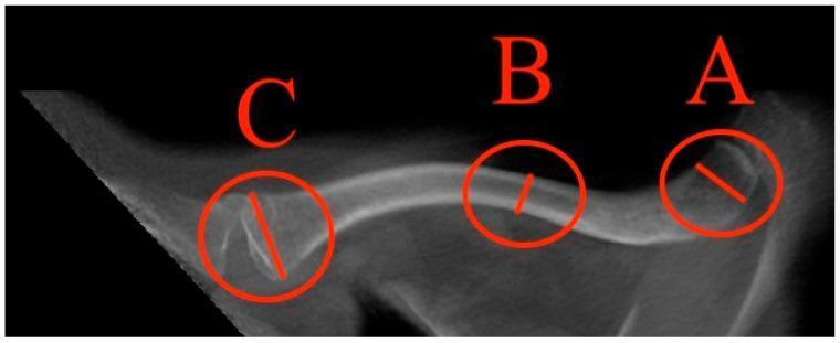
An anatomical diagram and diagnostic-style schematic of a human clavicle viewed from a superior perspective. The image illustrates standardized morphometric measurement points used in clinical imaging and forensic medicine. Three specific regions are highlighted with red circles and labeled with letters: 'A' indicates the maximal lateral diameter near the acromial end; 'B' represents the minimal diameter located at the midshaft (middle third) of the bone; and 'C' identifies the maximal medial diameter at the sternal end. The clavicle displays its characteristic 'S'-shaped double curvature, with a convex anterior medial portion and a concave anterior lateral portion. The visualization highlights structural differences, showing increased width and trabecular density at the articular ends compared to the more slender, cortical-dense midshaft. This diagram is utilized for teaching bone morphology, quantifying clavicular dimensions in orthopedic research, and establishing normative reference values for population studies.
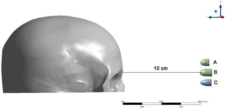
This forensic and clinical engineering visualization depicts a 3D finite element (FE) model setup for ballistic impact simulation on a human skull. The image shows a lateral (right side) view of a craniofacial model with a designated impact point at the glabella (forehead). Three specific ammunition types are positioned at a firing distance of 10 cm, indicated by a horizontal measurement line. Bullet A represents a .380-caliber bullet, Bullet B is a .40-caliber Smith & Wesson (S&W) flat-point bullet, and Bullet C is a 9×19-mm Luger round-nose bullet. The model highlights the different nose morphologies—rounded and flat—of the projectiles. A 3D coordinate system (X, Y, Z axes) and a millimeter scale bar at the bottom provide spatial calibration. This diagram is used in forensic pathology and trauma research to analyze kinetic energy transfer, von Mises stress distribution, and resulting fracture patterns in gunshot wounds to the frontal bone.
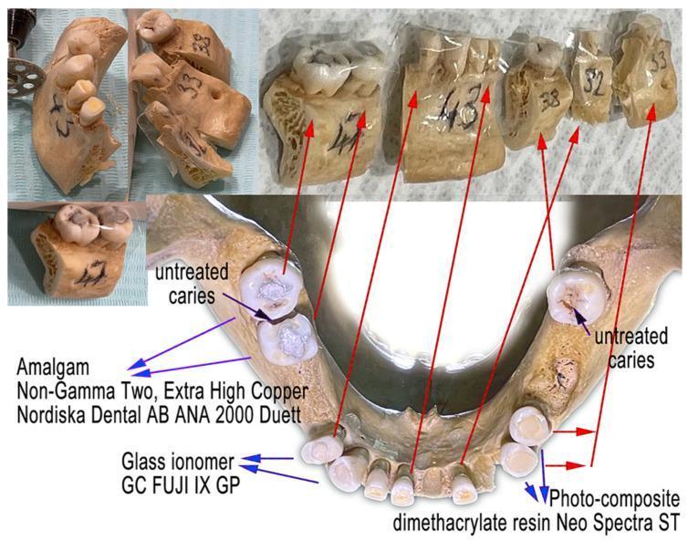
A clinical photograph and forensic diagram illustrating a human mandible used in dental material research. The image displays the mandible in several states: as a whole anatomical structure and subsequently divided into five labeled segments (47, 43, 38, 32, and 33) for experimental degradation studies. Key educational features include the identification of three types of dental restorative materials: silver-toned Amalgam (Nordiska Dental AB ANA 2000), white Glass Ionomer (GC FUJI IX GP), and tooth-colored Photo-composite (dimethacrylate resin Neo Spectra ST). Red arrows link the partitioned segments to their original locations in the dental arch. Additionally, the image points out pathological features such as untreated dental caries, visible as dark occlusal discolorations. The content serves to demonstrate forensic odontological methods, the visual morphology of different dental fillings, and the structural integrity of mandibular bone and dentition under experimental conditions.
Hymen in Forensic Medicine
Definition & Anatomy
- In children: appears as a taut membrane when thighs are separated
- During puberty: enlarges and appears as a series of folds
- May be rigid/fibrous, or elastic and easily distensible
- Supplied by blood vessels that bleed on rupture
- In virgins: admits only the tip of the little finger
Types of Hymen
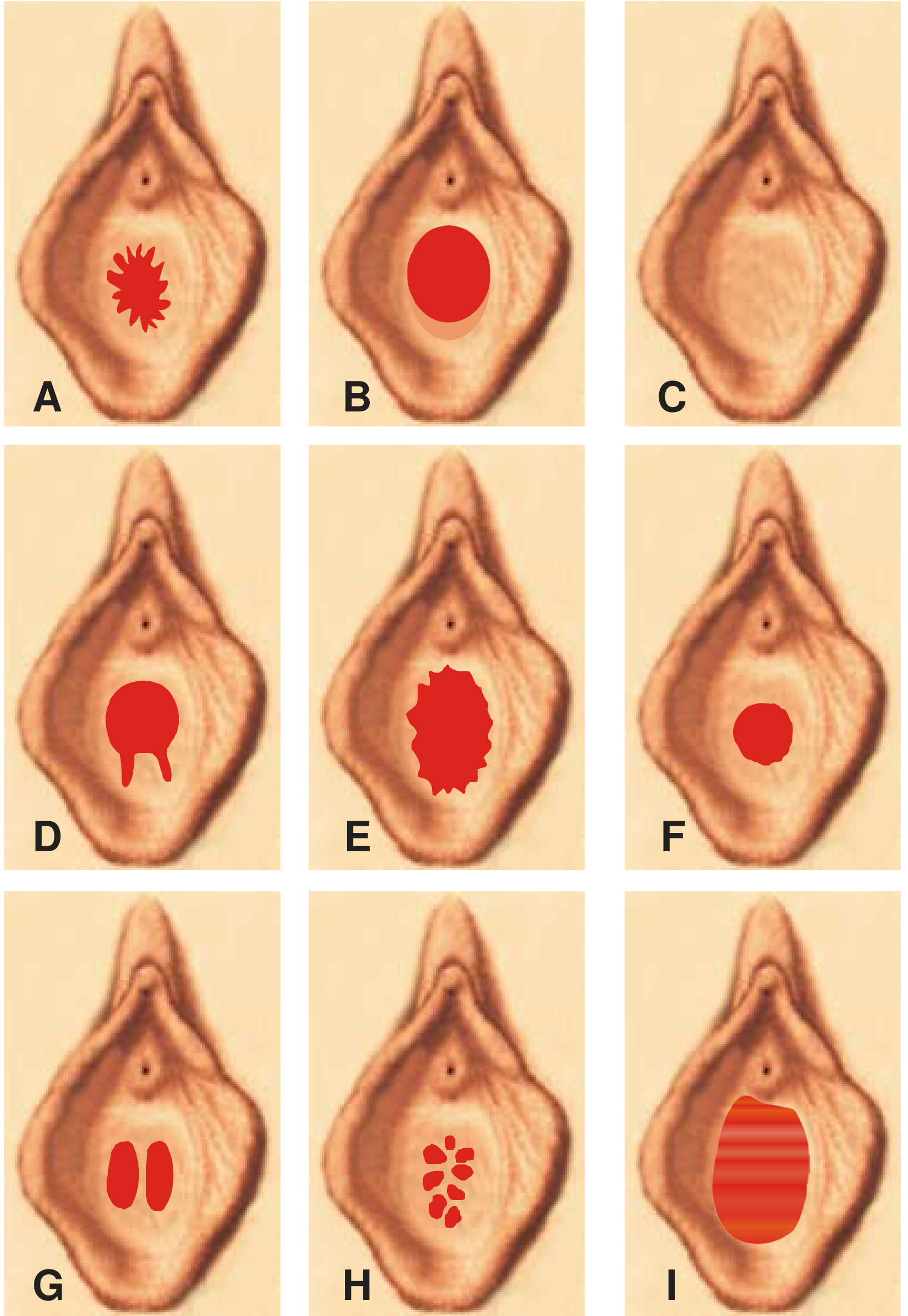
| # | Type | Description |
|---|---|---|
| 1 | Semilunar / Crescentic | Opening placed anteriorly; hymenal tissue present at lateral and posterior margins of vaginal opening |
| 2 | Annular | Oval opening situated centrally; encircles the entire vaginal opening |
| 3 | Infantile | Small linear opening in the midline |
| 4 | Cribriform | Multiple small hymenal openings |
| 5 | Vertical | A vertical hymenal opening |
| 6 | Septate | Two lateral openings side by side |
| 7 | Imperforate | No hymenal opening at all |
| 8 | Fimbriated / Notched (Frilly-edged) | Free margin presents natural notches - must be differentiated from traumatic tears |
Fimbriated vs. Ruptured Hymen - Key Differentiation
| Feature | Fimbriated Hymen | Ruptured Hymen |
|---|---|---|
| Location of notches | Anteriorly; do NOT extend to the base | Posteriorly (5 o'clock and 7 o'clock positions); extend to the point of attachment |
| Symmetry | Bilaterally symmetrical | Asymmetrical |
| Mucous membrane | Intact over notches | Not intact |
| Cause | Natural, congenital | Sexual intercourse or other causes |
Hymen in Relation to Sexual Intercourse
- In the majority of cases, hymen ruptures during the first act of coitus
- However, certain types will NOT rupture even with repeated acts:
- Annular hymen (fringe-like)
- Loose, folded, and elastic type
- Thick, tough, and fleshy type
- Present posteriorly, usually at 5 o'clock and 7 o'clock positions
- May sometimes be in the midline
- Usually, not more than two tears are found
- Raw, red, swollen, and painful to touch
- Bleed on touching (within 1-2 days)
- Heal in 5-6 days
- Within 8-10 days: shrunken, appear as granular tags of tissue
Causes of Rupture of Hymen (Non-Sexual)
- Accidental - Fall on a projecting object or slipping on furniture/fence (accompanied by other body injuries; forcible thigh separation in children does NOT rupture hymen unless perineum ruptures; riding, dancing, jumping do NOT rupture hymen)
- Masturbation - Use of long objects; causes enlarged clitoris and dilated vaginal orifice
- Foreign body - Introduction of hygroscopic materials (e.g., sola pith) into vagina
- Ulceration - Diphtheria, fungal infections
- Scratching - Due to poor personal hygiene
- Surgical causes - Medical procedures
- Sanitary tampons - Can cause rupture when used to absorb secretions
Forensic Significance - Diagnosis of Virginity
- "Virgo intacta" - term applied when the hymen remains intact despite acts of sexual intercourse
- Intact hymen can be found in married women and even in prostitutes
- Similarly, a virgin's hymen may be ruptured by non-sexual causes
- Hymen intact and well-stretched
- Edges distinct and regular
- Vaginal orifice is small - admits only terminal phalanx of a finger
- Hymen intact
- Hymenal orifice lets one or more fingers pass easily
- Hymen is relaxed and undulates