
Diff btw sympathetic parasympathetic and all kind of nerve fibers basic understanding of cns
Reading File
Finding Sources
Finding Sources
Searching PubMed
"autonomic nervous system" AND physiology
Searching the Web
autonomic nervous system sympathetic parasympathetic nerve fibers CNS overview NCBI
Reading File
Reading File
Reading File
Finding Sources
1. Basic map of the nervous system
Nervous system
- CNS = brain + spinal cord
- PNS = nerves and ganglia outside the brain and spinal cord
PNS has two main functional parts:
- Sensory (afferent) fibers: carry information to the CNS.
- Motor (efferent) fibers: carry commands from the CNS to the body.
A quick memory aid:
- Afferent arrives at CNS
- Efferent exits CNS
2. Somatic vs autonomic nervous system
| Feature | Somatic nervous system | Autonomic nervous system (ANS) |
|---|---|---|
| Main role | Voluntary movement | Automatic body functions |
| Target | Skeletal muscle | Cardiac muscle, smooth muscle, glands |
| Motor pathway | One neuron from CNS to muscle | Two neurons: preganglionic + postganglionic |
| Examples | Moving arm, walking | Heart rate, sweating, digestion, pupil size |
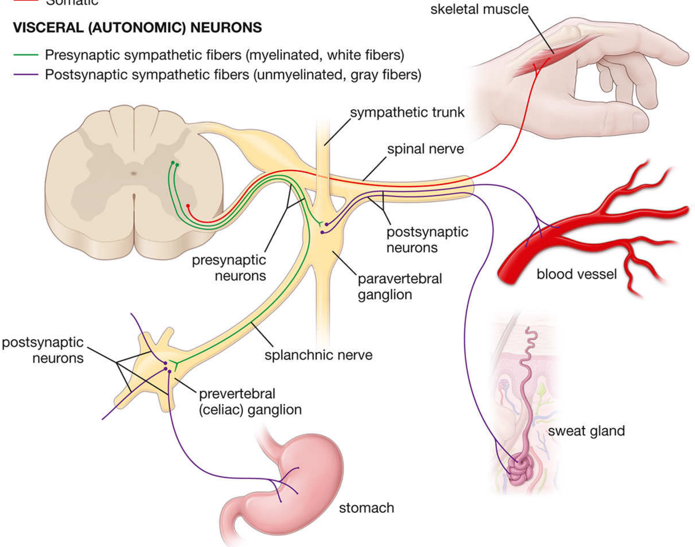
In the ANS, the first neuron starts in the CNS and synapses in a peripheral ganglion. The second neuron goes from the ganglion to the organ. Neuroanatomy through Clinical Cases, p. 262.
3. Sympathetic vs parasympathetic
Both are divisions of the autonomic nervous system. They often have opposite effects, but they also work together to keep the body balanced.
| Feature | Sympathetic | Parasympathetic |
|---|---|---|
| Main idea | “Fight or flight” | “Rest and digest” |
| Origin from CNS | Thoracolumbar: T1-L2 spinal cord | Craniosacral: brainstem cranial nerves + S2-S4 |
| Ganglia location | Near spinal cord | Near or inside the target organ |
| Preganglionic fiber | Usually short | Usually long |
| Postganglionic fiber | Usually long | Usually short |
| Main postganglionic transmitter | Usually norepinephrine | Acetylcholine |
| Typical overall action | Prepares body for stress/activity | Conserves energy, digestion, recovery |
The sympathetic outflow arises from T1-L2, while parasympathetic outflow arises through cranial nerves and S2-S4. Histology: A Text and Atlas, pp. 987-989; Neuroanatomy through Clinical Cases, p. 262.
Effects on organs
| Organ | Sympathetic | Parasympathetic |
|---|---|---|
| Pupil | Dilates | Constricts |
| Heart | Faster, stronger beat | Slower beat |
| Lungs | Bronchodilation | Bronchoconstriction |
| Gut | Less movement and secretion | More movement and secretion |
| Bladder | Promotes urine retention | Promotes urination |
| Sexual function | Ejaculation | Erection |
Important exception: sympathetic nerves to sweat glands release acetylcholine, not norepinephrine.
Parasympathetic cranial nerves
- CN III: pupil constriction and accommodation
- CN VII: lacrimal and salivary glands
- CN IX: parotid salivary gland
- CN X (vagus): heart, lungs, and most abdominal organs
About 75% of parasympathetic fibers travel in the vagus nerve. Guyton and Hall Textbook of Medical Physiology, p. 755.
4. The major types of nerve fibers
A. By direction and function
1. Sensory fibers: toward CNS
- Somatic sensory: skin, muscles, joints
- Touch, pain, temperature, vibration, position
- Visceral sensory: internal organs
- Stretch of bladder, gut pain, blood pressure, chemical state
2. Motor fibers: away from CNS
- Somatic motor: CNS to skeletal muscle
- Autonomic/visceral motor: CNS to smooth muscle, heart, and glands
B. Standard “fiber modalities”
| Abbreviation | Full name | What it carries | Example |
|---|---|---|---|
| GSA | General somatic afferent | Sensation from skin, muscle, joints | Pain from a cut, touch, muscle position |
| GVA | General visceral afferent | Sensation from organs | Bladder stretch, gut pain |
| GSE | General somatic efferent | Motor to skeletal muscle | Moving the tongue or arm |
| GVE | General visceral efferent | Autonomic motor to organs/glands | Heart rate, gut movement, sweating |
| SSA | Special somatic afferent | Vision, hearing, balance | Optic and vestibulocochlear nerves |
| SVA | Special visceral afferent | Smell and taste | Olfactory sensation, taste |
| SVE | Special visceral efferent | Motor to muscles derived from pharyngeal arches | Facial expression, chewing, swallowing |
For a basic CNS understanding, the most important four are GSA, GVA, GSE, and GVE.
Key point: visceral sensory fibers may travel alongside sympathetic or parasympathetic nerves, but a sensory fiber itself is not called “sympathetic” or “parasympathetic.” Those terms mainly describe autonomic motor (GVE) pathways.
5. Fiber types by myelin and speed
Myelin
Myelin is insulation around an axon. It makes electrical signals travel faster.
| Type | Myelin | Speed | Typical function |
|---|---|---|---|
| A fibers | Myelinated | Fast | Motor control, touch, position, sharp pain |
| B fibers | Lightly myelinated | Medium | Preganglionic autonomic fibers |
| C fibers | Unmyelinated | Slow | Slow pain, temperature, postganglionic autonomic fibers |
A-fiber subtypes
| Fiber | Main role |
|---|---|
| A-alpha | Skeletal muscle motor; proprioception |
| A-beta | Fine touch, pressure, vibration |
| A-gamma | Motor supply to muscle spindles |
| A-delta | Fast, sharp pain and cold sensation |
Pain example
If you touch something hot:
- A-delta fibers give immediate sharp pain: “Ouch!”
- C fibers give slower, dull/burning pain that lasts.
Autonomic fiber rule
- Preganglionic sympathetic fibers: myelinated B fibers. They form the “white rami.”
- Postganglionic sympathetic fibers: unmyelinated C fibers. They form the “gray rami.”
- Parasympathetic preganglionic fibers are also myelinated, and postganglionic fibers are unmyelinated.
Histology: A Text and Atlas, p. 987.
6. One-line summary
- CNS decides and coordinates.
- Sensory/afferent fibers bring information to the CNS.
- Motor/efferent fibers carry commands out.
- Somatic motor controls skeletal muscle through one neuron.
- Autonomic motor controls organs through two neurons.
- Sympathetic = activity/stress response.
- Parasympathetic = rest, digestion, recovery.
- Myelinated fibers conduct faster than unmyelinated fibers.
The NCBI autonomic-system overview also describes the two-neuron autonomic pathway and the craniosacral parasympathetic outflow.
Create a comparison table of all nerve fiber types
Reading File
Finding Sources
Finding Sources
Searching PubMed
"nerve fibers" AND classification
Searching the Web
nerve fiber classification A alpha beta gamma delta B C Erlanger Gasser table physiology
Comparison table: nerve fiber types
Nerve fibers can be classified in three different ways:
- By direction/function: sensory or motor
- By anatomical modality: GSA, GVA, GSE, etc.
- By size, myelin, and speed: A, B, C fibers and sensory groups I-IV
These classifications overlap. For example, an A-delta fiber is usually a GSA sensory fiber that carries fast pain/cold.
1. Functional and anatomical nerve fiber types
| Type | Full form | Direction | Carries | Main source/target | Example |
|---|---|---|---|---|---|
| GSA | General somatic afferent | To CNS | Pain, touch, temperature, vibration, proprioception | Skin, skeletal muscle, joints | Pain from a pinprick |
| GVA | General visceral afferent | To CNS | Stretch, chemical state, visceral pain | Internal organs, blood vessels | Bladder fullness; gut distension |
| GSE | General somatic efferent | From CNS | Motor command | Skeletal muscle | Moving the arm or tongue |
| GVE | General visceral efferent | From CNS | Autonomic motor command | Smooth muscle, heart, glands | Heart rate, pupil size, gut movement |
| SSA | Special somatic afferent | To CNS | Vision, hearing, balance | Eye and inner ear | Vision through CN II; hearing through CN VIII |
| SVA | Special visceral afferent | To CNS | Smell and taste | Nose and taste buds | Smell through CN I; taste through CN VII, IX, X |
| SVE | Special visceral efferent | From CNS | Motor command to pharyngeal-arch muscles | Muscles of face, chewing, pharynx, larynx | Facial expression via CN VII; chewing via CN V |
Remember
- Afferent = arrives at the CNS.
- Efferent = exits the CNS.
- Somatic = body wall, skin, skeletal muscle.
- Visceral = organs, vessels, glands.
2. Main nerve fibers by diameter, myelin, and conduction speed
| Fiber type | Myelin | Approx. diameter | Approx. conduction speed | Main function/examples |
|---|---|---|---|---|
| A-alpha | Thickly myelinated | 13-20 μm | 80-120 m/s | Skeletal muscle motor fibers; proprioception from muscle spindle and Golgi tendon organ |
| A-beta | Thickly myelinated | 6-12 μm | 35-75 m/s | Fine touch, pressure, vibration; some proprioception |
| A-gamma | Myelinated | 3-6 μm | 15-35 m/s | Motor supply to intrafusal fibers in muscle spindles |
| A-delta | Thinly myelinated | 1-5 μm | 5-30 m/s | Fast, sharp pain; cold; crude touch |
| B fibers | Lightly myelinated | 1-3 μm | 3-15 m/s | Preganglionic autonomic fibers |
| C fibers | Unmyelinated | 0.2-1.5 μm | 0.5-2 m/s | Slow, dull/burning pain; warmth; itch; postganglionic autonomic fibers |
Ranges differ slightly among textbooks and references, but the order is always the same: A-alpha fastest and largest, C fibers slowest and smallest.
3. Sensory fiber groups I-IV and their relation to A/B/C fibers
| Sensory group | Corresponding fiber type | Myelin | Main sensation/function |
|---|---|---|---|
| Group Ia | A-alpha | Thick | Primary muscle spindle ending: detects muscle stretch and rate of stretch |
| Group Ib | A-alpha | Thick | Golgi tendon organ: detects muscle tension |
| Group II | A-beta | Thick | Fine touch, pressure, vibration; secondary muscle spindle endings |
| Group III | A-delta | Thin | Fast pain, cold, some mechanosensation |
| Group IV | C fibers | None | Slow pain, heat, itch, chemical pain |
High-yield relation
| Sensation | Main fiber |
|---|---|
| Motor to skeletal muscle | A-alpha |
| Proprioception | A-alpha and A-beta |
| Vibration and fine touch | A-beta |
| Sharp pain and cold | A-delta |
| Dull/burning pain, heat, itch | C |
| Preganglionic sympathetic/parasympathetic | B |
| Postganglionic sympathetic/parasympathetic | C |
4. Autonomic nerve fibers
| Feature | Sympathetic | Parasympathetic |
|---|---|---|
| CNS origin | T1-L2 spinal cord | Brainstem and S2-S4 spinal cord |
| Preganglionic fiber | Myelinated B fiber | Myelinated B fiber |
| Postganglionic fiber | Unmyelinated C fiber | Unmyelinated C fiber |
| Preganglionic neurotransmitter | Acetylcholine | Acetylcholine |
| Postganglionic neurotransmitter | Usually norepinephrine | Acetylcholine |
| Ganglia | Near spinal cord | Near or within target organ |
| Fiber length | Short preganglionic, long postganglionic | Long preganglionic, short postganglionic |
Exception: sympathetic fibers to sweat glands are postganglionic sympathetic fibers but release acetylcholine.
5. Simplest exam memory chart
| Fiber | Think of it as |
|---|---|
| A-alpha | Motor and position |
| A-beta | Touch and vibration |
| A-delta | Fast pain and cold |
| B | Before ganglion |
| C | Chronic/slow pain and after ganglion |
Mnemonic:
A = fast, B = autonomic before ganglion, C = slow pain and autonomic after ganglion.
A = fast, B = autonomic before ganglion, C = slow pain and autonomic after ganglion.
The functional categories and the GSA/GVA/GSE/GVE terminology are described in Neuroanatomy through Clinical Cases, p. 262. The autonomic two-neuron arrangement and sympathetic/parasympathetic origins are described in Histology: A Text and Atlas, pp. 987-989.
This is a shared conversation. Sign in to Orris to start your own chat.