
Black eye disease
Reading File
"Black eye disease" is a colloquial term that can refer to a few different things - most commonly periorbital ecchymosis (bruising around the eye from trauma), but also sometimes used for conditions like black eye syndrome in animals or fungal orbital infections.
Searching sources...
Finding Sources
Finding Sources
Searching the Web
black eye disease periorbital ecchymosis causes treatment
Good - found relevant textbook entries. Let me now read the Kanski's Ophthalmology section (most focused on the clinical entity) and the forensic medicine section for images.
Reading File
Reading File
Reading File
Now let me fetch the clinical images and get more detail on management from the Family Medicine textbook.
Loading Image
Loading Image
Reading File
I now have comprehensive information from multiple authoritative textbooks. Here is a full clinical overview:
Black Eye (Periocular Haematoma / Periorbital Ecchymosis)
A "black eye" is the common name for bruising, swelling, and discolouration of the tissues around the eye following blunt trauma. Medically, it is termed periocular haematoma (focal blood collection) and/or periocular ecchymosis (diffuse bruising), often with accompanying oedema.
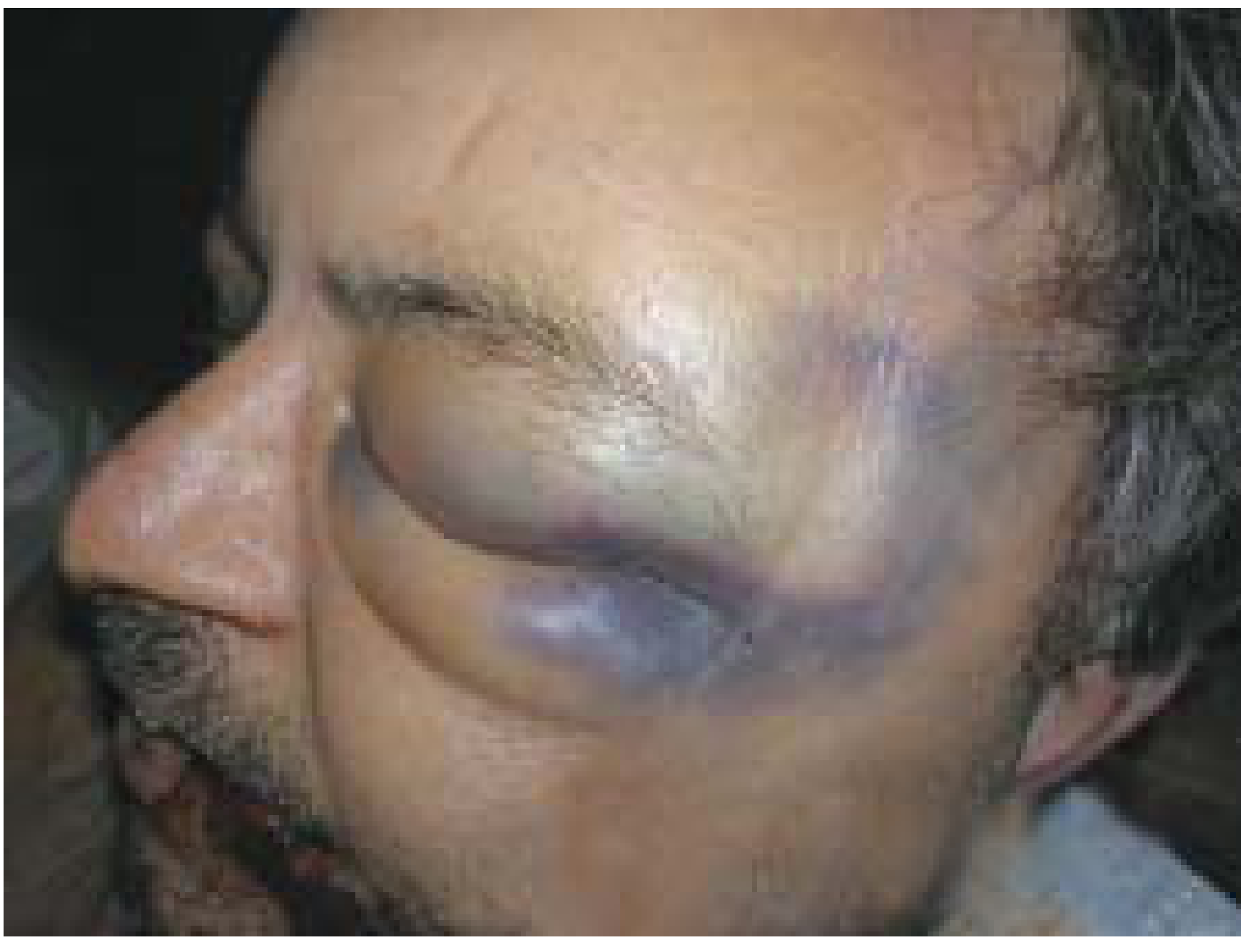
Figure: Black eye with extensive periorbital bruising and swelling following head injury - P.C. Dikshit, Textbook of Forensic Medicine
Mechanism
The periorbital tissues are very loose and highly vascular. Blunt trauma to the eye, forehead, or surrounding facial bones causes bleeding into this loose connective tissue space. Blood tracks easily within these tissues, producing the characteristic dark discolouration. The injury may arise from:
- Direct impact to the eye or orbit (fist, ball, projectile, fall)
- Indirect/remote injury - blood tracking from a skull base fracture into the orbital space (see below)
- Surgical/iatrogenic - e.g., post-rhinoplasty or post-sinus surgery periorbital ecchymosis is listed as a minor complication of endoscopic sinus procedures
Types and Their Significance
1. Simple Traumatic Black Eye (most common)
The most common blunt injury to the eyelid or forehead. Generally innocuous when isolated, but always requires exclusion of serious underlying injury - Kanski's Clinical Ophthalmology.
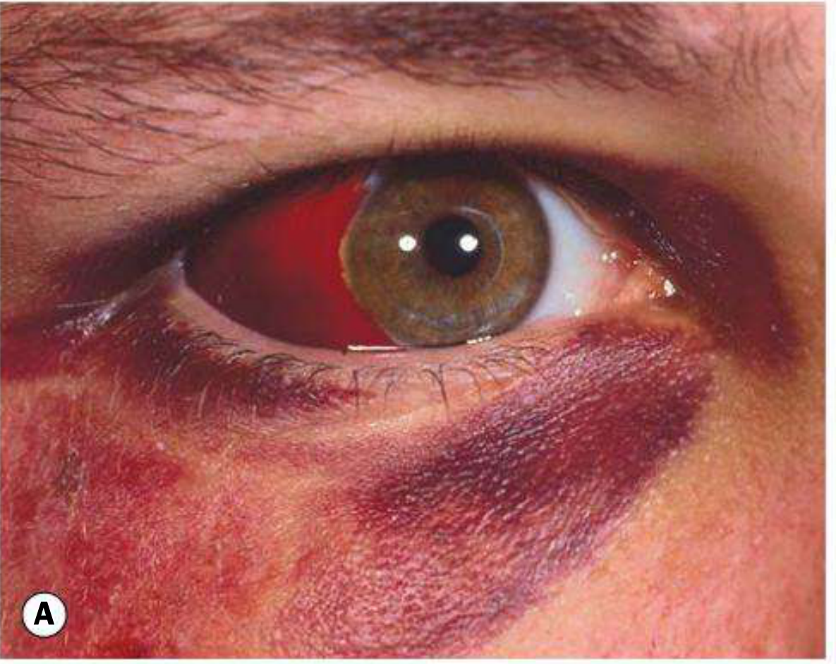
Figure: Periocular haematoma with associated subconjunctival haemorrhage - Kanski's Clinical Ophthalmology
2. "Raccoon Eyes" / "Panda Eyes" (Bilateral Periorbital Ecchymosis)
A critically important sign. Bilateral ring haematomas around both orbits are a classic indicator of anterior skull base (anterior cranial fossa) fracture. Blood from the fractured orbital plate of the ethmoid (cribriform area) tracks anteriorly into the periorbital space. May be accompanied by:
- CSF rhinorrhoea (blood-tinged nasal discharge)
- Anosmia (damage to olfactory nerve)
As noted in the Washington Manual and ROSEN's Emergency Medicine: "Periorbital ecchymosis ('raccoon eyes')... [is] indicative of a basilar skull fracture."
3. Spectacle Haemorrhage
In forensic medicine, unilateral or bilateral "spectacle haemorrhage" refers specifically to the black eye pattern following fracture of the anterior cranial fossa - the blood "spectacles" follow the contour of the orbital rim.
What Must Be Excluded (Red Flags)
According to Kanski's and the Textbook of Family Medicine, a black eye is generally innocuous BUT the following must be actively excluded:
| Serious Condition | Key Feature |
|---|---|
| Globe/orbital injury | Examine globe BEFORE lids swell shut |
| Orbital roof fracture | Subconjunctival haemorrhage with no visible posterior limit |
| Basilar skull fracture | Bilateral "panda/raccoon eyes," Battle's sign, CSF leak, hemotympanum |
| Ruptured globe | Severe pain, bleeding, constant blurred vision |
| Traumatic hyphema | Blood in anterior chamber - ocular emergency |
| Retinal detachment | Flashing lights, floaters, visual field loss |
| Traumatic mydriasis | Irregular, dilated pupil after trauma - suggests severe blunt injury |
Clinical tip from Kanski's: "It is critical not to apply any force on the globe itself until its integrity has been confirmed." If globe integrity is uncertain, urgent CT, MRI, or bedside ultrasonography (with care not to press on the globe) is indicated.
Assessment
- History: Mechanism, velocity, immediate vision changes (flashing lights = retinal irritation; persistent blur = serious injury), time elapsed
- Visual acuity - check before swelling worsens
- Globe inspection - look for lacerations, deformity
- Pupil - size, shape, reactivity
- Subconjunctival haemorrhage - if present without posterior limit = orbital roof fracture until proven otherwise
- Lid exam - check for lacerations, even small ones require exploration
- Imaging: CT orbit/head if any suspicion of orbital fracture, skull base fracture, or globe injury
Treatment
For an isolated, uncomplicated black eye:
- Cold compresses in the first 24-48 hours to reduce swelling
- Analgesics as needed
- No specific ophthalmologic treatment required
- Monitor for development of any red-flag symptoms
Urgent/specialist referral if:
- Severe pain, vision changes, diplopia
- Suspected globe rupture, hyphema, or retinal pathology
- Bilateral raccoon eyes (neurosurgical emergency - skull base fracture)
- CT scan findings of orbital fracture or intracranial injury
Prognosis
An uncomplicated black eye typically resolves over 1-2 weeks as blood is reabsorbed. The discolouration progresses through purple/blue → green → yellow as haemoglobin breaks down (biliverdin → bilirubin). Serious associated injuries (hyphema, retinal detachment, skull fracture) determine overall prognosis.
Sources:
- Kanski's Clinical Ophthalmology: A Systematic Approach, 10th ed. - Periocular haematoma
- Textbook of Family Medicine, 9e - Blunt Eye Injuries
- P.C. Dikshit, Textbook of Forensic Medicine and Toxicology - Black eye in head injury
- Parikh's Textbook of Medical Jurisprudence, Forensic Medicine and Toxicology - Skull base fractures
- The Washington Manual of Medical Therapeutics - Physical examination
- ROSEN's Emergency Medicine: Concepts and Clinical Practice
This is a shared conversation. Sign in to Orris to start your own chat.